Causes of renal failure in patients with decompensated
cirrhosis and its impact in hospital mortality
Grazielle Cerqueira de Carvalho,* Catarina de Andrade Regis,* Jamile Rosário Kalil,* Liv Aparício Cerqueira,* Daniel Silva Barbosa,* Marina Pamponet Motta,* Marília da Silva Nery,*
Maria Alice Pires Soares,* Claudio Celestino Zollinger,* Paulo Lisboa Bittencourt*
* Unit of Gastroenterology and Hepatology of the Portuguese Hospital of Salvador, Bahia, Brazil.
ABSTRACT
Background. Renal failure (RF) is reported to occur in 11-49% of the patients with decompensated end-sta-ge liver disease (ESLD) and has been associated with increased mortality, particularly in the occurrence of hepatorenal syndrome (HRS) type 1. Aims. To evaluate the frequency and outcome of RF in patients ad-mitted to the hospital due to decompensated ESLD and to assess the impact of the underlying cause of RF on survival. Material and methods. Four hundred and six patients (65% males, mean age 62 ± 12 years) with decompensated ESLD were evaluated for the occurrence of RF (defined as serum creatinine ³ 1.5 mg/mL). The underlying cause of RF was reckoned in each subject and compared to outcome. Results. Renal failu-re was observed in 39% of the patients at admission and in 10% of the subjects during hospitalization. Mor-tality was significantly higher in subjects with RF (26 vs. 1%, p < 0.000001). Hypovolemia, bacterial infections, parenchymal kidney diseases and HRS were identified as causes of RF in, respectively, 40, 32, 15 and 12% of the cases. Mortality was significantly higher in those subjects with HRS type 1 and bacterial in-fections, when compared to other causes of RF. Conclusions. Renal failure occurs in nearly half of the pa-tients with decompensated ESLD. It is most commonly caused by hypovolemia and bacterial infections. Occurrence of RF has an adverse impact in patient survival, particularly in those subjects with bacterial infections and HRS type 1, prone to develop progressive renal dysfunction despite intensive medical care.
Key words. Bacterial infections. End-stage liver disease. Hepatorenal syndrome. Liver cirrhosis. Renal failure.
Correspondence and reprint request: Paulo Lisboa Bittencourt Rua Professor Clementino Fraga 220, apto 1901
Ondina, Salvador-BA, Brazil Ph.: 00 55-71-2033457 Fax: 0055-71 2033456 E-mail: [email protected]
Manuscript received: June 27, 2011. Manuscript accepted: August 20, 2011.
INTRODUCTION
Renal failure (RF) is reported to occur in 11% of cirrhotic patients admitted to the hospital due to
up-per gastrointestinal bleeding (UGB),1 in 27-34% of
the patients with infections2-4 and in 40-49% of
criti-cally-ill subjects with cirrhosis admitted to the
in-tensive care unit (ICU).5,6 In those subjects, RF was
associated either with increased intra-hospital mor-tality, as well as shortened survival after hospital
discharge.5,6 Likewise, the occurrence of RF in
out-patients with end-stage liver disease (ESLD) has
been also associated with decreased survival7,8 and
creatinine levels has been incorporated in the Mo-del for End-Stage Liver Disease (MELD), designed to predict severity of ESLD and mortality in the
li-ver transplantation waiting list.9
The pathogenesis of RF in cirrhosis is
multifacto-rial.8,10 In this regard, distinct causes, not mutually
exclusive, were associated with renal impairment in patients with ESLD, including bacterial infections, hepatorenal syndrome (HRS) types 1 and 2, hypovo-lemia, parenchymal kidney diseases, and
nephrotoxi-city due to drugs or contrast agents.8,10 Hepatorenal
syndrome type 1 is the result of intense renal vaso-constriction due to worsening of the circulatory dys-function due to ESLD and is associated with a poor
prognosis,10,11 particularly in non-responders to
standard therapy with albumin and splancnic
vaso-constrictors.10,11 Bacterial infections, either
sponta-neous bacterial peritonitis,2,3,12 as well as other
infections4,12 can also alter the splancnic and
syste-mic circulation of cirrhotics and lead to RF that may regress after control of the infection episode or
Recently, the prognosis of RF in subjects with ESLD was shown to vary depending on the cause of
renal impairment and severity of liver disease.13 In
this respect, RF due to parenchymal kidney diseases and hypovolemia were reported to have a much bet-ter prognosis, when compared to RF attributable to
bacterial infections or HRS type 1.13
The purpose of the present study was to investi-gate the prevalence of RF in patients with cirrhosis admitted to the hospital due to decompensated ESLD, as well as to reassess the impact of the cause of RF in patient’s survival.
MATERIAL AND METHODS
Patients with decompensated ESLD consecutively admitted to the Unit of Gastroenterology and Hepa-tology of the Portuguese Hospital of Salvador, Bahia, Brazil from January 2005 to December 2007 were retrospectively evaluated, employing a prospec-tively collected database. This unit is focused on the treatment of liver diseases and on the postoperative care of liver transplant patients and is a referral center for patients with ESLD. Decompensation of ESLD was defined by the occurrence of variceal bleeding, hepatic encephalopathy, infections and ten-se ascitis as well as any other acute clinical event requiring hospitalization. The diagnosis of ESLD was based on clinical, biochemical and echographic findings, as well as on liver histology, whenever a liver biopsy specimen was available. The etiology of ESLD and the reason for hospitalization was esta-blished in all patients. In the case of more than one cause for admission, the main cause was reckoned based on the following hierarchy: upper digestive bleeding, infection, hepatic encephalopathy, tense
ascitis and others.14 Clinical and biochemical
para-meters associated with ESLD were sought in each patient. Severity of ESLD was assessed by the
Child-Pugh score (CPS) and the MELD score.9,15
Renal failure was arbitrarily defined as the pre-sence of creatinine levels equal or higher than 1.5 mg/mL either at admission or during
hospitaliza-tion.16 Renal failure was classified in distinct
groups including: bacterial infections, hypovole-mia, parenchymal or parenchymal kidney diseases,
HRS and contrast or drug-induced RF.8,10,13
Hypo-volemia was assumed as a cause for renal impair-ment in the presence of gastrointestinal hemorrhage or dehydration ascribed to gastrointes-tinal fluid losses or diuretic usage as well as in the occurrence of RF remission with the administration of intravenous saline. Prior diagnosis of
parenchy-mal kidney disease, past history of use of drugs or contrast agents was elicited in each patient as part of a protocol. Bacterial infections were sought in every hospitalized patient by urine analysis and culture and blood cultures. Ascitic fluid analysis and culture were performed in every patient with ascitis, whereas chest X rays were obtained whenever there was a clinical suspicion of respiratory tract infection. Renal failure was attributed to infection whenever the infection episode was suspected or documented within 48 h of the elevation of creatinine levels. Those subjects with presumed infections were initially treated with empiric antibiotics based on the most probable site of infection. Tailoring of antimicrobial therapy was subsequently based upon laboratory and radiology data as well as culture and sensitivity results.
The diagnosis of HRS types 1 and 2 were defined according to the non-revised criteria of the
Interna-tional Ascitis Group.16 Briefly, HRS-1 was suspected
in all subjects based on the presence of creatinine levels greater than 1.4 mg/mL either at admission or during hospitalization doubling to levels higher than 2.5 mg/mL within two weeks in the absence of shock, hypovolemia, bacterial infections or known exposure to nephrotoxic agents or a recognizable etiology for chronic renal failure. Failure to attain creatinine levels under 1.4 mg/mL 24 h after volume expansion with at least 1,500 mL of cristalloids or 72 h after institution of antibiotics with apparent control of infection were indicative of the presence of HRS, that was confirmed after exclusion of possible causes of intrinsic renal diseases by 24 h urine pro-tein excretion levels lower than 500 mg/day, as well as by the absence of renal parenchyma abnormali-ties at the ultrasound. Therapy with terlipressin and high-dose albumin was considered for all pa-tients with HRS-1. Outcomes for those papa-tients
were previously described elsewhere.11
ab-sence of reversal, three distinct intervals of creatini-ne levels were created for comparisons:
• Under 2 mg/mL. • Between 2-3 mg/dL. • Above 3 mg/mL.
Stabilization was considered whenever the creati-nine level at discharge remain below or in the same interval of the creatinine level at the diagnosis of RF in the absence of reversal, whereas progression was considered whenever the creatinine levels at dis-charge remains above the interval of the creatinine at diagnosis of renal impairment.
The study was approved by the Ethics Commit-tee in Research of the Portuguese Hospital of Sal-vador, Bahia.
Statistical analysis
The differences between groups of patients were compared using either the Mann-Whitney test or the Fisher exact probability test, when appropriate. A p value < 0.05 was considered significant. Clinical data are presented in text and tables as mean and standard deviation.
RESULTS
Four hundred and six patients (65% males, mean age 62 ± 12 years) with the diagnosis of de-compensated ESLD were evaluated. Clinical and laboratory parameters of those subjects are descri-bed in table 1. Most of the subjects had ESLD due to either alcoholic liver disease or hepatitis C with a mean CPS and MELD score of 10 ± 2 and 18 ± 7, respectively.
Based on the aforementioned criteria, 159 (39%) patients had RF at admission and 39 (10%) developed RF during the course of hospitalization. The main causes of RF are depicted on table 2. Bacterial
infec-Table 1. Clinical and laboratory features of patients with de-compensated end-stage liver disease.
Clinical and Laboratory Features n = 406
Male sex 65%
Mean age 63 ± 12 years Etiology of cirrhosis
Alcoholic liver disease 38% Hepatitis C 25% Cryptogenic or unknown 23%
Hepatitis B 6%
Other etiologies 8% Causes of decompensation
Tense ascitis 31%
Infections 23%
Hepatic encephalopathy 21% Variceal bleeding 20%
Other 5%
Mean Child-Pugh score 10 ± 2 Mean MELD score 18 ± 7
Table 2. Types of RF observed in cirrhotics admitted to the ICU (n = 198).
Bacterial infections 80 (40) Hypovolemia 64 (32) Parenchymal kidney disease 30 (15) Hepatorenal syndrome type 2 18 (9) Hepatorenal syndrome type 1 6 (3)
Numbers in parentheses are percentages.
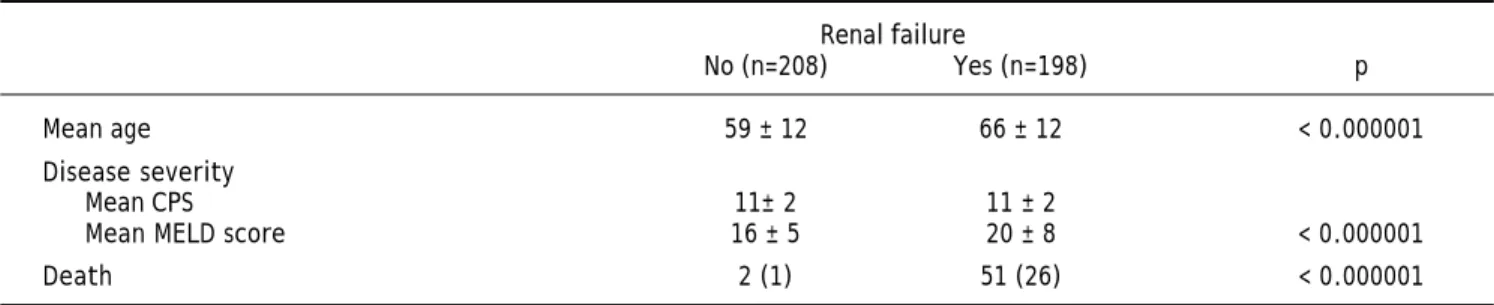
Table 3. Clinical and laboratory features of cirrhotic patients according to the presence of RF. Renal failure
No (n=208) Yes (n=198) p
Mean age 59 ± 12 66 ± 12 < 0.000001
Disease severity
Mean CPS 11± 2 11 ± 2
Mean MELD score 16 ± 5 20 ± 8 < 0.000001
Death 2 (1) 51 (26) < 0.000001
Numbers in parentheses are percentages.
tions and hypovolemia were responsible for most of the cases, whereas HRS was seen in only 12% of the patients with RF.
Patients with RF either at admission or during the course of hospitalization were older (66 ± 12
years vs. 59 ± 12 years, p < 0.000001) and, as
sub-jects with RF when compared to patients without RF (26 vs. 1%, p < 0.000001), particularly in res-pect to RF due to HRS type 1 and bacterial infections (Table 4). Overall, patients with RF, with exception of those with parenchymal kidney disease, had hig-her mortality, when compared to their counterparts without RF. In regard to the cause of RF, mortality in the group of patients with HRS type 1 was signi-ficantly higher, when compared to other groups with the exception of HRS type 2 (Table 4). On the other hand, subjects with RF due to hypovolemia and parenchymal kidney diseases had lower mortali-ty, when compared to their counterparts with HRS type 1 and with bacterial infections. In addition, subjects with parenchymal kidney diseases have lo-wer mortality even when compared to their counter-parts with HRS type 2 (Table 4).
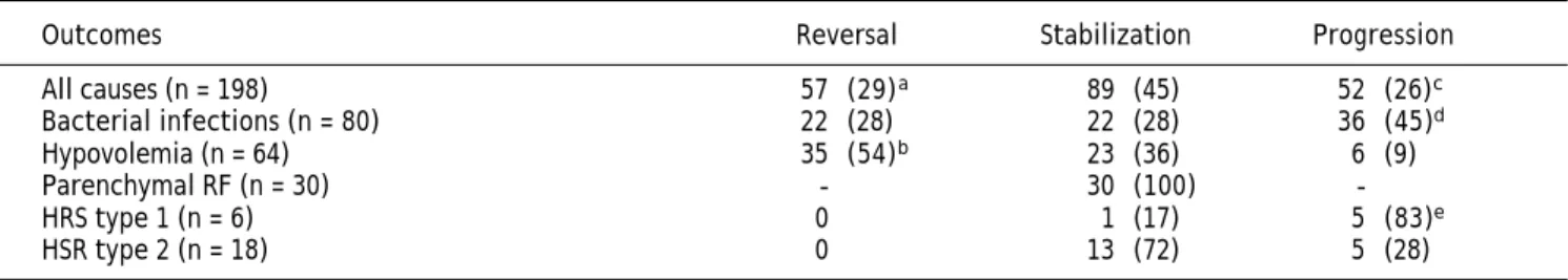
Reversal of RF, as previously defined, was seen in 57 (29%) of the affected subjects, whereas stabiliza-tion and progression was observed in, respectively, 89 (45%) and 52 (26%) of the cases. The outcome of RF was shown to vary markedly according to the cause of renal impairment. When compared to all other causes of RF, reversal was seen more often in subjects with hypovolemia (54 vs. 29%, p = 0.0003),
whereas progression was more frequently associated with HRS type 1 (83 vs. 26%, p = 0.007) and bacte-rial infections (45 vs. 26%, p = 0.004) (Table 5).
DISCUSSION
In the present investigation, RF was observed in 39% of the patients admitted to the hospital due to decompensated ESLD and in 10% of those subjects during the course of hospitalization. It is impor-tant to highlight that our study was retrospective, but our data is quite similar to other reports which described RF in 11-34% of patients hospitalized due to ESLD based on abnormalities in serum
creatini-ne levels.1-4,12 Higher frequencies of RF were
repor-ted by other investigators when cirrhotic subjects admitted to the ICU were evaluated using the RI-FLE system, which has a better performance for es-tablishing RF when compared to serum creatinine levels, because it takes into account changes in
creatinine levels, as well as in urinary output.17
In this regard, RF was disclosed in 40-49% of those critically ill subjects with ESLD using this scoring system.5,6
MELD score and age were recognized as risk fac-tors for RF in the present investigation. Higher MELD scores were invariably associated with the
occurrence of RF in several studies,12,18,19 but older
age was only associated with RF in one prospective study that assessed the frequency of RF in
outpa-tients with ESLD.20
The causes of RF encountered in the current stu-dy were:
• Bacterial infections (40%). • Hypovolemia (32%).
• Parenchymal kidney disease (15%). • HRS type 2 (9%) and type 1 (3%).
Other studies have reported variable frequencies of the aforementioned etiologies for RF in
hospitali-Table 5. Outcomes of RF observed at admission or during hospitalization in cirrhotic patients according to the cause of renal im-pairment.
Outcomes Reversal Stabilization Progression All causes (n = 198) 57 (29)a 89 (45) 52 (26)c
Bacterial infections (n = 80) 22 (28) 22 (28) 36 (45)d
Hypovolemia (n = 64) 35 (54)b 23 (36) 6 (9)
Parenchymal RF (n = 30) - 30 (100)
-HRS type 1 (n = 6) 0 1 (17) 5 (83)e
HSR type 2 (n = 18) 0 13 (72) 5 (28)
Numbers in parentheses are percentages. abp = 0.0003. cdp = 0.004. cep = 0.007.
Table 4. Mortality of cirrhotic patients in the ICU according to the occurrence and underlying cause of RF.
n Mortality No renal failure 208 2 (1)a
All causes 198 52 (26)b
HRS type 1 6 5 (83)c
Bacterial infections 80 30 (38)d
HRS type 2 18 6 (33)e
Hypovolemia 64 10 (16)f
Parenchymal kidney diseases 30 1 (4) g
zed subjects with cirrhosis,13,18 as well as in
outpa-tients.20 Martin-Llahi, et al.13 have investigated the
causes of RF in hospitalized patients with ESLD and have found quite similar results with infections, hypovolemia, HRS and parenchymal kidney disease accounting, respectively, for 46, 32, 13 and 9% of the cases. Lower frequencies of RF were reported by
Montoliu, et al.20 analyzing a different type of
co-hort of cirrhotic outpatients, who were prospectively followed for 41 ± 3 months, after the onset of asci-tis. The authors have found RF due to hypovolemia, infections and HRS in 27, 14 and 7.6% of those sub-jects, respectively. When compared to other studies,
only Schepke et al.18 have found HRS type 1 as the
main cause of RF in hospitalized patients with ESLD. This heterogeneity is due mainly to selection bias, but also due to the different criteria employed to ascertain the cause of RF. In the present study we have used the non-revised criteria of the Inter-national Ascitis Club for the diagnosis of HRS type
116 and it is clear that a great proportion of subjects
with RF due to infections would be reclassified as patients with HRS type 1, based on the current
employed criteria.21
Mortality was significantly associated with the oc-currence of RF. In this respect, 26% of those sub-jects with RF vs. 1% of their counterparts without RF died in the current investigation. Several repor-ts have disclosed reduced long-term survival of
patients with ESLD after the onset of RF.7,8,20
In hospitalized subjects with decompensated ESLD, mortality was associated with the occurrence of RF,1,7,12,18 as well as with its severity, when
asses-sed by the RIFLE score.5,6,22 In patients admitted to
the ICU, the prognosis is even more dismal, with
re-ported mortality rates of 65-81%,5,6,23-25 rising to
more than 90% in subjects requiring either dialysis
and/or inotropic support.26 It has to be pointed out,
however, that RF in those subjects is part of the multiple organ dysfunction syndrome due to either septic shock and/or liver failure which carry an
omi-nous prognosis in patients with ESLD.27,28
In the present investigation, even though retros-pective, mortality ascribed to RF was shown to vary according to its cause. In this respect, hypovolemia and renal parenchymal diseases were associated with lower mortality when compared to bacterial in-fections and HRS. Moreover, RF due to hypovole-mia and bacterial infections were shown to regress in approximately one third of the patients. These re-sults were also observed in other studies, where hig-her mortality was restricted to those subjects with
HRS-1 and bacterial infections.13,20 Hepatorenal
syn-drome type 1 was shown to carry a dismal prognosis and has been associated with a high risk of
mortali-ty that was independent of the MELD score.18,19
Bac-terial infections, particularly when severe or uncontrolled, were also associated with progression either to HRS and/or acute tubular necrosis due to
septic shock and high mortality rates.3,4,12
In summary, renal failure occurs in nearly half of the patients admitted to the hospital with decom-pensated ESLD, particularly in older subjects and in patients with more advanced liver disease assessed by the MELD score. Hepatorenal syndrome type 1 is infrequently seen and most of the cases of RF are due to bacterial infections and hypovolemia. The oc-currence of RF had an adverse impact in patient survival, particularly in those subjects with bacte-rial infections and HRS type 1, prone to develop pro-gressive renal dysfunction despite intensive medical care. These findings, altogether, have to be taken into consideration when assessing prognosis in sub-jects with cirrhosis and RF.
ABBREVIATIONS
• RF: renal failure.F
• ESLD: end-stage liver disease.S DL
• HRS: hepatorenal syndrome.R
• UGH: upper gastrointestinal bleeding.
• ICU: intensive care unit.U
• MELD: model for end-stage liver disease.E D
• CPS: Child-Pugh score.:
REFERENCES
1. Cárdenas A, Ginès P, Uriz J, Bessa X, Salmerón JM, Mas A, Orte-ga R, et al. Renal failure after upper Orte-gastrointestinal bleeding in cirrhosis: incidence, clinical course, predictive factors, and short-term prognosis. Hepatology 2001; 34: 671-6.
2. Sort P, Navasa M, Arroyo V, Aldeguer X, Planas R, Ruiz-del-Arbol L, Castells L, et al. Effect of intravenous albumin on renal impairment and mortality in patients with cirrhosis and spontaneous bacterial peritonitis. N Engl J Med 1999; 341: 403-9.
3. Follo A, Llovet JM, Navasa M, Planas R, Forns X, Francito-rra A, Rimola A, et al. Renal impairment after spontaneous bacterial peritonitis in cirrhosis: incidence, clinical cour-se, predictive factors and prognosis. Hepatology 1994; 20: 1495-501.
4. Terra C, Guevara M, Torre A, Gilabert R, Fernández J, Martín-Llahí M, Baccaro ME, et al. Renal failure in patients with cirrhosis and sepsis unrelated to spontaneous bacte-rial peritonitis: value of MELD score. Gastroenterology
2005; 129: 1944-53.
6. Cholongitas E, Senzolo M, Patch D, Shaw S, O’Beirne J, Bu-rroughs AK. Cirrhotics admitted to intensive care unit: the impact of acute renal failure on mortality. Eur J Gas-troenterol Hepatol 2009; 21: 744-50.
7. D’Amico G, Garcia-Tsao G, Pagliaro L. Natural history and prognostic indicators of survival in cirrhosis: a systema-tic review of 118 studies. J Hepatol 2006; 44: 217-31. 8. Garcia-Tsao G, Parikh CR, Viola A. Acute kidney injury in
cirrhosis. Hepatology 2008; 46: 2064-77.
9. Kamath P, Kim WR; Advanced Liver Disease Group. The model for endstage liver disease. Hepatology 2007; 45: 797-805.
10. Solà E, Ginès P. Renal and circulatory dysfunction in cirr-hosis: current management and future perspectives. J He-patol 2010; 53(6): 1135-45.
11. Kalil JR, Cerqueira LA, Barbosa DS, Kalil JR, Cerqueira LA, Barbosa DS, Motta MP, et al. Poor outcomes with treat-ment of hepatorenal syndrome type 1 with splanchnic va-soconstrictors and albumin: report of seven cases and review of the literature. Arq Gastroenterol 2009; 46: 214-8.
12. Fasolato S, Angeli P, Dallagnese L, Maresio G, Zola E, Maz-za E, Salinas F, et al. Renal Failure and Bacterial Infections in Patients with Cirrhosis: Epidemiology and Clinical Fea-tures. Hepatology 2007; 45: 223-9.
13. Martín-Llahí M, Guevara M, Torre A, Fagundes C, Restuc-cia T, Gilabert R, et al. Prognostic importance of the cau-se of renal failure in patients with cirrhosis.
Gastroenterology 2011; 140: 488-96.
14. Fernandez J, Navasa M, Gomez J, Colmenero J, Vila J, Arroyo V, Rodés J. Bacterial infections in cirrhosis: epide-miological changes with invasive procedures and norfloxa-cin prophylaxis. Hepatology 2002; 35: 140-8.
15. Murray KF, Carithers RL Jr. AASLD practice guidelines: evaluation of the patient for liver transplantation. Hepa-tology 2005; 41: 1407-32.
16. Arroyo V, Ginès P, Gerbes AL, Dudley FJ, Gentilini P, Laffi G, Reynolds TB, et al. Definition and diagnostic criteria of refractory ascites and hepatorenal syndrome in cirrhosis. International Ascites Club. Hepatology 1996; 23: 164-76. 17. Uchino S, Kellum JA, Bellomo R, Doig GS, Morimatsu H,
Mor-gera S, Schetz M, et al. Acute Renal Failure in Critically Ill Patients: A Multinational, Multicenter Study. JAMA 2005; 294: 813-8.
18. Schepke M, Appenrodt B, Heller J, Zielinski J, Sauerbruch T. Prognostic factors for patients with cirrhosis and kid-ney dysfunction in the era of MELD: results of a prospec-tive study. Liver Int 2006; 26: 834-9.
19. Alessandria C, Ozdogan O, Guevara M, Restuccia T, Jiménez W, Arroyo V, Rodés J. MELD Score and Clinical Type Predict Prognosis in Hepatorenal Syndrome: Rele-vance to Liver Transplantation. Hepatology 2005; 41: 1282-9.
20. Montoliu S, Ballesté B, Planas R, Alvarez MA, Rivera M, Mi-quel M, Masnou H, et al. Incidence and prognosis of diffe-rent types of functional renal failure in cirrhotic patients with ascites. Clin Gastroenterol Hepatol 2010; 8: 616-22
21. Salerno F, Gerbes A, Ginès P, Wong F, Arroyo V. Diagnosis, prevention and treatment of hepatorenal syndrome in cirrhosis. Gut 2007; 56: 1310-8.
22. Jenq CC, Tsai MH, Tian YC, Lin CY, Yang C, Liu NJ, Lien JM, et al. RIFLE classification can predict short-term prognosis in critically ill cirrhotic patients. Intensive Care Med 2007; 33: 1921-30.
23. Fang JT, Tsai MH, Tian YC, Jenq CC, Lin CY, Chen YC, Lien JM, et al. Outcome predictors and new score of critically ill cirrhotic patients with acute renal failure. Nephrol Dial Transplant 2008; 23: 1961-9.
24. Du Cheyron D, Bouchet B, Parienti JJ, Ramakers M, Char-bonneau P. The attributable mortality of acute renal failu-re in critically ill patients with liver cirrhosis. Intensive Care Med 2005; 31: 1693-9.
25. Arabi Y, Ahmed QAA, Haddad S, Aljumah A, Al-Shimemeri A, et al. Outcome predictors of cirrhosis patients admitted to the intensive care unit. Eur J Gastroenterol Hepatol
2004; 16: 333-9.
26. Juneja D, Gopal PB, Kapoor D, Raya R, Sathyanarayanan M. Profiles and outcome of patients with liver cirrhosis requiring mechanical ventilation. J Intensive Care Med
2011; 24 [Epub ahead of print].
27. Austin MJ, Shawcross DL. Outcome of patients with cirrhosis admitted to intensive care. Curr Opin Crit Care
2008; 14: 202-7.