Minimally invasive treatment of Mirizzi’s syndrome:
is there a safe way? Report of a case series
Giuseppe Piccinni, Andrea Sciusco, Giuseppe Massimiliano De Luca, Angela Gurrado, Alessandro Pasculli, Mario Testini
Unit of Endocrine, Digestive and Emergency Surgery; Department of Biomedical Sciences and
Human Oncology, Section of General and Oncologic Surgery, University Medical School of Bari “Aldo Moro”. Bari, Italy.
ABSTRACT
Mirizzi’s syndrome (MS) is a rare complication of the inveterate biliary lithiasis. Diagnostic and therapeutic standardization is still missing, especially since laparoscopic cholecystectomy has become the gold stan-dard approach for symptomatic cholelithiasis. Our study is a retrospective analysis based on a case-series. It considered 370 cholecystectomies performed from 2006 to 2011. We selected 11 patients affected by MS (2.97%). We divided them according to Csendes’ classification. Endoscopic Retrograde Cholangio-Pancrea-tography (ERCP) was used for biliary drainage when the patient suffered jaundice and/or cholangitis and, preoperatively, to confirm the suspicion of MS obtained through Magnetic Resonance Cholangio-Pancrea-tography (MRCP). We found it useful to exploit nasobiliary drainage (NBD) for intra-operative check of the biliary tree. In all 5 patients of the type 1 group MS was discovered intraoperatively and treated with Lapa-roscopic Sub-total Cholecystectomy (LSC). One patient suffered from biliary leakage, solved with NBD posi-tioning. The type 2 group was made up of 2 women and 1 man. All of them were preoperatively submitted to ERCP and NBD positioning. Two underwent LSC and one was converted to laparotomy. The type 3 was represented by a 63-year-old woman suffering from recurrent cholangitis. She was submitted to MRCP, ERCP and then underwent LSC. The 2 patients affected by type 4 underwent open biliary reconstruction. In conclusion, every attempt should be made to identify MS prior to LCS since it will allow NBD insertion by ERCP. Once LCS is initiated, if MS is identified intra-operatively, we can provide the most practical sur-gical options.
Key words. Cholecystobiliary fistula. Laparoscopic cholecystectomy. Sub-total cholecystectomy.
Correspondence and reprint request: Giuseppe Piccinni, M.D.
Sezione di Chirurgia Generale ed Oncologica, Dipartimento di Scienze Biomediche ed Oncologia Umana.
Universitá di Bari, Policlinico, Piazza G. Cesare 11, 70124, Bari, Italy. Tel.: +39 348 3311213. Fax +39 080 5595452
E-mail: [email protected]
Manuscript received: March 09, 2013. Manuscript accepte: October 01, 2013.
INTRODUCTION
In 1948 the Argentinian surgeon Pablo Luis Mir-izzi described the case of a patient showing a partial obstruction of the common hepatic duct, due to the extrinsic compression of a gallstone lodged in the cystic duct or infundibulum, causing the asso-ciated inflammation.1 This condition was named after him, even though Kehr and Ruge had already described it in the early 1900’s.2,3
Mirizzi’s syndrome (MS) is a rare complication of the inveterate biliary lithiasis, with a prevalence among patients with colelithiasis between 0.5 and 1.4%.4,5 that can rise to 2.7% in some ethnic groups, such as Navajo Native Americans.6 The overall inci-dence of MS is low, reported in 0.7-2.53% of all pa-tients undergoing cholecystectomy.7,8 In spite of intermittent symptoms and signs,9 the clinical pres-entation of MS can be outlined in 4 possibilities.10-12
• Obstructive jaundice (76% of cases).
• History of recurrent acute cholecystitis and/or cholangitis (35.3% of cases).
• Acute abdomen, due to a biliary peritonitis. • Asymptomatic or paucisymptomatic.
unexpected conditions and a difficult operation, just like observed in MS type 1.
In 1982 McSherry provided a better description of the syndrome thanks to the use of Endoscopic Ret-rograde Cholangio-Pancreatography (ERCP) and distinguished between MS type 1, characterized by the extrinsic compression of the common bile duct (CBD) due to a trapped gallstone in the infundibu-lum or in the cystic duct and subsequent inflamma-tion, and MS type 2, characterized by the presence of a cholecystocholedochal fistula.14 In 1989 Csend-es further distinguished MS type 2 into 3 subtypCsend-es: type 2, with the cholecystocholedochal fistula in-volving 1/3 of the CBD diameter; type 3, with the fistula involving 2/3 of the CBD diameter; type 4, with the fistula involving the whole CBD diameter.15 In 2012 Beltràn introduced a new classification in-cluding a fifth case with complex cholecystobiliary fistulas and associated cholecystoenteric fistulas.16
Until the end of the eighties, iatrogenic lesions of the CBD were a well defined17 and rather rare enti-ty, with an incidence between 0.2 and 0.5%.18 In the early nineties the rapid development of laparoscopic cholecystectomy suddenly determined an abrupt in-crease of CBD iatrogenic lesion incidence.19 Accord-ing to Csendes, one of the most important factors involved in the increase of morbidity due to laparo-scopic cholecystectomy is the dissection of Calot’s triangle (hepatocystic triangle), in patients with ex-isting or previous acute cholecystitis.15 As guide-lines haven’t yet been laid down, a diagnostic and therapeutic algorithm should be carefully planned for MS, with the aim of reducing the probability of encountering an unexpected complicated intraopera-tive anatomic situation. Our study is a retrospecintraopera-tive analysis based on a case-series which aims at stand-ardizing the MS diagnostic and therapeutic ap-proach, while suggesting some technical notes to complete cholecystectomy with laparoscopy in known and unexpected cases.
MATERIAL AND METHODS
Our experience considered 370 cholecystectomies, 315 laparoscopic ones (85.14%) and 55 open ones (14.86%), performed from 2006 to 2011 at the Unit of Endocrine, Digestive and Emergency Surgery, De-partment of Biomedical Sciences and Human Oncol-ogy, Section of General and Oncologic Surgery, University Medical School of Bari “A. Moro”.
We retrospectively analyzed 11 patients affected by MS (2.97% of all cholecystectomies), 6 females and 5 males between 43 and 70 years old (average
age: 63). We divided them into 4 groups according to Csendes’ classification. We examined the diagnostic approach that had been advocated and conducted since patient admittance. All patients admitted for cholecystitis were initially studied with UltraSound (US). If they presented an associate obstructive jaundice or cholangitis, a Computed Tomography (CT) scan was performed and then they underwent ERCP, sphincterotomy with stenting of the CBD and/or nasobiliary drainage (NBD) placement. Mag-netic Resonance Cholangio-Pancreatography (MRCP) was performed in all patients with a histo-ry of recurrent acute cholecystitis without obstruc-tive jaundice, with increased levels of hepatic enzymes (AST/ALT) and in cases of cholangitis or pancreatitis. On the contrary patients admitted with cholecystitis, first episode, without signs of CBD ob-struction, were directly forwarded to laparoscopic cholecystectomy. When MS was discovered by MRCP, patients underwent ERCP, considered as a mere therapeutic procedure, in order to place a stent or an NBD. We found it useful to exploit this device intraoperatively for the identification of the CBD and to perform an intraoperative cholangiography. In our experience it allowed postoperative T-tube placement to be avoided. The only efficient diagnos-tic strategy for MS type 1 was laparoscopy.
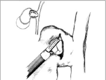
All patients affected by MS type 1 were intro-duced to laparoscopic cholecistectomy whereas pa-tients affected by MS type 2 and 3 were scheduled for laparoscopic subtotal cholecystectomy (LSC) broadly speaking 2 to 7 days after the endoscopic drainage. In the end MS type 4 was submitted to an open approach. The approach to cholecystectomy was the same even in the case of MS recognized in-traoperatively (MS type 1) (Figure 1): indeed when
Calot’s triangle (hepatocystic triangle) dissection was too difficult and it was impossible to recognize and dissect the cystic duct and artery contained in the inflammatory tissue by using the “critical view of safety” as described by Strasberg,20 we decided to perform the LSC as described below. Conversion to laparotomy was reserved to those cases with dense adhesions to colon and/or duodenum.
RESULTS
According to Csendes’ classification we identified: 5 cases of type 1, 3 cases of type 2, 1 case of type 3 and 2 cases of type 4. In all 5 patients of the type 1 group MS were discovered and confirmed intraopera-tively. This group was treated with LSC: after
iden-tification and opening of the gallbladder fundus we remove gallstones in a bag. Then, by using an “inside approach” as described by Hubert19 (Fig-ures 2 and 3), we explored the Hartmann’s pouch with the optic and detached the gallbladder starting from the fundus and until the rear of the infundibu-lum, where later the stump was closed by applying a linear endoscopic stapler at the Hartmann’s pouch (Figures 4 and 5). Drainage was left in the abdomen in all cases. In the cases of MS type 1 discovered in-traoperatively we never needed a T-Tube, in the oth-er cases the preopoth-erative ERCP and NBD placement avoided the intra-operative T-Tube placement.
One patient suffered from biliary leakage (20%), solved by sphincterotomy and NBD positioning.
Figure 2. Opening of the infundibulum and extraction of a gallstone.
Figure 3. Intraoperative view.
Figure 4. Subtotal cholecystectomy applying an Endo-GIA at Hartmann’s pouch.
The type 2 group was made up of 2 women aged 54 and 68 presenting obstructive jaundice, and 1 man aged 70 with transient jaundice. All of them were preoperatively submitted to ERCP with sphinc-terotomy, NBD placement and diagnosis of MS type 2. Two of them underwent LSC without complica-tions. In one case (33%) we converted the procedure to laparotomy because a dense inflammatory tissue around the gallbladder involved the duodenum. Then we performed a subtotal cholecystectomy, with closure of the biliary defect using the gallbladder in-fundibulum wall.
The type 3 group was represented by a 63 year old woman with cholelithiasis suffering from recur-rent cholangitis. She was submitted to MRCP and MS type 3 was diagnosed. She underwent ERCP with sphincterotomy and NBD positioned in order to grant an intraoperative guide to identify the CBD. Two days later she was submitted to LSC with in-traoperative cholangiography through the NBD to check the correct position of the stapler and the complete closure of the stump. The postoperative course was complication free.
The type 4 group was made up of a 70 year old man, and a 60 year old woman. The man, with re-current acute cholecystitis and a diagnosis of MS type 4 obtained through MRCP, was submitted to open cholecystectomy and reconstruction of the CBD with resection of the fistula, mobilization of the duodeno-pancreatic bloc and end-to-end anasto-mosis over a T-tube drainage. The postoperative course was uneventful and the T-tube was removed after 3 months. The woman, with a preoperative di-agnosis of cholelitiasis and jaundice (bilirubin level: 4 mg/dL) underwent ERCP and obtained a diagnosis of MS type 4: a NBD was positioned. She was then submitted to open cholecystectomy and
hepatic-jeju-nal anastomosis without biliary drainage. The post-operative course was uneventful.
Summarizing, as can be seen from the table 1, among the 9 LSCs performed, we reported only one case of biliary leakage (11.11%) and an overall conversion rate of 11.11%.
DISCUSSION
There is no consensus on the management of MS, in terms of both diagnostic and surgical choices. Neither in the classification nor in the definition of the various anatomo-pathologic pictures can we see a uniformity of view. Some authors still prefer to use McSherry’s classification, while a great deal re-mains to be done from a physiopathological view-point in order to define the first grade of MS in all classifications. In clinical practice the anatomic modification of the biliary tract due to Mirizzi’s syn-drome predisposes to iatrogenic lesions of the CBD during laparoscopic cholecystectomy and, in addi-tion, the chronic inflammation and subsequent pe-rivisceral fibrosis represents an obstacle to the safe dissection of Calot’s triangle (hepatocystic triangle). Because of its nonspecific characteristics at imag-ing, there is no optimal diagnostic technique for MS. Becker, et al., as well as for Erben, et al., showing that preoperative recognition for MS using imaging is problematic, inconsistent and limited: indeed MS is documented preoperatively in only 50% of cas-es.21,22 However, in common practice, the assess-ment of a diagnostic flowchart must consider US as the first step in the assessment of biliary lithiasis even though its sensitivity for MS is estimated to be between 8.3% and 27%.23-25
A CT scan does not show any additional informa-tion compared to US, but it is, in our opinion,
essen-Table 1. Results.
Csendes’ classification
Mirizzi I Mirizzi II Mirizzi III Mirizzi IV
No. of pts 5 3 1 2
M/F 3/2 1/ 2 0/1 1/1
MRCP 0 0 1 1
ERCP 1(post-op) 2 1 1
NB Drainage 1 2 1 1
Laparoscopic 5 3 1 0
Laparotomic 0 0 0 2
Conversion Rate 0 33%(1) 0 0
Biliary Leakage 1 0 0 0
Bleeding 0 0 0 0
Other complications 0 0 0 0
tial in order to differentiate MS from a malignant bilio-pancreatic obstruction.26-28
ERCP is mandatory in all cases of MS arising with obstructive jaundice, whereas MRCP, with more or less the same diagnostic accuracy as ERCP, is recommended in all cases of clinical suspicion of MS without obstructive jaundice. However, MRCP is weak in localizing a cholecystocholedochal fistula.13 Moreover it is our opinion that all non-invasive methods, US and CT and MRCP, must be systemati-cally used with clinical good sense to improve pre-op-erative diagnostic data. At the moment there is no consensus about the use of one versus another diag-nostic tool.
According to a recent systematic review by Antoni-ou, et al., the mean preoperative diagnosis rate of MS is 66.1%, with most authors reporting ERCP as the favorite diagnostic procedure; this tool has a satisfac-tory mean sensitivity rate of 76.2%.29
Many different ways of managing MS have been described in literature. Both on the diagnostic and on the therapeutic side there is a lack of defined guidelines. The anatomo-pathologic aspect of the bil-iary tract in MS can question the appropriateness of a laparoscopic cholecystectomy, because of the in-creased risk of even not immediately recognizable ia-trogenic lesions of the CBD. Therefore, it is often mandatory to recur to traditional open surgery tech-niques. Approaching the literature and looking for the best technique in “open” surgery for MS, a sub-total cholecystectomy with an anterograde dissec-tion, opening of the fundus, removal of the gallstone, reconstruction of the CBD using the re-maining portion of the gallbladder and placing T-tube drainage in the CBD, was one of the first options to be described.30,31 Sometimes the only ap-propriate “open” procedure is the hepatic-jejunal anastomosis with Roux en Y loop after the removal of the gallbladder and part of the hepato-choledo-chus. The surgical procedures that take into consid-eration Calot’s triangle (hepatocystic triangle) dissection, like total laparoscopic cholecystectomy, are penalized by a higher conversion rate than open surgery and a higher risk of CBD lesions or jejunal lacerations.9,32 On the contrary, subtotal laparo-scopic cholecystectomy, using an Endo-GIA, is more frequently completed without conversion26,33-34 and its complications are represented mainly by biliary leakage.31
From 1997 to 2003 Waisberg, et al., analyzed 8 patients with MS and successfully applied a subtotal cholecystectomy with the placement of a T-tube drainage to a patient with MS type 2, while in two
patients with MS type 1 and 3 they performed a cholecystectomy and side-to-side choledochoduode-nostomy. The same procedure, despite leaving the gallbladder in situ, was performed in a patient with MS type 4. In the remaining 4 cases, all MS type 1, they applied a total cholecystectomy.35
From 1995 to 1999 Schafer, et al. analyzed 13,000 laparoscopic cholecystectomies and found 39 cases of MS, classified according to McSherry into 34 MS type 1 and 5 MS type 2 ones. MS was preoperatively supposed only in 18 cases out of 39. ERCP played a key role in the preoperative assessment, being per-formed in each patient showing obstructive jaun-dice, while MRCP was performed only in few cases. The conversion rate was 74% in all patients with MS type 2 and 24 patients with MS type 1. As re-gards the surgical procedure, the dissection of Calot’s triangle (hepatocystic triangle) was always performed. This procedure was followed by: a total cholecystectomy in 23 cases; a T-tube implantation in 13 cases; hepatic-jejunal anastomosis with Roux en Y loop in 3 cases. In 5 cases a subtotal cholecys-tectomy was performed, because of the impossibility of a complete removal. The intraoperative complica-tion rate was 7.7%: one CBD lesion, one jejunal lac-eration, one incident of local bleeding. The overall complication rate was 10.3%.36
From 1994 to 2005 Gomez, et al. considered 33 patients with MS. A laparoscopic cholecystectomy with classical dissection of the cystic duct was cho-sen to be the appropriate treatment for MS type 1, although their conversion rate was 56%. Regarding MS type 2 they performed the hepatic-jejunal anas-tomosis with Roux en Y loop. The Authors stressed the importance of MRCP in the preoperative assess-ment, conferring to ERCP a merely preoperative in-terventional role, when necessary. They also indicated how an intraoperative cholangiography could improve the chances of completing laparoscop-ic cholecystectomy without conversion.27
From 2003 to 2005 Sinha, et al. performed 889 laparoscopic cholecystectomies, with the aim of es-tablishing an appropriate alternative to conversion to open surgery in complicated cases, and in 28 cas-es they carried out a subtotal cholecystectomy, with-out the dissection of Calot’s triangle (hepatocystic triangle) and without the ligation of the cystic duct. The conversion rate was reduced to 0.3%, with the additional advantage of conservative management of bile leaks, thanks to postoperative ERCP with endo-scopic drainage.37
cholecystecto-mies, suggesting a minimally invasive approach: opening fundus, removing the gallstones, exploring the potential fistula, and subtotal cholecystectomy performed with Endo-GIA if necessary. T-tube drainage was not always used.34
From 1991 to 2001 Yeh, et al. had previously analyzed 11 patients with MS: they had already successfully utilized an Endo-GIA to close the cystic duct stump in 2 of them.28
In 1995, Strasberg described a method to iden-tify the hidden cystic duct during a laparoscopic cholecystectomy in order to reduce the risk of ia-trogenic lesions of the CBD: the “critical view of safety”,20 consisting of the dissection of the apex of Calot’s triangle (hepatocystic triangle) be-tween the infundibulum and the hepatic border, showing two and only two structures entering the gallbladder (cystic duct and artery). This ap-proach is, in our opinion, really useful to avoid CBD damage, especially in difficult situations, like MS, when the inflammatory process has al-tered the normal anatomy of the gallbladder pedi-cle. Another alternative to the classic Calot’s triangle dissection was described by Hubert et al. for severe cholecystitis: in their “inside ap-proach” they suggested the incision of the ven-tral/peritoneal surface of the gallbladder, from the fundus to Hartmann’s pouch, the evacuation of its content and its dissection from the liver bed from inside and outside, enabling an easier deter-mination of the precise limits of the gallbladder wall and a safer dissection.19
CONCLUSION
Our policy is that ERCP is mandatory in cases of obstructive jaundice, pancreatitis and MS di-agnosed through MRCP. With this procedure we are able to perform a sphincterotomy and to place an NBD, considered to be very useful intra-operatively to identify the CBD and to perform an intraoperative colangiography. MS type 4 should be treated directly with a laparotomy. According to our experience, the surgical approach for MS type 1, 2, and 3 on the other hand should be laparoscopic, performing a subtotal cholecystecto-my as described before. The dissection of Calot’s triangle (hepatocystic triangle) in patients with MS should be prescribed. Laparoscopic cholecys-tectomy is a widespread and successful minimally invasive approach to cholelithiasis, but it should not, therefore, be considered a simple technique to treat MS. Moreover, it is desirable to achieve
a widespread knowledge of the various clinical presentations of MS as well as managing a flow-chart , like the one we propose (Figure 6), capa-ble of permitting a totally endoscopic approach in almost all cases.
ACKNOWLEDGEMENTS
We would like to thank Prof Malcolm CLARK (B.A.) for the English version.
FUNDING
Not supported.
ETHICAL APPROVAL
No needed.
COMPETING INTEREST
No benefits in any form have been recived or will be received from a commercial party related directly or indirectly to the subject of this article.
REFERENCES
1. Mirizzi PL. Sindrome del conducto hepatico. J Int Chirur 1948; 8: 731-3.
Figure 6. Management flow-chart of Mirizzi’s syndrome. Without jaundice
Cholecystitis With jaundice
US/CT/MRCP
US/CT ERCP/NBD
SURGERY
MIRIZZI I MIRIZZI II/III
Laparoscopic subtotal
2. Kehr H. Die in meiner klinik geobte Technik der Gallenstein operationen miteinem Hinweis. Auf die Indikation und die Dau-ererfolge. Munchen. J.F.: Lehmann’s Verlag; 1905, p. 209-83.
3. Ruge E. Deitrage Zur Chinurgishen Anatonmie der grossen Gallenwege (Ductus hepaticus, choledochus und pancreati-cus). Arch Clin Chir 1908; 78: 47.
4. Bower TC, Nagorney DM. Mirizzi syndrome. HPB Surg 1988; 1: 67-76.
5. Mishra MC, Vashishtha S, Tandon R. Bilio-biliary fistula: pre-operative diagnosis and management implications. Surgery 1990; 108: 835-9.
6. Curet MJ, Rosendale DE, Congilosi S. Mirizzi syndrome in a Native American population. Am J Surg 1994; 168: 616-21. 7. Blumgart LH. Surgery of the liver and biliary tract.
Edin-burgh: Churchill Livingstone; 1988, p. 1026.
8. Hunt DR. Another paper on Mirizzi syndrome? Aust N Z J Surg 2004; 74: 826.
9. Johnson LW, Sehon JK, Lee WC, Zibari GB, McDonald JC. Mi-rizzi syndrome: experience from a multi-institutional re-view. Am Surg 2001; 67: 11-14.
10. Al-Akeely MH, Alam MK, Bismar HA, Khalid K, Al-Teimi I, Al-Do-ssary NF. Mirizzi syndrome: ten years experience from a teaching hospital in Riyadh. World J Surg 2005; 29: 1687-92.
11. Kwon AH, Inui H. Preoperative diagnosis and ef?cacy of la-paroscopic procedures in the treatment of Mirizzi syndro-me. J Am Coll Surg 2007; 204: 409-15.
12. Berry MG, Allardice JT. Spontaneous biliary peritonitis: two unusual cases. Grand Rounds 2004; 4: 13-15.
13. Safioleas M, Stamatakos M, Revenas C, Chatziconstantinou C, Safioleas C, Kostakis A. An alternative surgical approach to a difficult case of Mirizzi syndrome: a case report and review of literature. World J Gastroenterol 2006; 12: 5579-81.
14. McSherry CK, Ferstenberg H, Virshup M.The Mirizzi syndro-me: suggested classification and surgical therapy. Surg Gastroenterol 1982; 1: 219-25.
15. Csendes A, Diaz J, Burdiles P, Maluenda P, Nava O. Mirizzi syndrome and cholecystobiliary fistula: a unifying classifica-tion. Br J Surg 1989; 76: 1139-43.
16. Beltràn MA. Mirizzi syndrome: History, current knowledge and proposal of a simplified classification. World J Gas-troenterol 2012; 18: 4639-50.
17. Martin RF, Rossi RL. Bile duct injuries. Spectrum, mecha-nism of injury, and their prevention. Surg Clin North Am 1994; 74: 781-803.
18. Lillemoe KD, Martin SA, Cameron JL, Yeo CJ, Talamini MA, Kaushal S, Coleman J et al. Major bile duct injuries during laparoscopic cholecystectomy. Follow-up after combined surgical and radiologic management. Ann Surg 1997; 225: 459-68.
19. Hubert C, Annet L, van Beers BE, Gigot JF. The “inside approach of the gallbladder” is an alternative to the classic Calot’s triangle dissection for a safe operation in severe cholecystitis. Surg Endosc 2010; 24: 2626-32.
20. Strasberg SM, Hertl M, Soper NJ. An analysis of the pro-blem of biliary injury during laparoscopic cholecystec-tomy. J Am Coll Surg 1995; 180: 101-5.
21. Erben Y, Benavente-Chenhalls LA, Donohue JM, Que FG, Kendrick ML, Reid-Lombardo KM, Farnell MB, et al. Diagno-sis and treatment of Mirizzi syndrome: 23-year Mayo Cli-nic experience. J Am Coll Surg 2011; 213: 114-19. 22. Becker CD, Hassler H, Terrier F. Preoperative diagnosis
of the Mirizzi syndrome: limitations of sonography and computed tomography. AJR Am J Roentgenol 1984; 143: 591-6.
23. Tan KY, Chng HC, Chen CY, Tan SM, Poh BK, Hoe MN. Miri-zzi syndrome: noteworthy aspects of a retrospective stu-dy in one centre. ANZ J Surg 2004; 74: 833-7.
24. England RE, Martin DF. Endoscopic management of Mirizzi’s syndrome. Gut 1997; 40: 272-6.
25. Abou-Saif A, Al-Kawas FH. Complications of gallstone disea-se: Mirizzi syndrome, cholecystocholedochal fistula, and gallstone ileus. Am J Gastroenterol 2002; 97: 249-54. 26. Yeh CN, Jan YY, Chen MF. Laparoscopic treatment for
Miri-zzi syndrome. Surg Endosc 2003; 17: 1573-8.
27. Gomez D, Rahman SH, Toogood GJ, Prasad KR, Lodge JPA, Guillou PJ, Menon KV. Mirizzi’s syndrome-results from a lar-ge western experience. HPB 2006; 8: 474-9.
28. Piccinni G, Testini M, Angrisano A, Lissidini G, Gurrado A, Memeo R, Basile F, et al. Nutritional support in patients with acute pancreatitis. Front Biosci (Elite Ed) 2012; E4: 1999-2006.
29. Antoniou SA, Antoniou GA, Makridis C. Laparoscopic treat-ment of Mirizzi syndrome: a systematic review. Surg En-dosc 2010; 24: 33-9.
30. Sandblom P, Tabrizian M, Rigo M, Fluckiger A. Repair of com-mon bile duct defects using the gallbladder or cystic duct as a pedicled graft. Surg Gynecol Obstet 1975; 140: 425-32. 31. Ransom KJ. Laparoscopic management of acute cholecys-titis with subtotal cholecystectomy. Am Surg 1998; 64: 955-7.
32. Baer HU, Matthews JB, Schweizer WP, Gertsch P, Blumgart LH. Management of the Mirizzi syndrome and the surgical implications of cholecystocholedochal fistula. Br J Surg 1990; 77: 743-5.
33. Beldi G, Glattli A. Laparoscopic subtotal cholecystectomy for severe cholecystitis. Surg Endosc 2003; 17: 1437-9. 34. Rohatgi A, Singh KK. Mirizzi syndrome: laparoscopic
mana-gement by subtotal cholecystectomy. Surg Endosc 2006; 20: 1477-81.
35. Waisberg J, Corona A, de Abreu IW, Farah JF, Lupinacci RA, Goffi FS. Benign obstruction of the common hepatic duct (Mirizzi syndrome): diagnosis and management. Arq Gas-troenterol 2005; 42: 13-18.
36. Schafer M, Schneiter R, Krähenbühl L. Incidence and mana-gement of Mirizzi syndrome during laparoscopic cholecys-tectomy. Surg Endosc 2003; 17: 1186-90.