The ability of 17
β
-estradiol to attenuate intrahepatic
vasoconstriction to endothelin-1 in female rats is lost in cirrhosis
Hsin-Ling Ho,*, Fa-Yauh Lee,*,||, Shao-Jung Hsu,*,§,||,** Sun-Sang Wang,‡,|| I-Fang Hsin,†,§,||Hui-Chun Huang*,|| Jing-Yi Lee*,§ Han-Chieh Lin*,|| Shou-Dong Lee||,¶
* Division of Gastroenterology, Department of Medicine. † Endoscopy Center for Diagnosis and Treatment. ‡ Department of Medical Affair and Planning, Taipei Veterans General Hospital, Taipei, Taiwan.
§ Department and Institute of Pharmacology. || Faculty of Medicine. National Yang-Ming University School of Medicine, Taipei, Taiwan.
¶ Division of Gastroenterology, Department of Medicine, Cheng Hsin General Hospital, Taipei, Taiwan. ** National Yang-Ming University Hospital, Yilan, Taiwan.
These authors contributed equally to the paper.
ABSTRACT
Background and rationale. The control of Endothelin-1 (ET-1)-mediated intrahepatic vasoconstriction in cirrhosis is beneficial for the alleviation of relevant complications. Cirrhosis is accompanied by hypogonad-ism and altered sex hormone status. Besides, sex hormones have vasoactive effects, but it is unknown if they influence vascular function in cirrhosis. This study aimed to investigate the roles of sex hormones in hepatic vascular reactions to ET-1 in cirrhosis. Liver cirrhosis was induced in Spraque-Dawley male and fe-male rats with common bile duct ligation (BDL). Sham-operated (Sham) rats were controls. On the 43rdday after operations, intrahepatic vascular concentration-response curves to ET-1 were obtained with the fol-lowing preincubatioins: 1) vehicle; 2) 17β-estradiol; 3) progesterone; 4) testosterone. Livers from sham and BDL rats were dissected for real-time polymerase chain reaction analysis of estrogen, progesterone and testosterone receptors. Results. Compared with sham males perfused with vehicle, sham females present-ed higher perfusion pressure changes to ET-1 which was reverspresent-ed only by 17β-estradiol. In cirrhosis, com-pared with males, 17β-estradiol no longer attenuated vascular responsiveness to ET-1 in females. In females, BDL rats had lower hepatic estrogen receptor α(ERα)mRNA expression than that in sham rats. Conclusions. The sham females showed a stronger intrahepatic vascular constrictive effect to ET-1 than sham males, which could be reversed by 17β-estradiol. However, the influence of 17β-estradiol was lost in cirrhotic females, which may be attributed, at least partly, to intrahepatic ERα down-regulation in females with cirrhosis.
Key words. Endothelin-1. Liver cirrhosis. Portal hypertension. Sex hormone.
Correspondence and reprint request: Hui-Chun Huang, M.D.
Division of Gastroenterology, Department of Medicine, Taipei Veterans General Hospital, No.201, Sec.2, Shih-Pai Road, Taipei, 11217, Taiwan. Tel.: +886-2-28712121, Ext. 2049. Fax: +886-2-28739318
E-mail:hchuang2@vghtpe.gov.tw
Manuscript received: September 01, 2014. Manuscript accepted: November 18, 2014.
INTRODUCTION
Cirrhosis of liver increases intrahepatic resist-ance through sinusoidal fibrosis and compression by regenerative nodules (fixed component) and to vaso-constriction (functional component). The increased hepatic resistance accompanied by an increased
por-tal inflow maintains the porpor-tal hypertensive status.1
In order to divert the heightened portal inflow and pressure, the portal-systemic collaterals develop gradually. After that, many severe complications su-pervene, including gastroesophageal hemorrhage and hepatic encephalopathy. The strategy of phar-macotherapy relies mainly on lowering portal in-flow, portal pressure and/or inducing collateral vasoconstriction.2 Therefore, to reduce intrahepatic
vascular resistance which subsequently ameliorate portal hypertension is a feasible way to control com-plications related to portal hypertension.
in its role in cirrhosis and portal hypertension.3 The
plasma levels of endothelin are increased in cirrho-sis, and correlate well with the severity of liver disease and portal pressure.4 On the other hand,
ad-ministration of endothelin antagonist reduces portal pressure.5 Furthermore, ET-1 directly induced
vasoconstriction on portal-systemic collateral circu-lation in portal hypertensive rats,6 suggesting the
role of ET-1 in regulating portal pressure via its modulation of both intrahepatic and collateral resistances.
Hypogonadism and feminization as well as in-creased circulatory concentrations of estradiol and decreased concentrations of testosterone have been noted in cirrhotic patients.7,8 It has been
re-ported that hypothalamic-pituitary dysfunction,9
changes in sex hormone binding proteins, hepatic sex hormone receptors, or in sex hormone metabo-lism may contribute to these derangements.10 In
humans with cirrhosis, there is also an increased conversion of weak androgens to estrogens.11 The
increased conversion of testosterone to estradiol in rats with portal vein bypass has been demon-strated as well.12
Numerous studies have established that gender and sex hormones are associated with the cardio-vascular and cerebral events. For instance, coro-nary artery disease and hypertension occur more frequent in men and postmenopausal women than premenopausal women.13 A stepwise decrease of
cer-ebrovascular reactivity from the 4th to 5th decades in women further suggests a possible influence of es-trogen.14 Additionally, intravenous estradiol
admin-istration in postmenopausal women elicits an increase in coronary blood flow and cross-sectional area and a decrease in coronary artery resistance.15
In fact, endothelial cells from female rat aorta re-lease more nitric oxide (NO) than those from male aorta.16 Furthermore, generation of superoxide
ani-ons, which increases NO metabolism, is also greater in aortas from male than in those from female rats,17 which may additionally explain the
differenc-es in the endothelium-dependent rdifferenc-esponsdifferenc-es observed between male and female animals.16
Although much evidence indicates the altered vascular reactivity to vasoactive mediators associat-ed with gender and sex hormones, the corresponding influences in the intrahepatic vascular bed of cirrho-sis have not been explored. Since the hepatic vascu-lar responsiveness to vasoactive agents and resistance play pivotal roles in portal pressure mod-ulation, this study was thus undertaken to investi-gate their participation on the hepatic vascular
reactions to ET-1 in sham and common bile duct li-gation (BDL)-induced cirrhotic rats.
MATERIAL AND METHODS
Animal model
Male Sprague-Dawley rats weighing 240-270 g at the time of surgery were used for experiments. The rats were housed in plastic cage and allowed free ac-cess to food and water. All rats were fasted for 12 h before the operation. Under ketamine anesthesia (100 mg/kg, intramuscularly), rats with secondary biliary cirrhosis were induced by BDL.18 The common bile
duct was exposed through a midline abdominal inci-sion. The common bile duct was catheterized by a PE-10 catheter and doubly ligated with 3-0 silk. The first ligature was made below the junction of the hepatic ducts and the second ligature above the entrance of the pancreatic duct. Then 10% formalin (~100 μl/100 g body weight) was slowly injected into the biliary tree to prevent the subsequent dilatation of the ligated re-sidual bile duct. The PE-10 catheter was then removed and the ligatures tightened, followed by section of the common bile duct between the ligatures. The incision was then closed and the animal allowed recovering. A high yield of secondary biliary cirrhosis was noted five weeks after the ligation.18,19 To avoid the coagulation
defects, BDL rats received weekly vitamin K injection (50 μg/kg intramuscularly).19 This study had been
ap-proved by Taipei Veterans General Hospital Animal Committee. All animals received humane care accord-ing to the criteria outlined in the “Guide for the Care and Use of Laboratory Animals” prepared by the National Academy of Sciences and published by the National Institutes of Health (NIH publication 86-23 revised 1985).
Systemic and portal hemodynamic measurements
In situ perfusion of liver
The in situ liver perfusion is a modification of the technique reported by Mittal, et al.21 The liver was
perfused in situ via portal vein, using a non-recircu-lating system. Briefly, the abdomen was opened and the portal vein was then cannulated with a 16-gauge Teflon catheter. The temperature around the per-fusion area was maintained at approximately 37 ± 0.5 °C. The liver was immediately perfused with Krebs solution. The perfusate was equilibrated with carbogen gas (95% O2-5% CO2) by a silastic membrane lung.22 Thereafter, the thorax was
opened and the supra-diaphragmatic part of inferior vena cava (IVC) was cut to ensure an adequate out-flow without any resistance. The liver was thus perfused with a constant flow rate of 40 mL/min through the portal vein to determine the cumula-tive concentration-response curves. To monitor and record continuously the pressure in the intrahepatic vasculature, a Spectramed DTX transducer at-tached to the Gould model RS 3400 recorder was connected to a side arm placed just proximal to the perfusion cannula.
The cumulative concentration-response curves of intrahepatic vessels were determined by using final concentration of 10-10, 10-9, 3 x 10⁻⁹, 10-8, 3 x 10⁻⁸
and 10-7M of ET-1 in the perfusate. After testing
ex-perimental agents, the contracting capability of the intrahepatic vessels were challenged with a 125 mM KCl solution at the end of experiments.
RNA extraction
and real-time quantitative PCR
Livers were isolated, cut, and stored in -80 °C un-til further analysis. Total RNA was extracted with the RNeasy Mini Kit (Qiagen GmbH, Hilden, Ger-many). One microgram of total RNA was reverse-transcribed with Superscript II reverse transcriptase and poly dT priming (Life Technologies, Rockville, MD). Quantitative RT-PCR was carried out on a LightCycler (LightCycler 480, Roche Diagnostics, Mannheim, Germany). The primers are:
• Hypoxanthine phosphoribosyl-transferase (HPRT): 5’-CTCATGGACTGATTATGGACAGGAC-3’(sense), 5’-GCAGGTCAGCAAAGAACTTAT AGCC-3’(antisense).
• ERα:
5’-CTGACAATCGACGCCAGAA-3’(sense) 5’-CAGCCTTCACAGGACCAGAC-3’ (antisense).
• ERβ:
5’-CTTGCCCACTTGGAAACATC-3’ (sense). 5’-CCAAAGGTTGATTTTATGGCC-3’ (antisense).
• PR-A+B:
5’-CTTTGTTTCCTCTGCAAAAATTG-3’ (sense). 5’-GTATACACGTAAGGCTTTCAGAAGG-3’ (an-tisense).
• PR-B:
5’-CAGACCAACCTGCAACCAGAA-3’ (sense). 5’-AGTCCTCACCAAAACCCTGGG-3’ (antisense).
• AR:
5’-ACCCTCCCATGGCACATTTT-3’ (sense). 5’-TTGGTTGGCACACAGCACAG-3’ (antisense).23,24
They are based on rat mRNA sequences (Gen-Bank Accession No. Y00102, U57439, L16922, M20133 and U06637 respectively). The first segment of the amplification cycle consisted of a dena-turation program. The second consisted of denatura-tion, primer annealing, elongation and quantification program repeated for 40 cycles. The third consisted of a melting curve program. The final consisted of a cooling program. Abundance of mRNA was determined by real-time RT-PCR normal-ized to abundance of HPRT mRNA. LightCycler analysis software (Roche Diagnostics, Mannheim, Germany) allowed the quantitative analysis.
Study protocol
On the 43rd day after sham or BDL surgeries, BW, MAP, PP, and HR were measured. After that, concentration-response curves of intrahepatic vascular bed to ET-1 of male or female, sham or cirrhotic rats were obtained with the liver incubat-ed in the following agents since 30 min prior to ET-1 administration until the end of the perfusion study:
• Vehicle.
• Estradiol (1 μM). • Progesterone (1 μM). • Testosterone (1 μM).
Drugs
Endothelin-1, 17-β estradiol, progesterone, testo-sterone and the reagents for preparing Krebs solu-tion were purchased from Sigma (Sigma Chemical Co, St. Louis, MO, USA). All the solutions were freshly prepared on the days of experiment.
Data analysis
The results are expressed as mean ± SEM. The changes in perfusion pressure (mmHg) over baseline were calculated for each concentration in each prep-aration. Statistical analyses were performed using the unpaired Student’s t-test, one-way ANOVA or two-way repeated measures ANOVA as appropriate. Results are considered statistically significant at a two-tailed P value less than 0.05.
RESULTS
Baseline hemodynamics
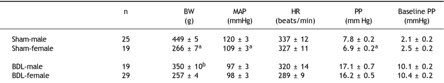
Table 1 depicts the BW, MAP, PP, HR and base-line PP in females or males underwent sham or BDL surgery. In sham-operated rats, the female sham rats showed lower BW, MAP and PP than those of the male rats. The baseline PP was not sig-nificantly different between the two groups. In BDL rats, there was no significant difference in MAP, PP, HR and baseline PP except that female BDL rats had lower body weights than male BDL rats.
Before the perfusion experiments, baseline BW and hemodynamic parameters were not significantly different among sham or BDL rats receiving vehicle, 17β-estradiol, progesterone or testosterone perfusion (P > 0.05).
Effects of sex and sex hormones on intrahepatic vascular concentration-response
relationships to ET-1 in sham rats
When preincubated with vehicle, the responses to ET-1 were higher in female rats at the concen-trations of 10-9 M (6.5 ± 1.2 vs. 3.7 ± 0.3 mmHg,
P = 0.025) and 3 x 10⁻⁹ M (15.0 ± 1.1 vs. 10.7 ±
0.8 mmHg, P = 0.013) whereas 17β-estradiol pre-incubation elicited lower perfusion pressure changes to ET-1 at the concentrations of 3x10⁻⁸ M
(19.0 ± 0.4 vs. 23.7 ± 1.5 mmHg, P = 0.02) in female rats compared with those in male rats (Figure 1). The perfusion pressure changes caused by testosterone and progesterone were not signifi-cantly different between male and female rats (P > 0.05) (Figure 1). Two-way repeated meas-ures ANOVA shows that compared with vehicle preincubation, 17β-estradiol elicited a lower per-fusion pressure changes to ET-1 in female sham rats (P = 0.011) (Figure 2B). On the other hand, there was no significant difference of perfusion pressure changes in male sham rats perfused with vehicle, 17β-estradiol, progesterone, or testoster-one (P > 0.05) (Figure 2A).
Effects of sex and sex hormones on intrahepatic vascular concentration-response
relationships to ET-1 in rats with BDL-induced cirrhosis
When preincubated with vehicle, the responses to ET-1 were higher in female cirrhotic rats at the concentrations of 10-9 M (11.6 ± 1.1 vs. 8.3 ± 0.9
mmHg, P = 0.046) and 3 x 10⁻⁹ M (19.4 ± 1.5 vs.
15 ± 0.6 mmHg, P = 0.025) and 10-8 M (28.6 ±
1.7 vs. 23.7 ± 1.2 mmHg, P = 0.042). 17β-estradiol
Table 1. Body weight and baseline hemodynamics of sham-operated and bile duct ligated rats.
n BW MAP HR PP Baseline PP
(g) (mmHg) (beats/min) (mm Hg) (mmHg) Sham-male 25 449 ± 5 120 ± 3 337 ± 12 7.8 ± 0.2 2.1 ± 0.2 Sham-female 19 266 ± 7a 109 ± 3a 327 ± 11 6.9 ± 0.2a 2.5 ± 0.2
BDL-male 19 350 ± 10b 97 ± 3 320 ± 14 17.1 ± 0.7 10.1 ± 0.2
BDL-female 29 257 ± 4 98 ± 3 289 ± 9 16.2 ± 0.5 10.4 ± 0.2
BW: body weight. MAP: mean arterial pressure. HR: heart rate. PP: portal pressure. Baseline PP: baseline perfusion pressure. BDL: bile duct ligation.
Figure 1. Concentration-response curves to ET-1 in intrahepatic vascular beds of male vs. female sham-operated rats pre-in-cubated with vehicle, testosterone, progesterone or 17β-estradiol, expressed as absolute increase over baseline value. Female rats pre-incubated with vehicle showed a stronger vasoconstrictive response to ET-1 but a weaker response to ET-1 when being pre-incubated with 17β-estradiol (* indicates a P < 0.05).
Figure 2. Concentration-response curves to ET-1 in intrahepatic vascular beds of sham-operated male (A) and female (B) rats pre-incubated with vehicle, testosterone, progesterone or 17β-estradiol, expressed as absolute increase over baseline value. As compared with vehicle, 17β-estradiol perfusion elicited lower perfusion pressure changes to ET-1 in female rats.
Perfusion pressure change (
Δ
mmHg)
25
20
15
10
5
0
-10 -9 -8 -7
ET-1 (Log M) Male
Female * P < 0.05
Vehicle
Male Female * P < 0.05
17β-estradiol
Perfusion pressure change (
Δ
mmHg)
30 25 20 15 10 5 0
-10 -9 -8 -7
ET-1 (Log M)
Perfusion pressure change (
Δ
mmHg)
25
20
15
10
5
0
-10 -9 -8 -7
ET-1 (Log M)
Perfusion pressure change (
Δ
mmHg)
25
20
15
10
5
0
-10 -9 -8 -7
ET-1 (Log M)
Vehicle Estradiol Progesterone Testosterone P > 0.05
Perfusion pressure change (
Δ
mmHg)
30 25 20 15 10 5 0
-10 -9 -8 -7
ET-1 (Log M)
Vehicle Estradiol Progesterone Testosterone
P = 0.011 vehicle vs. estradiol
Perfusion pressure change (
Δ
mmHg)
25
20
15
10
5
0
-10 -9 -8 -7
ET-1 (Log M) Male
Female P > 0.05
Male Female P > 0.05
Progesterone Testosterone
Figure 3. Concentration-response curves to ET-1 in intrahepatic vascular beds of male vs. female BDL rats pre-incubated with vehicle, testosterone, progesterone or 17β-estradiol, expressed as absolute increase over baseline value. Female rats pre-incu-bated with vehicle showed a stronger vasoconstrictive response to ET-1 but not significantly modified by 17β-estradiol (*indi-cates a P < 0.05).
Figure 4. Concentration-response curves to ET-1 in intrahepatic vascular beds of cirrhotic male (A) and female (B) rats pre-incubated with vehicle, testosterone, progesterone or 17β-estradiol, expressed as absolute increase over baseline value. There was no significant difference among these groups.
Female Male * P < 0.05
Vehicle
Perfusion pressure change (
Δ
mmHg)
40
30
20
10
0
-11 -10 -9 -8 -7
ET-1 (Log M)
Female Male P > 0.05
17β-estradiol
Perfusion pressure change (
Δ
mmHg)
-11 -10 -9 -8 -7
ET-1 (Log M) 40
30
20
10
0
Progesterone Testosterone
Perfusion pressure change (
Δ
mmHg)
40
30
20
10
0
-11 -10 -9 -8 -7
ET-1 (Log M)
Perfusion pressure change (
Δ
mmHg)
-11 -10 -9 -8 -7
ET-1 (Log M) 40
30
20
10
0 Female
Male * P < 0.05
Female Male P > 0.05
Perfusion pressure change (
Δ
mmHg)
40
30
20
10
0
-11 -10 -9 -8 -7
ET-1 (Log M)
Perfusion pressure change (
Δ
mmHg)
-11 -10 -9 -8 -7
ET-1 (Log M) 40
30
20
10
0 Vehicle
17-β-estradiol Progesterone Testosterone P > 0.05
preincubation abrogated the differences that the perfusion pressure changes were not significantly different between female and male cirrhotic rats (P > 0.05) (Figure 3). Vehicle, 17β-estradiol,
proges-terone and testosproges-terone did not induce significantly different hepatic perfusion pressure changes to ET-1 in either male (P > 0.05) (Figure 4A) and female (P > 0.05) (Figure 4 B) cirrhotic rats.
Figure 6. Intrahepatic estrogen receptors (ERα, ERβ), androgen receptor (AR), and progesterone receptor (PR-A+B, PR-B) mRNA expressions in male sham and BDL rats. Significant down regulations of ERα, AR and PR-A+B in BDL rats are noted.
Figure 5. Intrahepatic estrogen receptors (ERa, ERb), androgen receptor (AR), and progesterone receptor (PR-A+B, PR-B)
Hepatic sex hormone receptors expressions in female or male rats with sham or BDL surgeries
Figure 5 reveals the ERa, ERb, AR, PR-A+B and PR-B of female rats underwent sham or BDL sur-geries. Female rats with BDL-induced cirrhosis had significantly lower hepatic mRNA expression of ERα (ERα/HPRT: 0.0347 ± 0.0066 vs. 0.1180 ± 0.0158, P < 0.001 ) and higher expression of AR (AR/HPRT: 0.1115 ± 0.0126 vs. 0.0712 ± 0.0105, P = 0.022).
Figure 6 depicted the ERα, ERβ, AR, PR-A+B and PR-B of male rats underwent sham or BDL surger-ies. Male rats with BDL-induced cirrhosis had sig-nificantly lower hepatic mRNA expression of ERα (ERα/HPRT: 0.0485 ± 0.0086 vs. 0.1971 ± 0.0204, P < 0.001), AR (AR/HPRT: 0.0834 ± 0.0180 vs. 0.1731 ± 0.0163, P = 0.001), and PR-A+B (PR-A+B/ HPRT: 0.0007 ± 0.0001 vs. 0.0197 ± 0.0088, P = 0.049).
DISCUSSION
In this study, female sham rats had significantly lower MAP and PP than those of the male rats. Compatible with our current finding, Khoury and colleagues found that men had higher blood pres-sure than did women by ambulatory blood prespres-sure monitoring on 131 men and women.25 In an animal
study, male spontaneously hypertensive rats (SHR) have higher blood pressure than do females of simi-lar ages.26 Androgens, such as testosterone, have
been implicated in the mechanism. Interestingly, the baseline hepatic perfusion pressure was similar be-tween male and female sham rats. Since we used fixed flow rate perfusion system, this indicates that the intrahepatic resistance was not influenced by gender. Furthermore, PP is the net effect exerted mainly by portal inflow and hepatic resistance, so the lower PP in females may be related to systemic hypotension with reduced portal inflow. On the oth-er hand, in cirrhotic rats, the hemodynamic parame-ters were not statistically different between females and males. Liver cirrhosis has been characterized by hyperdynamic circulation with systemic arterial hy-potension. Our result implies that this overwhelm-ing phenomenon may not be overcome by the potential influence of gender on hemodynamics.27
It has been noted that estradiol administration to isolated vascular tissue for 30 minutes attenuated large coronary arterial contractions to ET-1.28
Su-dhir, et al. found that intracoronary estradiol re-duced the vasoconstrictor response to intracoronary ET-1 in pigs.29 The attenuating effects of estradiol
on ET-1–induced vasoconstriction in the portal venous system following trauma-hemorrhage in the rats have also been demonstrated.30 Regarding
the relevant studies on liver, estrogen receptors have been identified in rat liver sinusoidal endotheli-al cells (SEC), supporting the ability of estradiol in regulating intrahepatic microcirculation.31
In the current study, female rats, either sham or cirrhotic, showed higher intrahepatic vascular re-sponsiveness to ET-1 than male rats. Regarding the gender-related vascular reactions to ET-1, regional differences should be considered. It has been indicat-ed that the hindquarter vasoconstrictor effects elicit-ed by ET-1 were significantly higher in male than female rats.32 On the other hand, compatible with
our results, femoral and brachial arteries from female pigs developed greater contractile force in response to ET-1 than those from male pigs.33 The
similar finding was found in coronary arteries of pigs and the difference was not related to estrogen levels in female pigs, which is probably due to differ-ences in the regulation of intracellular calcium.34
Barber, et al. have also reported that coronary arteries from female pigs generate more forceful contractions in response to ET-1 compared with male pigs and that this difference was mediated by increased affinity of the endothelin receptors in cor-onary vascular smooth muscle of female pigs.35
However, Jiang, et al. did not detect a difference of rabbit coronary arterial responses to ET-1 from male and nonpregnant female rabbits unless estradi-ol was added to the organ chamber.28 The
vasocon-striction increment made by ET-1 on rat tail artery segments was also similar in both genders.36
Al-though the different conclusions might result from different experimental models in different species and vascular beds, further exploration for the mech-anism in intrahepatic circulation is required.
circulatory concentrations of estradiol have been noted in cirrhotic patients.7,8 In humans with
cir-rhosis, there is also an increased conversion of weak androgens to estrogens,11 which might be
responsi-ble for, at least partly, the ERαdown-regulation in cirrhotic liver. In fact, we also found a significant down-regulation of intrahepatic ERa expression in cirrhotic males, although 17β-estradiol had failed to exert changes in vascular responsiveness in sham males.
The vascular action characteristic of progesterone is relatively indefinite. In isolated vessels, progester-one reverses the beneficial effects of estrogens.38 In
the study reported by Lamping, et al.,39 progesterone
alleviates the vasoconstrictive effect of ET-1, but is weaker than that of estradiol. In this study, proges-terone did not induce different hepatic perfusion pres-sure changes to ET-1 in either male or female, sham or cirrhotic rats and irrespective of intrahepatic PR expressions. The influence of progesterone in intra-hepatic circulation may be negligible.
The mechanism of the vascular effects of testo-sterone seems rather complex. Testotesto-sterone relaxed coronary microvessels preconstricted with ET-1 sim-ilarly to estradiol and progesterone whereas it had no direct effect on the dose-response curves to ET-1.39 Furthermore, relaxation of coronary
microves-sels to testosterone was not mediated by release of vasodilator prostaglandins or NO.39 Studies of the
testosterone effect on rabbit coronary arteries sug-gest that it is independent of endothelium and is not mediated by stimulation of guanylate cyclase.40 Yue,
et al. had also found an acute vasodilatory effect of testosterone in the rabbit aorta and coronary artery in vitro that is endothelium independent and may in-volve activation of vascular smooth muscle K+
chan-nels.40 In contrast, others demonstrated that
testosterone induced acute vasorelaxation that is gender and androgen receptor independent and involves both endothelium-dependent (via NO) and -independent mechanisms of action.41 Similar
endothelium-dependent (via NO) and -independent vasodilatory effects of testosterone on canine coro-nary conductance and resistance arteries in vivo have also been reported.42 In the current study,
tes-tosterone did not induce different hepatic perfusion pressure changes to ET-1 in either male or female, sham or cirrhotic rats and was irrespective to intra-hepatic TR expressions. Likewise, testosterone may not elicit significant impacts on intrahepatic circula-tion.
To sum up, the sham and cirrhotic female rats ex-erted a stronger intrahepatic vascular constrictive
effect to ET-1 than their male counterparts, which was reversed by 17β-estradiol in sham but not in cirrhotic females. The reduced intrahepatic vascular response to 17β-estradiol in sham females was also abrogated in cirrhosis. This may be attributed to the down-regulation of intrahepatic ERα expression in cirrhosis.
ACKNOWLEDGEMENTS
We thank Chen Yi-Chou for his excellent techni-cal assistance. This study was supported by the grant from the National Science Council, Taiwan (96-2314-B-075-034-MY3). The experiments were as-sisted in part by the Animal Center of Department of Medical Research and Education at Taipei Veterans General Hospital.
ABBREVIATIONS
• AR: androgen receptor.
• BDL: common bile duct ligation.
• ER: estrogen receptor.
• ET-1: endothelin-1.
• HR: heart rate.
• IVC: inferior vena cava.
• NO: nitric oxide.
• MAP: mean arterial pressure.
• PP: portal pressure.
• PR: progesterone receptor.
REFERENCES
1. Bosch J, Pizcueta P, Feu F, Fernández M, Garciá-Pagán JC. Pathophysiology of portal hypertension. Gastroenterol
Clin Nor Am 1992; 21: 1-14.
2. D’Amico G, Pagliaro L, Bosch J. The treatment of portal hypertension: a meta-analytic review. Hepatology 1995; 22: 332-54.
3. Rockey DC. Vasoactive agents in intrahepatic portal hy-pertension and fibrogenesis: implications for therapy.
Gastroenterology 2000; 118: 1261-5.
4. Moller S, Gulberg V, Henriksen JH, Gerbes AL. Endothelin-1 and endothelin-3 in cirrhosis: relations to systemic and splanchnic haemodynamics. J Hepatol 1995; 23: 135-44. 5. Sogni P, Moreau R, Gomola A, Gadano A, Cailmail S, Calmus
Y, Clozel M, et al. Beneficial hemodynamic effects of bosentan, a mixed ET(A) and ET(B) receptor antagonist, in portal hypertensive rats. Hepatology 1998; 28: 655-9. 6. Chan CC, Wang SS, Lee FY, Chang FY, Lin HC, Chu
CJ, Chen CT, et al. Endothelin-1 induces vasoconstriction on portal-systemic collaterals of portal hypertensive rats.
Hepatology 2001; 33: 816-20.
7. Baker HW, Burger HG, de Kretser DM, Dulmanis A, Hudson B, O’Connor S, Paulsen CA, et al. A study of the endocrine manifestations of hepatic cirrhosis. Q J Med 1976; 45: 145-78. 8. Bannister P, Losowsky MS. Sex hormones and chronic liver
9. Farrell GC, Koltai A, Zaluzny L, Murray M. Effects of portal vein ligation on sex hormone metabolism in male rats: rela-tionship to lowered hepatic cytochrome P450 levels.
Gas-troenterology 1986; 90: 299-305.
10. Van Thiel DH, Gavaler JS, Schade RR. Liver disease and the hypothalamic pituitary gonadal axis. Semin Liver Dis 1985; 5: 35-45.
11. Gordon GG, Olivo J, Rafil F, Southren AL. Conversion of an-drogens to estrogens in cirrhosis of the liver. J Clin
Endo-crinol Metab 1975; 40: 1018-26.
12. Farrell G, Koltai A. Hepatic testosterone metabolism in male rats with portal bypass. Gastroenterology 1988; 95: 425-33.
13. Levy D. Kannel WB. Cardiovascular risks: new insights from Framingham. Am Heart J 1988; 116: 266-72.
14. Kastrup A, Dichgans J, Niemeier M, Schabet M. Changes of cerebrovascular CO2 reactivity during normal aging.
Stroke 1998; 29: 1311-4.
15. Reis SE, Gloth ST, Blumenthal RS, Resar JR, Zacur HA, Gerstenblith G, Brinker JA, et al. Ethinyl estradiol acutely attenuates abnormal coronary vasomotor re-sponses to acetylcholine in postmenopausal women.
Circu-lation 1994; 89: 52-60.
16. Kauser K, Rubanyi GM. Gender difference in bioassayable endothelium-derived nitric oxide from isolated rat aortae.
A J Physiol 1994; 267: H2311-H2317.
17. Brandes RP, Mugge A. Gender differences in the genera-tion of superoxide anions in the rat aorta. Life Sci 1997; 60: 391-6.
18. Franco F, Gigou M, Szekely AM, Bismuth H. Portal hyperten-sion after bile duct obstruction: effect of bile diverhyperten-sion on portal pressure in the rat. Arch Surg 1979; 114: 1064-7. 19. Kountouras J, Billing BH, Scheuer PJ. Prolonged bile duct
li-gation obstruction: a new experimental model for cirrho-sis in the rat. Br J Exp Pathol 1984; 65: 305-11.
20. Lee FY, Colombato LA, Albillos A, Groszmann RJ. Adminis-tration of ω-nitro-L-arginine ameliorates portal-systemic shunting in portal hypertensive rats. Gastroenterology 1993; 105: 1464-70.
21. Mittal MK, Gupta TK, Lee FY, Sieber CC, Groszmann RJ. Ni-tric oxide modulates hepatic vascular tone in normal rat liver. Am J Physiol 1994; 267: G416-G422.
22. Hamilton RL, Berry MN, Williams MC, Severinghaus EM. A simple and inexpensive membrane “lung” for small organ perfusion. J Lipid Res 1974; 15: 182-6.
23. Chen J, Rider DA, Ruan R. Identification of valid house-keeping genes and antioxidant enzyme gene expression change in the aging rat liver. J Gerontol 2006; 61: 20-7. 24. Okada A, Ohta Y, Inoue S, Hiroi H, Muramatsu M, Iguchi T.
Expression of estrogen, progesterone and androgen re-ceptors in the oviduct of developing, cycling and pre-im-plantation rats. J Mol Endocrinol 2003; 30: 301-15. 25. Khoury S, Yarows SA, O’Brien TK, Sowers JR. Ambulatory
blood pressure monitoring in a nonacademic setting: ef-fects of age and sex. Am J Hypertens 1992; 5: 616-23. 26. Ganten U, Schroder G, Witt M, Zimmerman F, Ganten D,
Stock G. Sexual dimorphism of blood pressure in spontane-ously hypertensive rats: effects of anti-androgen treat-ment. J Hypertens 1989; 7: 721-6.
27. Iwakiri Y, Groszmann RJ. The hyperdynamic circulation of chronic liver diseases: from the patient to the molecule.
Hepatology 2006; 43: S121-S131.
28. Jiang C, Sarrel PM, Poole-Wilson PA, Collins P. Acute ef-fect of 17 beta-estradiol on rabbit coronary artery con-tractile responses to endothelin-1. Am J Physiol 1992; 263: H271-H275.
29. Sudhir K, Ko E, Zellner C, Wong HE, Hutchison SJ, Chou TM, Chatterjee K. Physiological concentrations of estradi-ol attenuate endothelin 1-induced coronary vasoconstric-tion in vivo. Circulavasoconstric-tion 1997; 96: 3626-32.
30. Yokoyama Y, Toth B, Kitchens WC, Schwacha MG, Rue LW 3rd, Bland KI, . Estradiol’s effect on portal response to en-dothelin-1 after trauma-hemorrhage. Journal Surg Res 2004; 121: 25-30.
31. Sakamoto M, Uen T, Nakamura T, Hashimoto O, Sakata R, Kin M, Ogata R , et al. Estrogen upregulates nitric ox-ide synthase expression in cultured rat hepatic sinusoidal endothelial cells. J Hepatol 2001; 34: 858-64.
32. Tatchum-Talom R, Martel C, Labrie C, Labrie F, Marette A. Gender differences in hemodynamic re-sponses to endothelin-1. J Cardiovasc Pharmacol 2000; 36: S102-S104.
33. Laughlin MH, Schrage WG, McAllister RM, Garverick HA, Jones AW. Interaction of gender and exercise training: vasomotor reactivity of porcine skeletal muscle arteries.
J Appl Physiol 2001; 90: 216-27.
34. Miller VM, Barber DA, Fenton AM, Wang X, Sieck GC. Gen-der differences in response to endothelin-1 in coronary arteries: transcription, receptors and calcium regulation.
Clin Exp Pharmacol Physiol 1996; 23: 256-9.
35. Barber DA, Sieck GC, Fitzpatrick LA, Miller VM. Endothelin receptors are modulated in association with endogenous fluctuations in estrogen. Am J Physiol Heart Circ Physiol 1996; 271: H1999-H2006.
36. Fernandez N, Sanz E, Monge L, Martinez MA, Dieguez G, Garcia-Villalon AL. Potentiation by endothelin-1 and vaso-pressin of sympathetic vasoconstriction in male and fe-male rats. Clin Sci 2002; 103: 158s-61s.
37. Saceda M, Lippman ME, Chambon P, Lindsey RL, Ponglikitmongkol M, Puente M, Martin MB, et al. Regulation of the estrogen recep-tor in MCF-7 cells by estradiol. Molecular Endocrinol 1988; 2: 1157-62.
38. Miller VM, Vanhoutte PM. Progesterone and modulation of endothelium-dependent responses in canine coronary ar-teries. Am J Physiol 1991; 261: R1022-R1027.
39. Lamping KG, Nuno DW. Effects of 17 beta-estradiol on coronary microvascular responses to endothelin-1. Am J
Physiol 1996; 271: H1117-H1124.
40. Yue P, Chatterjee K, Beale C, Poole-Wilson PA, Collins P. Testosterone relaxes rabbit coronary arteries and aorta.
Circulation 1995; 91: 1154-60.
41. Costarella CE, Stallone JN, Rutecki GW, Whittier FC. Tes-tosterone causes direct relaxation of rat thoracic aorta.
J Pharmacol Exp Ther 1996; 277: 34-9.
42. Chou TM, Sudhir K, Hutchison SJ, Ko E, Amidon TM, Collins P, Chatterjee K. Testosterone induces dilation of canine coronary conductance and resistance arteries in vivo.