Otras secciones de este sitio:
! ! ! !
! Índice de este número
! ! ! !
! Más revistas
! ! ! !
! Búsqueda
Others sections in this web site:
! ! ! !
! Contents of this number !
! ! !
! More journals !
! ! ! ! Search Artículo:
Hepatocellular carcinoma. An overview
Copyright © 2006: Mexican Association of Hepatology ANNALS OF HEPATOLOGY
Number 1 January-March 2 0 0 6
Volume 5
MG
edigraphic.com
Annals of Hepatology 2006; 5(1): January-March: 16-24Annals of Hepatology
Concise Review
Hepatocellular carcinoma. An overview
Daniel Motola-Kuba;1 Daniel Zamora-Valdés;1 Misael Uribe;1 Nahum Méndez-Sánchez1
1Department of Biomedical Research and Liver Unit, Medica Sur Clinic & Foundation, Mexico City, Mexico.
Abbreviations: AFP: alfa-fetoprotein; CC: cryptogenic cirrhosis;
CEA: carcinoembrionary antigen; DN: dysplastic nodules; HBV: hepatitis B virus; HCC: hepatocellular carcinoma; HCV: hepatitis C virus;HH: hereditary hemochromatosis; IGF2r: insulin-like growth factor 2 receptor; LCD: large cell-dysplasia; MRI: magnetic resonance imaging; NAFLD: nonalcoholic fatty liver disease; NASH: nonalcoholic steatohepatitis; NK: natural killer cells; PBC: primary biliar cirrhosis; SCD: small cell-dysplasia; TGF-β: transforming growth factor β.
Address for correspondence: Nahum Méndez-Sánchez, M.D.,Ph.D.
Departments of Biomedical Research, Gastroenterology & Liver Unit, Medica Sur Clinic & Foundation, Puente de Piedra 150, Col. Toriello Guerra, Mexico City, Mexico. Phone: (+525) 606-6222, ext. 4215 Fax: (+525) 666-4031 and 606-1651; E-mail: nmendez@medicasur.org.mx
Manuscript received and accepted: 1 December, 2005.
and the fifth most common tumor worldwide. It has a high incidence in sub-Saharan Africa and Asia. The HCC 5-year survival rate is less than 5 per cent without treat-ment. Any chronic inflammatory liver disease has the po-tential to induce HCC, but the pathophysiological pro-cess found in up to 80 per cent of cases of the disease is
cirrhosis. Approximately90 to 95 percent of these tumors
are the biologic consequencesof persistent hepatitis B
virus (HBV) and hepatitis C virus(HCV) infections.
Cer-tain diseases other than chronic Hepatitis B or C are asso-ciated with increased HCC incidence; iron overload rhosis (hemochromatosis), long-standing alcoholic cir-rhosis, alpha1-antitrypsin deficiency, and tyrosinemia. The disease is often clinically silent until it is well ad-vanced or tumor diameter exceeds 10 cm. Given the poor prognosis and lack of effective therapies for hepatocellu-lar carcinoma, prevention programs are desperately need-ed. Surgical resection is the treatment of choice for pa-tients with HCC when the tumor is small and limited to one lobe of the liver. In cases where the tumor is larger or involves more than one lobe of the liver such that it can-not be removed, liver transplantation has also been per-formed. In either case, the cure rate averages only 20-30 per cent, which has somewhat limited the use of liver transplantation for this problem. Surveillance for HCC in patients with cirrhosis may lead to an improved survival in cohort studies.
The aim of this review is to analyze information re-garding epidemiology, risk factors, clinical manifesta-tion, diagnosis and actual treatment of HCC.
Epidemiology
Hepatocellular carcinoma (HCC) represents approxi-mately 6 per cent of all malignancies. It is the fifth most common malignancy in men and ninth in women, with an estimated 500,000 to 1 million new cases annually
around the world.1 Its incidence is low in the occidental
world and high in Southeast Asia and sub-Saharan Afri-ca, even though it has risen in the United States, Japan,
England, and France.2 HCC is considered a disease of
older persons, with a high incidence in people between 65 to 69 years old. However, the prevalence in young people has risen in recent years due to environmental
risk factors at birth.3 HCC is the most common primary
liver cancer (78 per cent of all primary liver cancer in the United States) with an incidence of 2.4 for every
Abstract
Hepatocellular carcinoma is a common malignancy affecting approximately one million people around the world every year. The incidence is low in the occiden-tal world and high in locations such as Southeast Asia and sub-Saharan Africa. Hepatocellular carcinoma primarily affects old people, reaching its highest preva-lence among those aged 65 to 69 years old. Chronic in-fection by the hepatitis B virus is the most common cause of this disease. Other important causes are cir-rhosis, chronic viral hepatitis (hepatitis C virus, and hepatitis B plus D viruses), alcohol abuse, obesity, hemochromatosis, alfa1-antitripsin deficiency, and tox-ins similar to aflatoxin. In most cases, hepatocellular carcinoma is asymptomatic and has a low life expect-ancy. This article presents a review of the most impor-tant epidemiological, diagnostic and treatment data about this disease.
Key words: Hepatocellular carcinoma, cancer, liver, tumor.
Introduction
D Motola-Kuba et al. Hepatocellular carcinoma 17
edigraphic.com
100,000 persons living in the United States from 1991-1995. Its incidence rises with age, as well as in high-risk populations including Hispanic groups, Native Americans and Asians. The three most important risk factors for HCC development in the United States are Hepatitis C virus infection (HCV), Hepatitis B virus in-fection (HBV) and cirrhosis caused by alcoholic liver disease. In people with HCV or HBV chronic liver dis-ease, HCC can develop in approximately 10 to 30
years.3 Some studies have shown that high alcohol
con-sumption (more than 80 g per day) and cirrhosis caused by alcohol consumption are strongly associated with HCC development even in the absence of viral
infec-tion.4,5 In one of these studies, high alcohol consumption
and viral hepatitis (primarily HCV infection) represented
63 per cent of HCC cases.4 In Brazil, the most common
causes of HCC were HCV and HBV infection.6 At this
time we have no HCC incidence studies in Mexico. In a study made in a general hospital in Mexico City of 12,556 cases of necropsy, an HCC prevalence of 0.59 percent (n = 73) was found. The age at death among these patients lay between 25 and 90 years, with a median of
65 years.7 In Spain, the most common risk factors for
HCC development in Child Pugh A-B cirrhotic patients were age of 54 years or older, low prothrombin activity,
low platelet count, and chronic HCV infection.8
Risk factors, predisponent conditions and pathogenesis
HCC etiology varies depending on the geographical location. As indicated in Table 1, in countries where HCC is endemic (sub-Saharan Africa, Asia and Alaska), the most common cause is HBV infection, but in low-risk countries the most common HCC cause is cirrhosis caused by
chron-ic viral infection or alcohol consumption.9
Cirrhosis
Independent of its cause, cirrhosis is considered a ma-jor clinical and histopathological risk factor for HCC de-velopment. Five per cent of all cirrhotic patients develop
HCC.9 In a Mexican study,10 the main causes of cirrhosis
among 1,486 patients were alcohol (587; 39.5 per cent), HCV (544; 36.6 per cent), cryptogenic (154; 10.4 per cent), primary biliar cirrhosis (PBC) (84; 5.7 per cent), HBV (75; 5.0 per cent). Cortes-Espinosa et al. found
cir-rhosis in 75 per cent of all HCC cases.7 We know that
cir-rhosis caused by alcohol consumption alone is an impor-tant risk factor for HCC development, but in a Japanese study, alcohol consumption was a co-factor to prior ex-posure to HBV infection in accelerating HCC
develop-ment.5 According to liver disease prevalence trends in
Mexico, nearly 2 million cases of chronic liver disease are expected between 2005 and 2050, with
alcohol-relat-ed liver disease the most important cause.11
Precursor histological injuries
Hepatocarcinogenesis is the development and pro-gression of a HCC chronic liver disease. Hepatocarcino-genesis is a multistep process characterized by the accu-mulation of poorly understood interacting genetic alter-ations. HCC coexists with a number of microscopically distinct lesions that are thought to be its precursors.
Regenerative nodules are characteristic lesions of the cirrhotic liver. They exhibit a lack of bile ducts and poor-ly organized hepatocytes surrounded by fibrosis and pro-liferating cholangiocytes. These lesions are arbitrarily classified as micro or macronodular (cut point 0.3 cm).
Regenerative nodules may present dysplastic foci, which are smaller than 1 mm and can only be recognized by microscopic studies. There are two types of dysplastic foci in cirrhotic livers, the small cell-dysplasia (SCD) and the large cell-dysplasia (LCD), according to the nucleo-cytoplasmic ratio of each one (high in SCD and normal in LCD). SCD are thought to be HCC precursor lesions that result from the proliferation of hepatocytes and oval cells. On the other hand, LCD apparently arise from per-sistent necroinflammation-induced senescent hepato-cytes and are therefore not considered to be HCC precur-sor lesions, although patients with LCD are at an
in-creased risk of HCC.12
Dysplastic nodules (DN) are macroscopically recogniz-able lesions that show atypical features microscopically, such as increased nucleocytoplasmic ratio, nuclear con-tour, thickness of hepatocellular plates and compression of adjacent hepatocytes. DN represents parts of a spectrum that is arbitrarily divided for the purposes of clinical utili-ty into low-grade or high-grade DN, according to the
pres-ence of cytological or structural atypia or both.13 The risk
of HCC in patients with high-grade DN is four-fold higher. By contrast, patients with only low-grade DN are not at a
significantly increased risk of HCC.14
Hepatitis B infection
Chronic HBV infection is well established as a risk factor for HCC development. In the United States, 25 per
cent of patients with HCC are chronic carriers of HBV.2
Sixty to ninety per cent of patients with HBV-related HCC have cirrhosis, but cirrhosis development is not
necessary for HCC development.9 HBV chronic infection
MG
edigraphic.com
the mobility and mortality of this infection as well as
risk for HCC development.15
Hepatitis C virus infection
HCV infection is recognized as a significant risk fac-tor for HCC development, with 6–75 per cent of HCC
cases having positive antibodies for HCV.9,16 In the
United States, HCV accounts for approximately 50 per
cent of HCC cases.15 Some studies identified genotype
1b with a high risk for HCC development.16 A number
of studies have demonstrated a direct relationship between HCC incidence and advanced stages of hepatic
fibrosis in chronic active hepatitis.15 Because of a
HCV-related nonspecific inflammatory process that induces hepatocyte proliferation associated with a rise in alanine-aminotransferase (ALT) levels, patients with high inflammatory and proliferation activity are more
prone to the progression to HCC.17 There is evidence of
a direct viral effect on carcinogenesis, such as HCV core protein inhibition of apoptosis. High incidence of HCC is seen in people with HCV infection and high alcohol
consumption.15
HVC/HBV infection
Co-infection of HBV in people with HCV infection el-evates HCC development risk. The mechanisms that cause this high incidence include augmented fibrosis,
and inflammation and high cellular re-change.15
Aflatoxins
Aflatoxin is a toxin produced by Aspergillus flavus
and A. parasiticus, which grow in foods like peanuts.9 It
causes alterations in the hepatocyte DNA (see genetic al-terations). It is related to HCC in countries where
infesta-tion of crops and animal feed is common.18 Aflatoxin
me-tabolism produces aflatoxin B1-8,9-epoxide, a toxic product that induces a G to T mutation of the p53 gene at codon 249 up-regulating insulin-like growth factor II that
leads to a reduction of apoptosis and HCC formation.19,20
Hereditary hemochromatosis
Hereditary hemochromatosis (HH) is an autosomic re-cessive disease in which an alteration in iron absorption, inducing deposition in the liver and other organs
oc-curs.9 HH is a significant risk factor for HCC
develop-ment. Its presence is associated with a 200 major risk for HCC. A case-control study demonstrated a 1.8 relative risk for HCC development in HH patients compared with
non-HH chronic liver disease patients.21 Iron toxicity in
the liver is produced by free radical formation, lipid per-oxidation of cell organs causing cell death with fibrosis
and cirrhosis.9
ααααα-1-antitripsin deficiency
Alpha-1-antitrypsin is the archetypal member of the serine proteinase inhibitor (or serpin) superfamily, play-ing an important role in the control of proteinases in-volved in the inflammatory, complement, coagulation
and fibrinolytic cascades.22α-1-antitripsin deficiency is
an autosomic recessive disease, with an abnormal
accu-mulation of α-1-antitripsin in the hepatocyte
endoplas-mic reticulum resulting in hepatic cells dysplasia and
cirrhosis.9 Although many [alpha]
1-antitrypsin deficiency
variants have been described, only two other mutants of
[alpha]1-antitrypsin have been associated with plasma
de-ficiency and hepatic inclusions: [alpha]1-antitrypsin
Si-iyama (53Ser->Phe) and [alpha]
1-antitrypsin Mmalton
(52Phe deleted).23
Wilson’s disease
Wilson’s disease is a heritable disease with mutations in the gene ATP7B and alteration in plasma copper circulation and its bile excretion. Excessive free copper can provoke
cytoplasmic injury, cirrhosis and sometimes HCC.9
Cryptogenic cirrhosis
In 1980, Ludwig et al. gave the name nonalcoholic steatohepatitis (NASH) to an advanced form of fatty liver disease, defining it as a well-recognized clinical-patho-logic syndrome primarily occurring in obese female pop-ulations with diabetes mellitus, with histological similar-ities to alcoholic liver disease in the absence of heavy
al-cohol consumption.24 Nonalcoholic fatty liver disease
(NAFLD) affects 10 to 24 per cent of the total population
in various countries.25 This prevalence is higher in
high-risk groups with a prevalence of 70 to 86 per cent in
obese and/or diabetic patients.26 NASH is estimated to
occur in 10 per cent of NAFLD patients. NASH has been posited as a possible cause of cryptogenic cirrhosis
(CC).27 Mortality trends in Mexico show a significant
as-sociation between increased prevalence of obesity and
increases in mortality caused by chronic liver disease.28
Patients with CC also develop HCC. There is increas-ing evidence that obesity and NAFLD are risk factors for HCC as the link between CC and nonalcoholic fatty liver
disease (NAFLD) in many patients is strengthened.29
Bu-gianesi et al.30 performed a case-control study in which
23 retrospectively identified patients with CC and HCC were compared to 641 age- and sex-matched patients with alcohol or viral cirrhosis and HCC. The prevalence of obesity and diabetes was higher in the CC patients. In addition, CC patients had higher glucose, cholesterol, and triglyceride plasma levels, and increased insulin re-sistance. Overweight patients with cryptogenic cirrhosis had a greater risk of developing HCC compared to lean
D Motola-Kuba et al. Hepatocellular carcinoma 19
edigraphic.com
Although NASH may progress to cirrhosis, it is not known if NASH has a role in the development of HCC. These data show that features suggestive of NASH are fre-quently observed in patients with CC-associated HCC. Some studies have confirmed that HCC may represent a late complication of CC cirrhosis in patients with
meta-bolic syndrome.31,32
Genetic alterations
Some genetic alterations have been associated with HCC development.
• p53, localized in chromosome 17p, is mutated in 30 per cent of HCC cases worldwide. This mutation pri-marily occurs either because of aflatoxins or HCV,
HBV chronic infection.33 A protein is produced by
p53 that recognizes injured DNA and controls cell
replication.34 B1-8,9-epoxid-aflatoxin is a toxic
prod-uct of aflatoxin metabolism and it is metabolized by the epoxid hydrolase and glutation-S-transferase. If this toxin is not metabolized, it combines with genom-ic structures to create mutations in p53, producing
toxic accumulation.9,34
• Insulin-like growth factor 2 receptor (IGF2r) and SMAD4 y SMAD2 genes. The primary function of IGF2r is the activation of the transforming growth factor
β (TGF-β) and the SMAD4 and SMAD2 intracellular
mediators of the TGF-β, resulting in growth inhibition
and apoptosis.33 Mutation and chromosomic deletion
of IGF2r occurs in 61 per cent of HCC cases associated
with other factors such as viral hepatitis and cirrhosis.34
• Table II summarizes the most important genes impli-cated in HCC.
PRIMARY SIGNS AND SYMPTOMS
The clinical presentation of HCC differs slightly in low and high incidence areas (Table III). There are three main clinical forms: a) right superior quadrant pain; b) worsening of general conditions in cirrhotic patients; c) asymptomatic (found as a result of screening, see figure
1).35 HCC at the beginning is asymptomatic, and when
the disease becomes symptomatic in most cases the
dis-ease is advanced and spread. Lungs, adrenal glands and
bone are the most common sites of metastasis.36
Diagnosis
Imaging
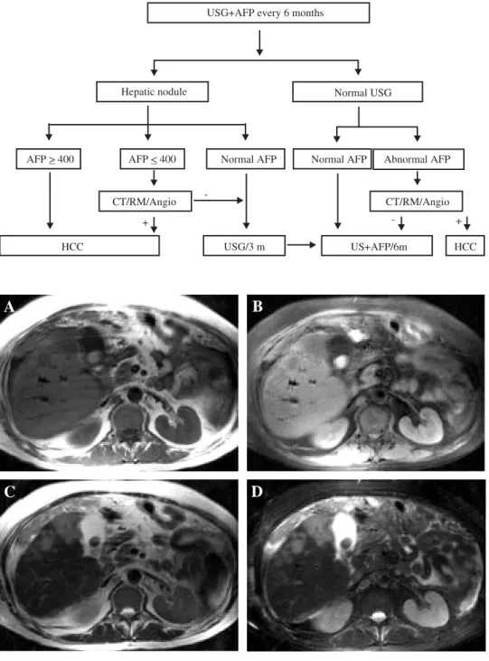
Hepatic ultrasound is useful in detecting HCC larger than 2 cm, but is a poor tool for detecting lesions smaller than 2 cm. As a result, computed tomography has re-placed ultrasound in HCC detection. Contrast tomogra-phy in HCC has three phases: before contrast infusion; arterial phase (2 to 40 seconds after infusion) where tu-mor presence is tu-more evident; portal vein phase (50 to 90 seconds after contrast infusion) where liver parenchyma is more evident. Magnetic resonance imaging (MRI) is useful for HCC image diagnostics because it can differ-entiate between regenerative nodules and early high
fat-containing HCC through T1-sequencing.37 For an
exam-ple of MRI imaging in HCC, exam-please see figure 2.
Alfa-fetoprotein
Alfa-fetoprotein (AFP) is the most important proteic component of fetal serum. It is synthesized in the
viscer-Table I. Risk factors for HCC development.
Major risk factors Minor risk factors
Cirrhosis Primary biliary cirrhosis
Male sex Thorotrast exposure
Older age Tobacco
HBV infection Vinylic exposure HCV infection Estrogen use Hemochromatosis, Wilson’s,
glucogenosis Androgen use
Aflatoxin exposure
Table II. Chromosomal localization of potential and candidate suppressor genes for HCC.
Chromosomic region Potential and candidate suppressor genes
1p36 p73 (functionally related to p53) 4 q Potential genes include albumin, alcohol
dehydrogenase (ADH3), fibrinogen, and UDP-glucoronyl-transferase
6q26-27 Insulin-like growth factor 2
8q21-22 PDGF-receptor beta-like tumor suppressor 13q12-q32 BRCA2 and retinoblastoma gene
17p13.1 p 5 3
Table III. HCC clinical manifestation in low and high incidence areas.
Low incidence High incidence
Symptoms areas1 areas2
Abdominal pain 53%-58% 62%-95%
Weight loss 19%-73% 19%-73%
Abdominal mass 33% < 33%
Anorexia 33% 47%-60%
Hematemesis 1%-19% 15%
Bone ache 3%-12%
Signs
Hepatomegaly 56%-74% 86%-98%
Ascitis 55%-61% 30%-51%
Splenomegaly 15%-48% 27%-57%
Fever 10% 38%
Jaundice 44% 25%
1. Low incidence areas = East Europe and United States of America
2. High incidence areas = Southeast Asia (Especially China, Thailand, and Korea) Modified from Hillebrand DJ, Sandowski SA. Hepatocellular carcinoma. Clin Fam
MG
edigraphic.com
al endoderm of the vitelin sac in the first part of fetal development, after which it is synthesized in the liver. AFP levels eventually diminish after birth to virtually undetectable levels, and only elevate under pathologi-cal conditions. AFP is a 591 aminoacid-glycoprotein,
with a weight of 70Kd.38 It has been known for more
than four decades that AFP expression becomes notable in patients with HCC. Other gastrointestinal tumors and benign liver diseases like hepatitis and cirrhosis also el-evate AFP levels. Approximately 10 per cent of HCC pa-tients have AFP levels greater than 1,000 ng/mL. The sensitivity and specificity of AFP varies according to its serum levels. AFP levels less than 500 ng/mL in pa-tients with chronic liver disease trigger an obligation to determine whether there is any other type of hepatothy. When image studies detect a hepatic mass in pa-tients with chronic liver disease and AFP levels > 500 ng/mL, a virtual diagnosis of HCC can be made. In most cases, AFP levels rapidly return to normal when HCC is completely resected. Such low AFP levels do not exclude recurrence because low AFP-producing
me-tastasis can persist.39
Carcinoembrionary antigen
Carcinoembrionary antigen (CEA) was first described in 1996. It is a member of the immunoglobulin family and it is important for some biological functions including:
a) Cellular adhesion: CEA plays an important role in Ca++ dependent cellular adhesion and in the process of metastasis. These actions are the result of a direct effect (tumoral membrane CEA union with Kupffer cell or he-patic sinusoidal receptors) or because tisular response modulation supporting cellular anklage. CEA and oth-er similar molecules react with their receptors,
facilitat-ing cytokine secretion (IL-1, IL-5, α-NTF) that
stimu-lates the expression of adhesion molecules and conse-quent retention in the liver of tumor cells.
b) Tumorigenicity: CEA contributes to tumorigenicity in three ways: 1) Cellular differentiation inhibition; 2) Immunity diminution — CEA diminishes tumoral cells and NK cells’ ratio with less tumoral lysis and a reduced activity of T and B lymphocytes; 3) Interrela-tions with Lewis blood group antigens, facilitating migration, tissue protection and differentiation of nor-mal tissue, neutrophil migration, tumoral differentia-tion and neoplasic disseminadifferentia-tion.
Other oncogenes relations: CEA relates and cooperates
with some oncogenes such as ras, mos, v-myc y bcl-2. Ki-ras cells have twice the amount of Catepsin B and tumor dissemination protease. CEA blocks cellular differentia-tion. CEA-positive cells have a higher multidrug-resistance gene (mdr1) expression and higher glutation-transferase pi (gst-pi) expression. All of these factors
con-tribute to low cell sensitivity to some drugs.40,41
c) Microorganism recognition and protection: Evidence of this phenomenon is first seen in the digestive tract.
Other serum markers
Serum PIVKA II (protein induced vitamin K absence) is elevated in one third of HCC cases, including some cases with normal AFP levels.
DIAGNOSTIC CRITERIA
HCC diagnostic criteria adopted by the European As-sociation for the Study of the Liver in Patients with Cir-rhosis in 2000 are:
Non-invasive methods
Radiological criteria: focal lesion ≥ 2 cm with arterial hypervascularization demonstrated with two different ra-diological diagnosis methods: Doppler-ultrasound, heli-coidal tomography, magnetic resonance and angiography.
Mixed criteria: AFP > 400 ng/dL + one suggestive
HCC image method.
Invasive methods
Histological diagnosis: Fine-needle aspiration biopsy.
Liver biopsy
Liver biopsy is an important element in HCC diagno-sis, but its utilization is controversial, particularly in pa-tients who can be cured by liver transplant or resection. Liver biopsy can be done with diverse methods: guided and surgical ultrasound or tomography. One of the risks of percutaneous aspiration is tumor extension in the
punction zone (1 per cent).37
Staging
After HCC diagnosis, staging of the carcinoma is im-portant to separate patients into different groups to de-termine the most adequate treatment modality, and mortality. The TNM system evaluates tumor size, ef-fects on lymphatic nodules and presence of metastasis. Although surgeons often use the TNM system for as-sessing the success of surgical resection and liver trans-plantation, it has been criticized for a lack of
prognos-tic value, and has been virtually abandoned.42 The
Oku-da system includes tumor size parameters and liver disease status. Although easily applicable, the Okuda
system is also outdated.2 The CLIP system (Table IV)
D Motola-Kuba et al. Hepatocellular carcinoma 21
edigraphic.com
showed the CLIP score more accurately defined HCC
patients with good and poor prognoses.43 In a
retrospec-tive study comparing the CLIP in a Japanese popula-tion, TNM scores confirmed the discriminatory ability
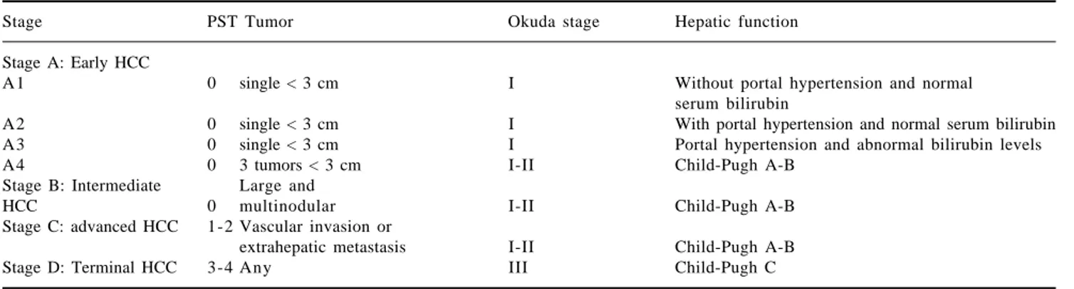
and predictive power of the CLIP score.44 The HCC
study clinic in Barcelona (BCLC) proposed an HCC classification (Table V) that has 4 principal stages, and divides patients into early (A), intermediate (B), ad-vanced (C), and terminal (D) stages. This system utiliz-es the clinical significance that HCC has in every pa-tient for normal daily activities. CLIP and BCLC sys-tems have been shown to provide more precise estimates of survival than the Okuda system. The BCLC system also appeared to be more accurate than the CLIP
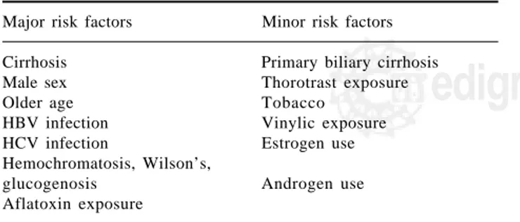
Figure 1. Recommended fo-llow-up algorithm for HCC. Modified by the authors from
J Hepatol 2001; 35: 421–30. USG+AFP every 6 months
Hepatic nodule Normal USG
HCC US+AFP/6m
CT/RM/Angio
USG/3 m +
-CT/RM/Angio Normal AFP
AFP < 400 Normal AFP Abnormal AFP
HCC +
-AFP > 400
B
D
C
A
Figure 2. MRI imaging of HCC. Axial images showing three separated nodules in the right lobe, with round bor-ders, satellite lesions, and a hypointense region in the cen-ter of the largest nodule, sug-gesting necrosis. A) T1/SE; B) T1/FAT SAT; C) T2; D) T2/ FAT SAT. Courtesy of Dr. Roberto Corona-Cedillo, Me-dica Sur, Mexico.
Table IV. CLIP staging system for HCC.
Points
Variables 0 1 2
Child-Pugh A B C
Tumor
morphology Single node Multiple Massive or nodules and Extension area Extension Extension ≥ 50% area ≤ 50% area ≤ 50%
AFP (ng/mL) < 400 ≥ 400 Portal vein
thrombosis No Yes
MG
edigraphic.com
score in identifying cases with better prognosis (small tumors, 3 cm in patients with well compensated liver
function and no portal hypertension).45
Treatment
Only 10 to 13 per cent of HCC patients can be cured with liver transplant, surgical resection and tumor ablation
therapies. Overall, liver transplants have low mortality.2
Non-surgical treatment
Arterial liver chemotherapy
Selective administration of chemotherapy in the he-patic artery is based on the idea that HCC irrigates from this artery so that the drug can be delivered direct to the tumor. The most common drugs used for this procedure are 5-fluorouracil and 5-fluorouracildesoxiribonucleosid. Unfortunately most patients with advanced HCC have as-sociated thrombocytopenia that contraindicates this pro-cedure. This procedure has not been demonstrated to
lower HCC mortality.46
Chemoembolization
Chemoembolization is the most commonly used
treat-ment for HCC that cannot be submitted to surgery.47 It is
based on the objective of tumor devascularization, in which the oxygen and nutrient supply to the tumor is blocked, resulting in tumor necrosis. The most common-ly used agents for this treatment are Gelfoam, polivinilic
acid, collagen, iodinized oil and angiotensine II.46,47
Ethanol percutaneous injection
Intratumoral injection of ethanol causes dehydrata-tion, intracellular coaguladehydrata-tion, necrosis, vascular
occlu-sion and tumor fibrosis. This technique has been used
primarily in small tumors, 3-5 cm.46
Radiation
Radiotherapy is not commonly used as a single treat-ment. It can be used on tumors with diameters smaller than 8 cm in patients with Child A and smaller than 5 cm
in patients with Child B.47
Cryosurgery
Cryosurgery has been used in patients with HCC and cirrhosis, with inadequate hepatic reserves and inade-quate or multifocal lesions. Survival is approximately 20
per cent in three years.47
Thermotherapy
Thermotherapy uses changes in temperature for tumor destruction. It is estimated that tumors as large as 9 cm
can be cured with this method.46
Systemic chemotherapy
The most common drugs used as palliative therapy are: 5-fluorouracil, doxorrubicin, interferon, cisplatine,
tamoxifen, and capecitabine.47 Interferon has been shown
to reduce incidence and recurrence of HCC in patients
with HCV, even in the absence of virological response.48
Surgical treatment
After decades of poorly defined decision-making cri-teria, surgery is the therapy of choice for HCC in selected
cases.49 Surgical options are liver resection and
trans-plantation. For the small group of non-cirrhotic patients and cirrhotic patients with acceptable residual liver
func-Table V. BCLC system for HCC staging.
Stage PST Tumor Okuda stage Hepatic function
Stage A: Early HCC
A1 0 single < 3 cm I Without portal hypertension and normal
serum bilirubin
A2 0 single < 3 cm I With portal hypertension and normal serum bilirubin
A3 0 single < 3 cm I Portal hypertension and abnormal bilirubin levels
A4 0 3 tumors < 3 cm I-II Child-Pugh A-B
Stage B: Intermediate Large and
HCC 0 multinodular I-II Child-Pugh A-B
Stage C: advanced HCC 1-2 Vascular invasion or
extrahepatic metastasis I-II Child-Pugh A-B
Stage D: Terminal HCC 3-4 Any III Child-Pugh C
Stage A y B: Require all criteria
D Motola-Kuba et al. Hepatocellular carcinoma 23
edigraphic.com
sustraídode-m.e.d.i.g.r.a.p.h.i.c cihpargidemedodabor
:rop odarobale FDP
VC ed AS, cidemihparG
arap
acidémoiB arutaretiL :cihpargideM
sustraídode-m.e.d.i.g.r.a.p.h.i.c
tion, liver resection is the first choice of treatment. Usual-ly, right hepatectomy induces greater decompensation than left hepatectomy. Indications for resection depend on tumor size, number and extrahepatic involvement
ac-cording to the Milano criteria.50 Patients with solitary
tu-mors of less than 5 cm, or up to three tutu-mors of less than 3 cm, without extrahepatic involvement, are candidates for resection, with a five-year survival up to 70 per cent
in some series.51 Abnormal serum bilirubin and portal
hy-pertension are the main clinical prognostic indicators of
survival after liver resection for HCC;52 these factors are
associated with a decrease in survival to less than 50 per cent at five years. Portal hypertension is suspected in pa-tients with less than 100,000 platelets/mm³ and sple-nomegaly in patients with ascites requiring diuretics, and confirmed by measuring hepatic vein pressure gradient or
finding esophageal varices in upper endoscopy.53 In
pa-tients with high bilirubin levels, low platelets or sple-nomegaly, transplant is the treatment of choice, although only 5 per cent of patients with HCC and cirrhosis are
chosen for this kind of treatment.49 The best histological
predictor of recurrence in operated patients is
microvas-cular invasion and additional tumor sites.54
Preoperative chemoembolization has not shown clear
benefits.55 Preoperative embolization of the hepatic
ar-tery and the portal vein of the affected hepatic lobes have probable benefits by inducing growth of the nonaf-fected lobes. However, this procedure carries the poten-tial risk of malignant hepatocytes being stimulated by
is-chemia to induce angiogenesis and tumor growth.56 In
some cases, ethanol ablation or thermoablation might be useful as a bridge to surgical resection or transplantation. Liver transplantation is indicated in patients with ad-vanced liver disease who meet the Milano criteria. MELD scores are currently used to allocate organ distribution, although MELD is considered a poor prognostic tool for HCC. Delays in proceeding could lead to disease pro-gression and a dismal prognosis. Twenty-two additional points are added to the MELD scores of patients with HCC if they meet surgical criteria, and a 10 per cent
in-crease is made for every three months of waiting.57 The
chance of five-year survival after liver transplantation is 60-70 per cent. Special postransplant management is a question of debate and frustration. Prevention of graft in-volvement during viral infection is mandatory, although not satisfactory; especially in Hepatitis C infected pa-tients in whom graft infection is observed in 90-100 per cent of cases. Some reports suggest that postoperative systemic chemotherapy (adriamycin) added to immuno-suppressive regimens reduces the risk of recurrence after
liver transplantation.58
Conclusions
HCC is a tumor that primarily affects patients of ad-vanced age. Its detection is very difficult because in most
cases it has an asymptomatic evolution and when symp-toms begin, most cases are already at an advanced stage with a low survival rate. Early detection is important in order to begin treatment as soon as possible and reduce mortality rates.
References
1. el-Serag HB. Epidemiology of hepatocellular carcinoma. Clin
Liver Dis 2001; 5: 87–107, vi.
2 . Marrero JA. Hepatocellular carcinoma. Curr Opin Gastroenterol 2003; 19: 243–9.
3 . El-Serag HB, Mason AC. Rising incidence of hepatocellular carcinoma in the United States. N Engl J Med 1999; 340: 7 4 5 – 5 0 .
4. Horie Y, Yamagishi Y, Kajihara M, Kato S, Ishii H. National survey of hepatocellular carcinoma in heavy drinkers in Japan.
Alcohol Clin Exp Res 2003; 27: 32S–6S.
5. Uetake S, Yamauchi M, Itoh S, Kawashima O, Takeda K, Ohata M. Analysis of risk factors for hepatocellular carcinoma in pa-tients with HBs antigen- and anti-HCV antibody-negative alco-holic cirrhosis: clinical significance of prior hepatitis B virus infection. Alcohol Clin Exp Res 2003; 27: 47S–51S.
6. Goncalves CS, Pereira FE, Gayotto LC. Hepatocellular carci-noma in Brazil: report of a national survey (Florianopolis, SC, 1995). Rev Inst Med Trop Sao Paulo 1997; 39: 165–70. 7. Cortes-Espinosa T, Mondragon-Sanchez R, Hurtado-Andrade H,
Sanchez-Cisneros R. Hepatocellular carcinoma and hepatic cirrhosis in Mexico: a 25 year necroscopy review. Hepatogastroenterology 1997; 44: 1401–3.
8 . Velazquez RF, Rodriguez M, Navascues CA, Linares A, Perez R, Sotorrios NG, Martinez I, et al. Prospective analysis of risk fac-tors for hepatocellular carcinoma in patients with liver cirrhosis.
Hepatology 2003; 37: 520–7.
9. Bailey MA, Brunt EM. Hepatocellular carcinoma: predisposing conditions and precursor lesions. Gastroenterol Clin North Am 2002; 31: 641–62.
10. Mendez-Sanchez N, Aguilar-Ramirez JR, Reyes A, Dehesa M, Juorez A, Castaneda B, Sanchez-Avila F, et al. Etiology of liver cirrhosis in Mexico. Ann Hepatol 2004; 3: 30–3.
11. Mendez-Sanchez N, Villa AR, Chavez-Tapia NC, Ponciano-Rodriguez G, Almeda-Valdes P, Gonzalez D, Uribe M. Trends in liver disease prevalence in Mexico from 2005 to 2050 through mortality data. Ann Hepatol 2005; 4: 52–5.
12. Libbrecht L, Desmet V, Roskams T. Preneoplastic lesions in hu-man hepatocarcinogenesis. Liver Int 2005; 25: 16–27. 13. Hytiroglou P. Morphological changes of early human
hepato-carcinogenesis. Semin Liver Dis 2004; 24: 65–75.
14. Borzio M, Fargion S, Borzio F, Fracanzani AL, Croce AM, Stroffolini T, Oldani S, et al. Impact of large regenerative, low grade and high grade dysplastic nodules in hepatocellular carci-noma development. J Hepatol 2003; 39: 208–14.
15. Kaplan DE, Reddy KR. Rising incidence of hepatocellular carci-noma: the role of hepatitis B and C; the impact on transplantation and outcomes. Clin Liver Dis 2003; 7: 683–714.
16. Zein NN, Poterucha JJ, Gross JB, Jr., Wiesner RH, Therneau TM, Gossard AA, Wendt NK, et al. Increased risk of hepatocellular carcinoma in patients infected with hepatitis C genotype 1b. Am
J Gastroenterol 1996; 91: 2560–2.
17. Tarao K, Rino Y, Ohkawa S, Tamai S, Miyakawa K, Takakura H, Endo O, et al. Close association between high serum alanine aminotransferase levels and multicentric hepatocarcinogenesis in patients with hepatitis C virus-associated cirrhosis. Cancer 2002; 94: 1787–95.
18. Bosch FX, Ribes J, Cleries R, Diaz M. Epidemiology of hepato-cellular carcinoma. Clin Liver Dis 2005; 9: 191–211, v. 19. Bressac B, Kew M, Wands J, Ozturk M. Selective G to T
MG
edigraphic.com
20. Lee YI, Lee S, Das GC, Park US, Park SM. Activation of theinsullike growth factor II transcription by aflatoxin B1 in-duced p53 mutant 249 is caused by activation of transcription complexes; implications for a gain-of-function during the for-mation of hepatocellular carcinoma. Oncogene 2000; 19: 3 7 1 7 – 2 6 .
21. Fracanzani AL, Conte D, Fraquelli M, Taioli E, Mattioli M, Losco A, Fargion S. Increased cancer risk in a cohort of 230 patients with hereditary hemochromatosis in comparison to matched con-trol patients with non-iron-related chronic liver disease.
Hepatology 2001; 33: 647–51.
22. Potempa J, Korzus E, Travis J. The serpin superfamily of pro-teinase inhibitors: structure, function, and regulation. J Biol Chem 1994; 269: 15957–60.
23. Parmar JS, Lomas DA. Alpha-1-antitrypsin deficiency, the serpinopathies and conformational disease. J R Coll Physicians
Lond 2000; 34: 295–300.
24. Ludwig J, Viggiano TR, McGill DB, Oh BJ. Nonalcoholic steatohepatitis: Mayo Clinic experiences with a hitherto unnamed disease. Mayo Clin Proc 1980; 55: 434–8.
25. Bellentani S, Saccoccio G, Masutti F, Croce LS, Brandi G, Sasso F, Cristanini G, et al. Prevalence of and risk factors for hepatic steatosis in Northern Italy. Ann Intern Med 2000; 132: 112–7. 26. Marceau P, Biron S, Hould FS, Marceau S, Simard S, Thung SN,
Kral JG. Liver pathology and the metabolic syndrome X in se-vere obesity. J Clin Endocrinol Metab 1999; 84: 1513–7. 27. Caldwell SH, Oelsner DH, Iezzoni JC, Hespenheide EE, Battle EH,
Driscoll CJ. Cryptogenic cirrhosis: clinical characterization and risk factors for underlying disease. Hepatology 1999; 29: 664–9. 28. Mendez-Sanchez N, Sanchez-Castillo CP, Villa AR, Madrigal H, Merino B, Garcia E, Lopez P, et al. The relationship of over-weight and obesity to high mortality rates from liver cirrhosis in Mexico. Ann Hepatol 2004; 3: 66–71.
29. Calle EE, Rodriguez C, Walker-Thurmond K and Thun MJ. Over-weight, obesity, and mortality from cancer in a prospectively studied cohort of U.S. adults. N Engl J Med 2003; 348: 1625–38. 30. Bugianesi E, Leone N, Vanni E, Marchesini G, Brunello F, Carucci P, Musso A, et al. Expanding the natural history of nonalcoholic steatohepatitis: from cryptogenic cirrhosis to hepatocellular car-cinoma. Gastroenterology 2002; 123: 134–40.
31. Ratziu V, Bonyhay L, Di Martino V, Charlotte F, Cavallaro L, Sayegh-Tainturier MH, Giral P, et al. Survival, liver failure, and hepatocellular carcinoma in obesity-related cryptogenic cirrho-sis. Hepatology 2002; 35: 1485–93.
32. Nair S, Mason A, Eason J, Loss G, Perrillo RP. Is obesity an independent risk factor for hepatocellular carcinoma in cirrho-sis? Hepatology 2002; 36: 150–5.
33. Ozturk M. Genetic aspects of hepatocellular carcinogenesis. Semin
Liver Dis 1999; 19: 235–42.
34. Macdonald GA. Pathogenesis of hepatocellular carcinoma. Clin
Liver Dis 2001; 5: 69–85.
35. Schafer DF, Sorrell MF. Hepatocellular carcinoma. Lancet 1999; 353: 1253–7.
36. Sohara N, Takagi H, Yamada T, Ichikawa T, Abe T, Itoh H, Mori M. Esophageal metastasis of hepatocellular carcinoma. Gastrointest
Endosc 2000; 51: 739–41.
37. Befeler AS, Di Bisceglie AM. Hepatocellular carcinoma: diagno-sis and treatment. Gastroenterology 2002; 122: 1609–19. 38. Ryder SD. Guidelines for the diagnosis and treatment of
hepato-cellular carcinoma (HCC) in adults. Gut 2003; 52 Suppl 3: iii1–8. 39. Johnson PJ. The role of serum alpha-fetoprotein estimation in the diagnosis and management of hepatocellular carcinoma. Clin
Liver Dis 2001; 5: 145–59.
40. Weissenberger C, Fiebig HH, Lutterbach J, Barke A, Momm F, Muller M, Witucki G, et al. Is there any correlation between MDR1, GST-pi-expression and CEA? Anticancer Res 2000; 20: 5139–44.
41. Taheri M, Saragovi U, Fuks A, Makkerh J, Mort J, Stanners CP. Self recognition in the Ig superfamily. Identification of precise subdomains in carcinoembryonic antigen required for intercel-lular adhesion. J Biol Chem 2000; 275: 26935–43.
42. Shouval D. HCC: what’s the score? Gut 2002; 50: 749–50. 4 3 . Levy I, Sherman M. Staging of hepatocellular carcinoma:
assessment of the CLIP, Okuda, and Child-Pugh staging sys-tems in a cohort of 257 patients in Toronto. Gut 2002; 50: 8 8 1 – 5 .
44. Ueno S, Tanabe G, Sako K, Hiwaki T, Hokotate H, Fukukura Y, Baba Y, et al. Discrimination value of the new western prognostic system (CLIP score) for hepatocellular carcinoma in 662 Japa-nese patients. Cancer of the Liver Italian Program. Hepatology 2001; 34: 529–34.
4 5 . Grieco A, Pompili M, Caminiti G, Miele L, Covino M, Alfei B, Rapaccini GL, et al. Prognostic factors for survival in patients with early-intermediate hepatocellular carcinoma undergoing non-surgical therapy: comparison of Okuda, CLIP, and BCLC staging systems in a single Italian centre.
Gut 2005; 54: 411–8.
46. Aguayo A, Patt YZ. Nonsurgical treatment of hepatocellular car-cinoma. Clin Liver Dis 2001; 5: 175–89.
47. Geschwind JF, Ramsey DE, Choti MA, Thuluvath PJ, Huncharek MS. Chemoembolization of hepatocellular carcinoma: results of a metaanalysis. Am J Clin Oncol 2003; 26: 344–9.
48. Yoshida H, Shiratori Y, Moriyama M, Arakawa Y, Ide T, Sata M, Inoue O, et al. Interferon therapy reduces the risk for hepatocel-lular carcinoma: national surveillance program of cirrhotic and noncirrhotic patients with chronic hepatitis C in Japan. IHIT Study Group. Inhibition of Hepatocarcinogenesis by Interferon Therapy.
Ann Intern Med 1999; 131: 174–81.
49. Rust C, Gores GJ. Locoregional management of hepatocellular carcinoma. Surgical and ablation therapies. Clin Liver Dis 2001; 5: 161–73.
50. Mazzaferro V, Regalia E, Doci R, Andreola S, Pulvirenti A, Bozzetti F, Montalto F, et al. Liver transplantation for the treatment of small hepatocellular carcinomas in patients with cirrhosis. N Engl
J Med 1996; 334: 693–9.
51. Bruix J, Llovet JM. Prognostic prediction and treatment strategy in hepatocellular carcinoma. Hepatology 2002; 35: 519–24. 52. Llovet JM, Fuster J, Bruix J. Intention-to-treat analysis of
surgi-cal treatment for early hepatocellular carcinoma: resection versus transplantation. Hepatology 1999; 30: 1434–40.
53. Bruix J, Castells A, Bosch J, Feu F, Fuster J, Garcia-Pagan JC, Visa J, et al. Surgical resection of hepatocellular carcinoma in cirrhotic patients: prognostic value of preoperative portal pres-sure. Gastroenterology 1996; 111: 1018–22.
54. Okada S, Shimada K, Yamamoto J, Takayama T, Kosuge T, Yamasaki S, Sakamoto M, et al. Predictive factors for postopera-tive recurrence of hepatocellular carcinoma. Gastroenterology 1994; 106: 1618–24.
55. Yamasaki S, Hasegawa H, Kinoshita H, Furukawa M, Imaoka S, Takasaki K, Kakumoto Y, et al. A prospective randomized trial of the preventive effect of pre-operative transcatheter arterial embolization against recurrence of hepatocellular carcinoma. Jpn
J Cancer Res 1996; 87: 206–11.
56. Farges O, Belghiti J, Kianmanesh R, Regimbeau JM, Santoro R, Vilgrain V, Denys A, et al. Portal vein embolization before right hepatectomy: prospective clinical trial. Ann Surg 2003; 237: 208–17.
57. Bruix J and Sherman M. Management of hepatocellular carci-noma. Hepatology 2005; 42: 1208–36.