a
RESEARCH ARTICLE
Cerebrospinal Fluid Control of Neurogenesis
Induced by Retinoic Acid During Early Brain
Development
M. I. Alonso,1,2C. Martı´n,1E. Carnicero,1,2D. Bueno,3and A. Gato1,2*
Embryonic-cerebrospinal fluid (E-CSF) plays crucial roles in early brain development including the con-trol of neurogenesis. Although FGF2 and lipoproteins present in the E-CSF have previously been shown to be involved in neurogenesis, the main factor triggering this process remains unknown. E-CSF contains all-trans-retinol and retinol-binding protein involved in the synthesis of retinoic acid (RA), a neurogene-sis inducer. In early chick embryo brain, only the mesencephalic-rombencephalic isthmus (IsO) is able to synthesize RA. Here we show that in chick embryo brain development: (1) E-CSF helps to control RA syn-thesis in the IsO by means of the RBP and all-trans-retinol it contains; (2) E-CSF has retinoic acid activity, which suggests it may act as a diffusion pathway for RA; and (3) the influence of E-CSF on embryonic brain neurogenesis is to a large extent due to its involvement in RA synthesis. These data help to
understand neurogenesis from neural progenitor cells.Developmental Dynamics :000–000, 2011. VC 2011
Wiley-Liss, Inc.
Key words:CSF; brain development; retinol binding protein; neural precursors; neural tube; neurogenesis
Accepted 5 April 2011
INTRODUCTION
At early stages of embryonic develop-ment, subsequent to the closure of the anterior neuropore, the architecture of the brain primordium is formed by a pseudo-monostratified neuroepithelium enclosing the brain vesicles, which are completely filled with embryonic cere-brospinal fluid (E-CSF). Recently, it has been shown that E-CSF plays an important role in brain development at both embryonic and foetal stages and performs several crucial functions (reviewed by Gato and Desmond, 2009).
It has previously been reported, in chick and rat embryos at early stages of development, that E-CSF exerts positive pressure against the neuroe-pithelial walls to generate an expan-sive force that drives morphogenesis (Gato et al., 1993; Desmond and Jacobson, 1977; Alonso et al., 1998, 1999; Desmond et al., 2005). However, in recent years new roles for E-CSF have been demonstrated in the behav-iour of neuroepithelial cell precursors, as it contributes to the regulation of the survival, proliferation, and neural differentiation of the neuroepithelial
progenitor cells during early (Gato et al., 2005) and late development (Mashayekhi et al., 2002; Owen-Lynch et al., 2003; Miyan et al., 2003), as well as collaborating with the isth-mic organiser in regulating mesence-phalic gene expression (Parada et al., 2005a). As a result, neurogenesis is one of the most relevant processes con-trolled by the action of E-CSF (Gato et al., 2005; Martı´n et al., 2006), and although it was recently demonstrated that FGF2 contained within chick E-CSF regulates cell proliferation and neurogenesis, the existence of another
Developmental Dynamics
1Departamento de Anatomı´a y Radiologı´a, Facultad de Medicina, Universidad de Valladolid, Valladolid, Spain
2Laboratorio de Desarrollo y Teratologı´a del Sistema Nervioso, Instituto de Neurociencias de Castilla y Leo´n (INCYL), Universidad de Valladolid, Valladolid, Spain
3Departament de Gene`tica, Facultat de Biologia, Universitat de Barcelona, Barcelona, Catalonia, Spain
Grant sponsor: Ministerio de Educacio´n y Ciencia; Grant number: BFU207/6516; Grant sponsor: Junta de Castilla y Leo´n; Grant number: GR195.
*Correspondence to: A. Gato, Departamento de Anatomı´a y Radiologı´a, Facultad de Medicina, Universidad de Valladolid, C/ Ramo´n y Cajal 7, E-47005-Valladolid, Spain. E-mail: gato@med.uva.es
DOI 10.1002/dvdy.22657
Published online in Wiley Online Library (wileyonlinelibrary.com).
VC2011 Wiley-Liss, Inc.
regulatory mechanism has been sug-gested (Martı´n et al., 2006; Parada et al., 2008a).
We recently showed that, at the be-ginning of primary neurogenesis, both avian and mammal E-CSF pro-teomes include a set of molecules whose roles during development in systems other than E-CSF are related to crucial biological functions (Dzie-gielewska et al., 1981; Gato et al., 2004; Zapaterra et al., 2007; reviewed by Parada et al., 2007). A remarkable finding that may contribute to an understanding of E-CSF’s overall effect on the neurogenesis of neuroe-pithelial precursors is the presence of retinol-binding protein (RBP) as well as all-trans retinol in both chick and rat E-CSF (Parada et al., 2005b, 2006, 2008b; Parvas et al., 2008). According to the literature, in systems other than E-CSF, RBP specifically binds to all-trans retinol, a member of the retinoid family of molecules, which is enzymatically metabolised into reti-noic acid (RA), a well-known morpho-gen that has crucial impact on CNS development (Moro et al., 1993; McCaffery and Dra¨ger, 2000; reviewed by Maden, 2002). Moreover, RA is known to be a neurogenic agent in both embryo and adult neural progeni-tor cells (Gonc¸alves et al., 2005; Wang et al., 2005; Jacobs et al., 2006).
RA synthesis requires the concur-rence of a precursor molecule, namely all-trans retinol, a carrier molecule such as RBP whose involvement has also been described in the delivery and uptake of all-trans retinol to the cells in which it is enzymatically metabolised in RA (Kawaguchi et al. 2007), and cells expressing the particu-lar enzymes involved in this metabo-lism, namely, retinaldehyde dehydroge-nases (RALDHs; Reijntjes et al., 2005) and retinol dehydrogenases (RDHs; Romand et al., 2008). At the earliest stages of chick embryo brain develop-ment, RALDH3 is located in the mesen-cephalic-rombencephalic isthmus (IsO), which is thought to be the RA source for mesencephalic neuroepithelium in chick embryos (Blentic et al., 2003; Par-ada et al., 2008b).
In this context, RA diffuses from the clusters of cells expressing these enzymes, such as those forming some of the well-known organising centres, to the target cells (Duester, 2008;
Blentic et al., 2003). When RA is taken up by the target cells, it binds to spe-cific nuclear receptors (RAR), regulat-ing a series of genes involved in neural differentiation and patterning of ante-rior-posterior and dorso-ventral axes (Kolm et al., 1997; Clotman et al., 1997; McCaffery and Dra¨ger, 2000; Begemann and Meyer, 2001; Diez del corral and Storey, 2001; Kudoh et al., 2002; Maden, 2002; McCaffery et al., 2003; Duester 2008).
In this report, we analyse the spe-cific role of the RBP-all-trans retinol system contained within E-CSF in the regulation of mesencephalic neuroepi-thelial cell neurogenesis in chick embryos at the beginning of primary neurogenesis, by means of RA synthe-sis in the IsO. Using functional analy-sis, we demonstrate that E-CSF is a source of all-transretinol for the IsO cells to synthesise RA, and that the presence of RBP in CSF is necessary for the efficient delivery of all-trans
retinol to IsO cells for such synthesis. Finally, we show that the former mechanism is involved in mesence-phalic neurogenesis during early brain development.
RESULTS
Retinoic Acid Synthesis by
the IsO Is Controlled by
Molecules Contained Within
E-CSF
In this study, we first aimed to dem-onstrate whether a relationship exists between E-CSF composition and RA synthesis in the IsO, as we previously described that in chick embryos (1) this fluid contains the precursor mole-cule for this morphogen, i.e., all-trans-retinol, as well as the carrier protein, RBP, and (2) IsO cells are the only cells in the early brain primor-dium neuroepithelium expressing a recognised RA-synthesising enzyme, RALDH3, at this developmental stage. As RA is a relatively unstable com-pound and is present in very low con-centrations in physiological conditions of biological samples, we employed a biological test to detect RA activity, based on the reporter F9-1.8 cell line culture. These cells, derived from a mouse teratocarcinoma, contain a RARE promoter coupled to the lacZ (b-galactosidase) reporter gene, and
are very sensitive to RA induction. To test the responsiveness of these cells in our culture conditions, we first cultured F9-1.8 cells in a chemically defined medium supplemented with several different concentrations of commercial RA, which has been shown, by Horton and Maden (1995), to be at a physiological level in various embryonic tissues (Fig. 1A).
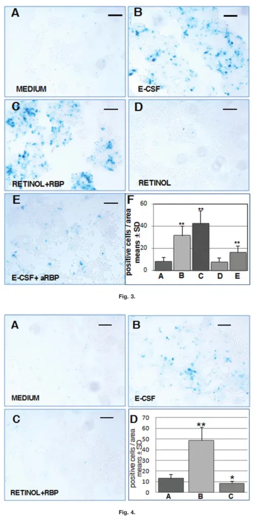
We then developed a co-culture technique of IsO cells and F9-1.8 re-porter cells to detect the production of RA by the former (Figs. 2A and 3). In order to test basalb-galactosidase ac-tivity, we co-cultured these two cell types with a chemically defined me-dium; few F9-1.8 blue cells (Fig. 3A; see also Fig. 3F for a plotted compari-son with other co-culture conditions) were seen, suggesting that in the ab-sence of E-CSF, IsO cells are not self-sufficient to synthesize retinoic acid. Conversely, when F9-1.8 and IsO cells were co-cultured in a defined medium supplemented with E-CSF (Fig. 3B and F), the number of F9-1.8 blue cells significantly increased, suggest-ing that E-CSF contains factors that activate the synthesis of retinoic acid by the IsO cells.
Due to the presence, as previously described, of all-trans retinol and RBP in the E-CSF, we then checked the role of these two molecules in RA synthesis by the IsO cells. When F9-1.8 and IsO cells were co-cultured in a defined medium supplemented with both commercial all-trans retinol and RBP (Fig. 3C and F), the number of blue F9-1.8 cells was significantly greater than for the basal level of the chemically defined medium (Fig. 3A and F), and, what is interesting, greater also than the cells cultured in the presence of E-CSF. However, when these cells were co-cultured with a defined medium supplemented only with all-trans retinol (Fig. 3D and F), the number of blue F9-1.8 cells was similar to that for the basal condition (Fig. 3A and F), which supports the involvement of RBP in this process.
To check this point, we then co-cul-tured F9-1.8 and IsO cells with a defined medium supplemented with E-CSF, to which antibodies specifi-cally recognising RPB were added to block its biological activity (Lide´n and Erickson, 2005). In this experiment (Fig. 3E and F), the number of blue
F9-1.8 cells was significantly lower than when only E-CSF, or alterna-tively retinolþRBP, was present, but also significantly higher than for the basal level (Fig. 3A and F). This result confirms the involvement of RBP con-tained in CSF, in RA synthesis by the IsO. Taken together, these results sug-gest that (1) the retinol contained within E-CSF is the substrate for IsO cells to synthesise RA, and (2) the RPB contained within E-CSF is needed for all-trans retinol to reach the IsO cells.
We then checked whether the F9-1.8-induced activity exerted by the IsO cells in the presence of E-CSF in the culture medium, or, alternatively, by retinolþRBP, was due specifically to IsO cells, and thus to the RA they synthesise by means of RALDH3. In order to do this, we co-cultured F9-1.8 reporter cells with dorsal mesence-phalic neuroepithelial cells not con-taining the IsO (Figs. 2B and 4). When these cells were co-cultured with the chemically defined medium (Fig. 4A; see also Fig. 4D for a plotted compari-son with other co-culture conditions), the number of blue F9-1.8 cells was very low, similar to the basal condition in which F9-1.8 cells were co-cultured with IsO cells together also with the defined medium (Fig. 3A and F). Sur-prisingly, when these cells were co-cul-tured with the defined medium supple-mented with E-CSF, the number of blue F9-1.8 cells significantly increased (Fig. 4B and D). In contrast, when these cells were co-cultured with the defined medium supplemented with commercial all-trans retinol þ RBP (Fig. 4C and D), the number of blue cells was not significantly differ-ent from those for the basal condition. Taken together, all such findings rule out unknown RALDH activity in mes-encephalic cells and strongly suggest the presence of retinoic acid within the E-CSF, directly capable of activating the reporter promoter of F9-1.8 cells, supporting the idea of CSF as a diffu-sion way for retinoic acid.
To further check whether E-CSF has a direct role in the reporter activ-ity of F9-1.8 cells independent from IsO cells, i.e., if RA activity is involved, we cultured F9-1.8 cells in the absence of neuroepithelial cells with a defined medium supplemented with E-CSF at several different con-centrations, thereby performing a
dose-response plot. As shown in Figure 1B, the number of blue F9-1.8 cells induced by E-CSF increased in paral-lel to their concentration in the culture medium, which strongly supports the presence of RA activity within this embryo fluid; in addition, they were similar to values plotted in our RA dose response, where the highest dose we used was 12% of CSF in the culture medium (compare Fig. 1A with B).
RBP Contained Within E-CSF
Contributes to Mesencephalic
Neurogenesis Regulation by
Controlling RA Synthesis
Since the involvement of RA had been previously noted in the induction of neurogenesis in specific regions of the central nervous system, we then aimed to demonstrate that the capacity of E-CSF to control RA syn-thesis in the IsO cells affects neuroe-pithelial neurogenesis.
In order to show the role of the spe-cific elements of the RBP/retinol-IsO-RA system contributing to primary neurogenic induction from the E-CSF, and so as to analyse the possible syn-ergistic involvement of other neuro-genic factors also contained within E-CSF (e.g., FGF2), an organotypic tissue culture of mesencephalicþIsO neuroepithelium was used. All the elements required to investigate neu-rogenesis activation by RA were included in this in vitro approach, i.e.,
raldh-3-expressing IsO cells, mesence-phalic neuroepithelium capable of responding to neurogenic RA induc-tion, and a chemically defined medium in which the different elements could be added or subtracted (see Fig. 6).
Organotypic mesencephalicþIsO neuroepithelium cultures were made under several different conditions to evaluate induced neurogenesis (Fig. 5), which was detected by beta3-tubu-lin immunostaining. As previously reported (Gato et al., 2005), the E-CSF present in the culture medium is capa-ble of sustaining neuroepithelial cell survival as well as activating prolifera-tion and neurogenesis at a level simi-lar to that for embryos developed in ovo. Thus, as a positive control, we used explants cultured with E-CSF-supplemented medium (Fig. 5A; see also Fig. 5H for a plotted comparison
between different culture conditions); this represented the maximum induc-tion of neurogenesis mediated by E-CSF under these culture conditions. As a negative control, we used explants cultured only in a chemically defined medium (Fig. 5B and H), which represented the basal neuro-genic capacity of the neuroepithelium alone, and/or due to the accumulated factors it might still contain after a 24-hr culture. This value was considered to be inherent to all culture conditions. Since the neurogenic role of FGF2 contained within E-CSF at this devel-opmental stage has also been previ-ously described (Martı´n et al., 2006), we checked neurogenic induction of FGF2 in this experimental model system. To achieve this, we cultured mesencephalicþIsO explants with a medium supplemented with FGF2 (Fig. 5C and H) and the number of beta3-tubulin expressing cells was significantly higher than in the nega-tive controls. Similarly, as it has been proposed that RA has neurogenic properties, we cultured explants with the medium supplemented with com-mercial all-trans RA (Fig. 5D and H), the number of beta3-tubulin-express-ing cells bebeta3-tubulin-express-ing similar to that for explants cultured in the presence of FGF2. Subsequently, to verify the potential synergistic effect of RA and FGF2, mesencephalicþIsO explants were cultured in a chemically defined medium supplemented with both RAþFGF2. In these explants (Fig. 5E and H), the number of beta3-tubulin-expressing cells was higher than those cultured in the medium supplemented only with RA or, alternatively, with FGF2, and similar to the explants cul-tured in the E-CSF-supplemented me-dium. Taken together, these results suggest that both FGF2 and RA are synergistically involved in primary neurogenesis induced by E-CSF in mesencephalic progenitor cells.
Following this, and to confirm the involvement of the RBP-retinol sys-tem contained in E-CSF in inducing primary neurogenesis through the retinol uptake of IsO cells and its sub-sequent conversion to RA, mesence-phalicþIsO explants were cultured with a medium supplemented with E-CSF, to which an antibody blocking RBP biological activity had been added (Fig. 5F and H). In these
explants, the number of beta3-tubulin-expressing cells was drastically reduced with respect to the positive controls, although there were more pos-itive cells than in the negative controls; this demonstrates the involvement of RBP in the induction of primary neuro-genesis and suggests that E-CSF
Fig. 3.
Fig. 4. Fig. 1. Dose–response of F9-1.8 cells.
A:Dose-response plot of F9-1.8 cells to differ-ent nM concdiffer-entrations of commercial RA. B:Dose-response plot of F9-1.8 cells cultured with different concentrations of E-CSF.
Fig. 2. Diagrams showing the co-culture of F9-1.8 cells with neuroepithelial cells.A: Co-culture of F9-1.8 cells with IsO cells. B: Co-culture of F9-1.8 cells with mesencephalic cells lacking the IsO.
contains other factors apart from RA involved in neural differentiation, such as, for example, FGF2.
Alternatively, explants were cul-tured with a medium supplemented with all-trans retinolþRBP to emulate physiological conditions (Fig. 5G and H). In these explants, the number of beta3-tubulin-positive cells was signifi-cantly higher than in the negative con-trols, and similar to that for explants cultured in the presence of RA, which suggests that RBP is needed to produce the maximum rate of RA synthesis in the IsO cells. Taken together, these results show that RBP from CSF indi-rectly contributes to neural mesence-phalic differentiation, promoting the conversion of retinol into RA in the IsO.
DISCUSSION
In this report, we have shown that E-CSF plays a role in regulating RA synthesis in the mesencephalic-rom-boencephalic isthmus (IsO) during early chick development, and that, to some extent, this is a means by which E-CSF controls neuroepithelial neuro-genesis. These results (summarized in Fig. 6) contribute to an under-standing of the general role of E-CSF during early brain development.
E-CSF Contributes to
Regulating RA Synthesis in
the IsO
The classic view is that IsO is a source of RA, mainly due to the expression of enzymes involved in RA synthesis,
i.e., RALDHs and RDHs (Reijntjes et al., 2005; Parada et al., 2008b; Romand et al., 2008). In mouse embryo models, the involvement of these enzymes in RA synthesis has been shown by using a set of knock-outs (Niederreither et al., 2002; Mic et al., 2002; Ribes et al., 2006; Molot-kova et al., 2007). In avians, however, no equivalent functional evidence exists, so further experimental approaches are needed. Our data, based on the co-culture of IsO cells and F9-1.8 cells engineered to monitor the presence of RA, indicate that in chick embryos, the IsO cells actively produce RA. Thus, at early stages of brain development, IsO can be consid-ered a local source for this morphogen. Therefore, due to our knowledge that IsO is an organising centre controlling the expression of genes involved in CNS regionalisation and patterning, some of its recognised capacities as an organising centre may be the result of their capacity to produce RA. However, it should be noted that RALDH3 is not expressed in mouse Iso, but at later stages of development RALDH2 is expressed in hindbrain meninges (McCaffery et al., 2006).
Hence, one of the main contribu-tions of this study is the demonstra-tion that E-CSF is involved in the con-trol of RA synthesis in the IsO. We demonstrate that E-CSF is also able to regulate the activity of one specific neuroepithelial functional area, the IsO, which indicates that E-CSF does not only have general roles for all neuroepithelial cells, but also
particu-lar roles for different populations of neuroepithelial cells.
In a previous study, we demon-strated that the presence of RBP and all-trans retinol in the E-CSF is dynamic and varies over time, and we suggested that their changes in con-centration may be related to their specific roles in RA synthesis (Parada et al., 2008b). The results presented in this report demonstrate that E-CSF regulation of RA synthesis in the IsO is exerted by means of RBP and the all-trans retinol it contains. It is interesting to point out that E-CSF not only acts as a retinol reservoir for the IsO, but also facilitates its uptake by means of RBP, which therefore may act as a control mechanism for RA synthesis, suggesting a new role for E-CSF on brain development.
The traditional opinion regarding RA diffusion into cells is that it is stored as a gradient in the extracellu-lar matrix. Our results strongly sug-gest the existence of another source of RA in the E-CSF. Our findings imply a previously undocumented means of possible RA supply to all brain cavity neuroepithelial cells during early stages of development, namely, via this embryonic fluid. This RA diffusion channel may act in parallel with other RA sources, such as, for example, RA synthesised by the frontonasal surface ectoderm, known to act on the prosen-cephalon neuroepithelium (Molotkova et al., 2007). This should be taken into account in order to understand the general role of RA on brain develop-ment. It is, therefore, interesting to note that in adult choroids plexus there is also the expression of RALDHs and RDHs, which suggests a similar diffu-sion method for RA in the adult brain, via CSF in the brain ventricles.
RA Is Involved in
Neurogenesis Induced
by E-CSF
As has been stated above, E-CSF plays a role in controlling embryonic neurogenesis. Some particular factors have also been associated with early brain neurogenesis, such as, for example, FGF2 and certain lipopro-teins, although it is unclear whether they act as direct activators of neuro-genesis or by indirect means. For
Fig. 3. E-CSF regulation of RA synthesis by the IsO cells (see also Fig. 6A).A:X-gal develop-ment of F9-1.8 cells after co-culture with IsO cells in chemically defined medium.B:X-gal devel-opment of F9-1.8 cells after co-culture with IsO cells in E-CSF supplemented medium.C:X-gal development of F9-1.8 cells after co-culture with IsO cells in defined medium supplemented with commercial all-trans retinol and RBP.D:X-gal development of F9-1.8 cells after co-culture with IsO cells in defined medium to which all-trans retinol has been added. E:X-gal develop-ment of F9-1.8 cells after co-culture with IsO cells in E-CSF suppledevelop-mented medium to which antiRBP commercial antibody has been added. F: Quantitative analysis of F9-1.8 blue cells under several different co-culture conditions. Foot letters of each bar correspond to letters of picture in this same figure. Asterisks denote values that differ significantly (P<0.05) from con-trols according to the two-tailed Student’st-test (see Table 1). Scale bar¼100mm.
Fig. 4. E-CSF regulation of RA synthesis by the IsO cells; co-culture of F9-1.8 cells with mes-encephalic cells lacking the IsO (see also Fig. 6B). A:X-gal development of F9-1.8 cells after co-culture with mesencephalic cells in chemically defined medium.B:X-gal development of F9-1.8 cells after co-culture with mesencephalic cells in E-CSF supplemented medium.C:X-gal de-velopment of F9-1.8 cells after co-culture with mesencephalic cells in defined medium supple-mented with commercial all-trans retinol and RBP. D:Quantitative analysis of F9-1.8 blue cells under several different co-culture conditions. Foot letters of each bar correspond to letters of picture in this same figure. Asterisks denote values that differ significantly (P<0.05) from con-trols according to the two-tailed Student’st-test (see Table 2). Scale bar¼100mm.
example, it has been suggested that FGF2 may act on neurogenesis by driving cells to terminal mitoses (Mar-tin et al., 2006), and that the action of lipoproteins in this cellular process is the supply to the neuroepithelial cells of the necessary building materials (Parada et al., 2008b). Moreover, the presence of other neurogenic factors in the E-CSF has been suggested to account for overall neurogenesis (Mar-tin et al., 2006; Parada at al., 2008a; Gato and Desmond, 2009). Our results show that mesencephalic neurogenesis
Fig. 5.
Fig. 6.
in chick embryos is also driven by RA activity, which comes from the IsO, which in turn is regulated by E-CSF via the RBP and the all-trans retinol it contains. This concurs with previous reports demonstrating that RA is one of the early signalling components in the chain of molecular events that results in fully differentiated neurons in specific regions of the central nerv-ous system (McCaffery and Dra¨ger, 2000). Here, data presented in this report offer a better understanding of the mechanisms regulating neurogen-esis from neural progenitor cells in the mesencephalon.
Furthermore, the presence of all molecular and enzymatic elements needed for RA activity taking place in adult brain has been demonstrated, especially in areas that are known to display neurogenesis from neural stem cells, like, for example, the hip-pocampus and the subventricular zone (McCaffery et al., 2006). In addi-tion, a direct relationship between RA and neurogenesis in these brain areas has been demonstrated (Jacobs et al., 2006 and Bonnet et al., 2008, for hip-pocampus; Wang et al., 2005, and Zhang et al., 2006, for the subventric-ular zone). In this regard, although
RA is not alone in inducing the differ-ent steps in neuronal differdiffer-entiation, it may act as a master control in cer-tain regions of the central nervous system by inducing the expression of a variety of factors that control differen-tiation (Joh et al., 1992; Kaplan et al., 1993; Afink et al., 1995; Xie et al., 1997; Sizemore et al., 1998; Yoshizawa et al., 1998; Maden, 2002; Duester, 2008). Consequently, the knowledge of the mechanisms involved in neurogen-esis activation from neural precursors in both embryonic and adult brain, may be a hidden key to developing neuro-regenerative strategies based on the expansion and differentiation of neural precursor populations in the adult brain.
EXPERIMENTAL
PROCEDURES
Obtaining Embryonic
Cerebrospinal Fluid
Fertile chicken eggs were incubated at 38C in a humidified atmosphere to obtain chick embryos at the desired developmental stages. Embryos were incubated to stage HH24 (Hamburger and Hamilton, 1951) to obtain E-CSF. After dissection of the embryos from the extra-embryonic membranes, E-CSF was aspirated as previously described (Gato et al., 2004). To minimize protein degradation, E-CSF samples were kept at 4C, aliquoted, lyophilized, and fro-zen at40C, until they were used.
Organotypic Cultures
Organotypic cultures of both mesence-phalicþ IsO neuroepithelial explants were produced as described by Gato et al. (2005). Briefly, Mesencephalicþ IsO neuroectodermal explants were cultured in vitro for 24 hr, between HH20 and HH23, fixed peripherally to small rectangles of Millipore (Billerica, MA) filters (0.8 mm pore size) with a tungsten needle, and then cultured with a chemically defined serum-free medium (basal medium; DMEM:F12; Sigma, St. Louis, MO) with 1% ascor-bic acid, or, alternatively, with this me-dium supplemented with various addi-tives, at 37C and with 5% CO2.
To generate different experimental conditions, the following additives were administered to 250ml of the ba-sal medium: 15% E-CSF; FGF2 (50 ng
Fig. 5. RBP regulation of RA neurogenic induction in vitro monitored by the presence of beta3-tubulin-positive cells. A:MesencephalicþIsO neuroepithelum explants cultured in vitro with E-CSF supplemented medium. Note the presence of beta3-tubulin-positive cells.B: Mesencepha-licþIsO neuroepithelum explants cultured in vitro with chemically defined medium. Note that the number of beta3-tubulin-positive cells is much lower than in explants cultured with E-CSF sup-plemented medium.C:MesencephalicþIsO neuroepithelum explants cultured in vitro with defined medium to which commercial FGF2 has been added. D: Mesencephalic neuroepithelumþIsO explants cultured in vitro with defined medium to which commercial RA has been added.E: Mes-encephalicþIsO neuroepithelum explants cultured in vitro with defined medium to which both commercial RA and FGF2 have been added.F:MesencephalicþIsO neuroepithelum explants cul-tured in vitro with E-CSF supplemented medium to which antibody to RBP has been added to block its biological activity.G:MesencephalicþIsO neuroepithelum explants cultured in vitro with defined medium to which commercial all-trans retinol and RBP has been added.H: Quantitative analysis of beta3-tubulin-positive cells under several different culture conditions. Foot letters of each bar correspond to letters of picture in this same figure. Asterisks denote values that differ significantly (P <0.05) from controls according to the two-tailed Student’st-test (see Table 3). Scale bar¼50mm. All images have the same orientation as A. bs, basal side; as, apical side.
Fig. 6. Diagram showing the proposed mechanism to CSF influence in neurogenesis via RA. Schematic diagram showing how E-CSF contributes to RA synthesis. Dorsal mesencephalon and mesencephalo-rombencephalic istmo (Iso) regions are represented in dark blue. RA requires the uptake of retinol, mediated by retinol binding protein, from E-CSF (Sky blue) to Iso cells which expresses RALDH3 (orange). We propose that RA diffuses (red) from the neuroepithelium or E-CSF to target cells to induce neurogenesis (green cells).
TABLE 2. Coculture of F9-1.8 Cells With Mesencephalic Cellsa
Medium E-CSF RETINOLþRBP
Means 13.10 4.82** 8.53*
SD 63.88 612.26 62.10
aValues and statistical significance plotted in graph bars of Figure 4.
*P<0.05. **P<0.001.
TABLE 1. Coculture of F9-1.8 Cells With Iso Cellsa
Medium E-CSF RETINOLþRBP RETINOL E-CSFþaRBP
Means 8.17 31.35* 42.52* 7.85 16.60* SD 63.55 68.91 611.29 63.52 65.67
aValues and statistical significance plotted in graph bars of Figure 3.
*P<0.001.
of human recombinant; Sigma); Reti-noic Acid (RA; 33 nM of all-trans reti-noic acid; Sigma); RA þ FGF2 (at stated concentrations); 15% E-CSF þ antibody to RBP (20 ng; Labvision, Fre-mont, CA) and all-trans retinol (340 nM; Fluka)þRBP (500 ng; Sigma).
Culture of F9-1.8 Cell Line
and Co-Culture With
Neuroepithelial Cells
F9-1.8 cell line was maintained as previously described (Schulz and Gais, 1989) on gelatine-coated tissue culture flasks in Advanced-DMEM (Gibco, Gaithersburg, MD) supple-mented with 7.5% heat-inactivated FBS, 44 nM NaHCO3, and 4 mg/ml
G418 (Sigma). The cells were plated for experimentation at a density of 3.0
104 cells/cm2 and cultured over-night at 37C in a 5% CO2
atmos-phere. This cell line contains the RARE promoter coupled to the lacZ
(b-galactosidase) reporter gene. For the co-culture of F9-1.8 cells with neuroepithelial cells, the surface ectoderm and most of the mesen-chyme were removed with tungsten needles and then 4 small pieces of IsO neuroepithelium (including the tip of the dorsolateral Iso fold, which encompassed the area of RALDH3 expression), or, alternatively, 3 small pieces of dorsolateral mesencephalic neuroepithelium (without RALDH expression), in order to obtain an equivalent amount of tissue, were micro-dissected and mechanically dis-aggregated in 4C Ringer and recov-ered in pellet following centrifuging. After re-suspending the cells in DMEM:F12:G418, they were added to the wells containing F9-1.8 cells (Fig. 6A and B). They were then co-cul-tured for 24 hr at 37C in a 5% CO2
atmosphere. To generate the different experimental conditions, the follow-ing additives were administered to
300 ml of the basal medium: RA (3.3, 33, 66, 133, and 266 nM of all-trans
retinoic acid; Sigma); E-CSF at 15%; E-CSF (15%) þ antibody to RBP (20 ng; Labvision); all-trans retinol (340 nM; Fluka, St. Louis, MO) þ RBP (500 ng; Sigma); andall-transretinol (at the concentration stated above). In some experiments, F9-1.8 cells were cultured in the absence of neuroepi-theial cells, in the basal medium sup-plemented with several different con-centrations of E-CSF (0.4, 1.2, 2, 6, and 12%).
Colour Development of
F9-1.8 Cells
After experiments with the F9-1.8 cells, these were fixed in 4% parafor-maldehide in PBS, washed in PBS, and subsequently in de-ionised water, and exposed to 20 mg/ml of X-gal solu-tion (Sigma) for 1 hr at 37C, as described by Sonneveld et al. (1999). Culture wells have 1.9 mm2, the F9-1.8-positive cells were counted in 20 different, randomly selected, areas of 0.55 mm2for each experimental con-dition (4 areas per each well, from 5 different wells). The results were expressed as means of positive cells/ area and standard deviation, and the significant differences were evaluated by a two-tailed Student’st-test.
Beta3-Tubulin Determination
In order to detect early neuronal dif-ferentiation, we monitored beta 3-tubulin (Tuj-1) expression. After orga-notypic culture, the explants were fixed in Carnoy for 20 min, dehy-drated in an alcohol series, passed through xylene, and embedded in par-affin. After the tissues had been cut transversally, they were de-paraffi-nized and subsequently incubated with a monoclonal antibody anti-beta3-tubulin at 1/500 (BAbCO) and
an anti-mouse antibody conjugated to FITC at 1/64 (Sigma) for 1 hr at RT. For visualization and photographing of the preparations, we used a confo-cal microscope (Zeiss LSM-310; Zeiss, Thornwood, NY). A quantitative anal-ysis of beta3-tubulin localization was performed by counting the number of neuroepithelial cells with immuno-stained cytoplasm in 20 1,900mm2 microscopic areas, taken from the central area of each explant and from 5 different explants. The average of each condition and the standard error were plotted, and their significance was tested by an unpaired two-tailed Student’st-test.
ACKNOWLEDGMENTS
The authors thank Dra.Sagrario Call-ejo, Pilar Martı´n, and Isabel Garcia for technical support, Dr. Ricard Albalat, from the University of Barcelona, for F9 1.8 cells, and Dr. David Rixham for language translation assistance.
REFERENCES
Afink GB, Niste´r M, Stassen BH, Joosten PH, Rademakers PJ, Bongcam-Rudloff E, Van Zoelen EJ, Mosselman S. 1995. Molecular cloning and functional char-acterization of the human platelet-derived growth factor alpha receptor gene promoter. Oncogene 10:1667–1672. Alonso MI, Gato A, Moro JA, Barbosa E. 1998. Disruption of proteoglycans in neural tube fluid by beta-D-xyloside alters brain enlargement in chick embryos. Anat Rec 252:499–508. Alonso MI, Gato A, Moro JA, Martin P,
Barbosa E. 1999. Involvement of sul-fated proteoglycans in embryonic brain expansion at earliest stages of develop-ment in rat embryos. Cells Tissues Organs 165:1–9.
Begemann G, Meyer A. 2001. Hindbrain patterning revisited: timing and effects of retinoic acid signalling. BioEssays 23:981–986.
Blentic A, Gale E, Maden M. 2003. Reti-noic acid signalling centres in the avian embryo identified by sites of expression of synthesising and catabolising enzymes. Dev Dyn 227:114–127.
TABLE 3. Mesencephalic1Iso Cells Explants Culturea
E-CSF Medium FGF-2 R.A. R.A.þFGF-2 E-CSFþaRBP RETINOLþRBP
Means 49.80 15.25* 33.62* 30.50* 51.58 21.68* 27.80* SD 66.79 62.50 66.37 66.45 64.94 67.48 65.30
aValues and statistical significance plotted in graph bars of Figure 5.
*P<0.001.
Bonnet E, Touyarot K, Alfos S, Pallet V, Higueret P, Abrous DN. 2008. Retinoic acid restores adult hippocampal neuro-genesis and reverses spatial memory deficit in vitamin A deprived rats. PLoS ONE 3:e3487.
Clotman F, Van Maele-Fabry G, Picard J. 1997. Retinoic acid induces a tissue-spe-cific deletion in the expression domain of Otx2. Neurotoxicol Teratol 19:163–169. Desmond ME, Jacobson AG. 1977.
Embry-onic brain enlargement requires cere-brospinal fluid pressure. Dev Biol 57: 188–198.
Desmond ME, Levitan ML, Haas AR. 2005. Internal luminal pressure during early chick embryonic brain growth: de-scriptive and empirical observations. Anat Rec 285:737–747.
Diez del corral R, Storey K. 2001. Markers in vertebrate neurogenesis. Nat Rev Neurosci 2:835–839.
Duester G. 2008. Retinoic acid synthesis and signaling during early organogene-sis. Cell 134:921–931.
Dziegielewska KM, Evans CA, Lai PC, Lorscheider FL, Malinowska DH, Moll-gard K, Saunders NR. 1981. Proteins in cerebrospinal fluid and plasma of fetal rats during development. Dev Biol 83: 193–200.
Gato A, Desmond ME. 2009. Why the embryo still matters: CSF and the neuro-epithelium as interdependent regulators of embryonic brain growth, morphogen-esis and histiogenmorphogen-esis. Dev Biol 327: 263–272.
Gato A, Moro JA, Alonso MI, Pastor JF, Represa JJ, Barbosa E. 1993. Chondroi-tin sulphate proteoglycan and embry-onic brain enlargement in the chick. Anat Embryol 188:101–106.
Gato A, Martı´n P, Alonso MI, Martı´n C, Pulgar MA, Moro JA. 2004. Analysis of cerebro-spinal fluid protein composition in early developmental stages in chick embryos. J Exp Zool 301:280–289. Gato A, Moro JA, Alonso MI, Bueno D,
De La Mano A, Martı´n C. 2005. Embry-onic cerebrospinal fluid regulates neu-roepithelial survival, proliferation, and neurogenesis in chick embryos. Anat Rec 284A:475–484.
Gonc¸alves MBCV, Boyle J, Webber DJ, Hall S, Minger SL, Corcoran J PT. 2005. Timing of the retinoid-signalling pathway determines the expression of neuronal markers in neural progenitor cells. Dev Biol 278:60–70.
Hamburger V, Hamilton HL. 1951. A se-ries of normal stages in the develop-ment of the chick embryo. J Morphol 88:49–92.
Horton C, Maden M. 1995. Endogenous distribution of retinoids during normal development and teratogenesis in the mouse embryo. Dev Dyn 202:312–323. Jacobs S, Lie DC, DeCicco KL, Shi Y,
DeLuca LM, Gage FH, Evans RM. 2006. Retinoic acid is required early during adult neurogenesis in the den-tate gyrus. PNAS 103:3902–3907. Joh T, Darland T, Samuels M, Wu JX,
Ad-amson ED. 1992. Regulation of epidermal
growth factor receptor gene expression in murine embryonal carcinoma cells. Cell Growth Differ 3:315–325.
Kaplan DR, Matsumoto K, Lucarelli E, Thiele CJ. 1993. Induction of TrkB by retinoic acid mediates biologic respon-siveness to BDNF and differentiation of human neuroblastoma cells. Eukaryotic Signal Transduction Group. Neuron 11: 321–331.
Kawaguchi R, Yu J, Honda J, Hu J, White-legge J, Ping P, Wiita P, Bok D, Sun H. 2007. A membrane receptor for retinol binding protein mediates cellular uptake of vitamin A. Science 315:820–825. Kolm PJ, Apekin V, Sive H. 1997.
Xeno-pus hindbrain patterning requires reti-noid signaling. Dev Biol 192:1–16. Kudoh T, Wilson SW, Dawid IB. 2002.
Distinct roles for Fgf, Wnt and retinoic acid in posteriorizing the neural ecto-derm. Development 129:4335–4346. Lide´n M, Erikson, U. 2005. Development
of a versatile reporter assay for studies of retinol uptake and metabolism in vivo. Exp Cell Res 310:401–408. Maden M. 2002. Retinoid signalling in
the development of central nervous sys-tem. Nat Rev Neurosci 3:843–853. Martin C, Bueno D, Alonso MI, Moro JA,
Callejo S, Parada C, Martı´n P, Carnicero E, Gato A. 2006. FGF2 plays a key role in embryonic cerebrospinal fluid trophic properties over chick embryo neuroepi-thelial stem cells. Dev Biol 297:402–416. Mashayekhi F, Draper CE, Bannister CM, Pourghasem M, Owen-Lynch PJ, Miyan JA. 2002. Deficient cortical development in the hydrocephalic Texas (H-Tx) rat: a role for CSF. Brain 125:1859–1874. McCaffery P, Dra¨ger U. 2000. Regulation
of retinoic acid signaling in the embry-onic nervous system: a master differen-tiation factor. Cytokine Growth FR 11: 233–249.
McCaffery PJ, Adams J, Maden M, Rosa-Molinar E. 2003. Too much of a good thing: retinoic acid as an endogenous regulator of neural differentiation and exogenous tera-togen. Eur J Neurosci 18:457–472. McCaffery P, Zhang J, Crandall JE. 2006.
Retinoic acid signaling and function in the adult hippocampus. J Neurobiol 66: 780–791.
Mic FA, Haselbeck RJ, Cuenca AE, Duester G. 2002. Novel retinoic acid generating activities in the neural tube and heart identified by conditional res-cue of Raldh2 null mutant mice. Devel-opment 129:2271–2282.
Miyan JA, Nabiyouni M, Zendah M. 2003. Development of the brain: a vital role for cerebrospinal fluid. Can J Physiol Pharmacol 81:317–328.
Molotkova N, Molotkov A, Duester G. 2007. Role of retinoic acid during fore-brain development begins late when Raldh3 generates retinoic acid in the ventral subventricular zone. Dev Biol 303:601–610.
Moro Balba´s JA, Gato A, Alonso Revuelta MI, Pastor JF, Represa JJ, Barbosa E. 1993. Retinoic acid induces changes in the rhombencephalic neural crest cells
migra-tion and extracellular matrix composimigra-tion in chick embryos. Teratology 48:197–206. Niederreither K, Vermot J, Fraulob V,
Chambon P, Dolle P. 2002. Retinalde-hyde dehydrogenase 2 (RALDH2)- inde-pendent patterns of retinoic acid synthesis in the mouse embryo. Proc Natl Acad Sci USA 99:16111–16116. Owen-Lynch PJ, Draper CE, Mashayekhi
F, Bannister CM, Miyan JA. 2003. Defec-tive cell cycle control underlies abnormal cortical development in the hydroce-phalic Texas rat. Brain 126:623–631. Parada C, Martı´n C, Alonso MI, Moro JA,
Bueno D, Gato A. 2005a. Embryonic cerebrospinal fluid collaborates with the isthmic organizer to regulate mesence-phalic gene expression. J Neurosci Res 82:333–345.
Parada C, Gato A, Bueno D. 2005b. Mamma-lian embryonic cerebrospinal fluid pro-teome has greater apolipoprotein enzyme pattern complexity than the avian pro-teome. J Proteome Res 4:2420–2428. Parada C, Gato A, Aparicio M, Bueno D.
2006. Proteome analysis of chick embryonic cerebrospinal fluid. Proteomics 6:312–320. Parada C, Parvas M, Bueno D. 2007.
Cer-ebrospinal fluid proteome from neural development to neurodegenerative dis-eases. Curr Proteomics 4:89–106. Parada C, Escola`-Gil JC, Bueno D. 2008a.
Low-density lipoproteins from embry-onic cerebrospinal fluid are required for neural differentiation. J Neurosci Res 86:2674–2684.
Parada C, Gato A, Bueno D. 2008b. All-trans retinol and retinol-binding pro-tein from embryonic cerebrospinal fluid exhibit dynamic behaviour during early central nervous system development. Neuroreport 19:945–950.
Parvas M, Parada C, Bueno D. 2008. A blood-CSF barrier function controls em-bryonic CSF protein composition and homeostasis during early CNS develop-ment. Dev Biol 321:51–63.
Reijntjes S, Blentic A, Gale E, Maden M. 2005. The control of morphogen signal-ling: Regulation of the synthesis catabo-lism of retinoic acid in the developing embryo. Dev Biol 285:224–237.
Ribes V, Wang Z, Dolle´ P, Niederreither K. 2006. Retinaldehyde dehydrogenase 2 (RALDH2)-mediated retinoic acid syn-thesis regulates early mouse embryonic forebrain development by controlling FGF and sonic hedgehog signaling. De-velopment 133:351–361.
Romand R, Kondo T, Cammas L, Hashino E, Dolle´ P. 2008. Dynamic expression of the retinoic acid-synthesizing enzyme retinol dehydrogenase 10 (rdh10) in the developing mouse brain and sensory organs. J Comp Neurol 508:879–892. Schulz WA, Gais G. 1989. Constitutive
c-myc expression enhances proliferation of differentiating F9 teratocarcinoma cells. Biochim Biophys Acta 1013:125–132. Sizemore N, Choo CK, Eckert RL, Rorke
EA. 1998. Transcriptional regulation of the EGF receptor promoter by HPV16 and retinoic acid in human ectocervical epithelial cells. Exp Cell Res 244:349–356.
Sonneveld E, van den Brink CE, van der Leede BJ, Maden M, van der Saag PT. 1999. Embryonal carcinoma cell lines sta-bly transfected with mRARbeta2-lacZ: sen-sitive system for measuring levels of active retinoids. Exp Cell Res 250:284–297. Wang T-W, Zhang H, Parent JM. 2005.
Retinoic acid regulates postnatal neuro-genesis in the murine subventricular zone-olfactory bulb pathway. Develop-ment 132:2721–2732.
Xie P, Cheung WM, Ip FC, Ip NY, Leung MF. 1997. Induction of TrkA receptor by retinoic acid in leukaemia cell lines. Neuroreport 8:1067–1070.
Yoshizawa M, Miyazaki H, Kojima S. 1998. Retinoids potentiate transforming growth factor-beta activity in bovine en-dothelial cells through up-regulating the expression of transforming growth factor-beta receptors. J Cell Physiol 176:565–573.
Zappaterra MD, Lisgo SN, Lindsay S, Gygi SP, Walsh CA, Ballif BA. 2007. A comparative proteomic analysis of human and rat embryonic cerebrospinal fluid. J Proteome Res 6:3537–3548. Zhang X, Klueber KM, Guo Z, Cai J, Lu
C, Winstead WI, Qiu M, Roisen FJ. 2006. Induction of neuronal differentia-tion of adult human olfactory neuroepi-thelial-derived progenitors. Brain Res 1073–1074:109–119.