The T-loop Extension of the Tomato Protein Kinase
AvrPto-dependent Pto-interacting Protein 3 (Adi3) Directs
Nuclear Localization for Suppression of Plant Cell Death
*
□SReceived for publication, February 23, 2010, and in revised form, April 5, 2010Published, JBC Papers in Press, April 6, 2010, DOI 10.1074/jbc.M110.117416
María J. Ek-Ramos‡, Julian Avila‡, Cheng Cheng‡, Gregory B. Martin§¶, and Timothy P. Devarenne‡1
From the‡Department of Biochemistry and Biophysics, Texas A&M University, College Station, Texas 77843, the§Boyce Thompson Institute for Plant Research, Ithaca, New York 14853, and the¶Department of Plant Pathology and Plant-Microbe Biology, Cornell University, Ithaca, New York 14853
In tomato (Solanum lycopersicum), resistance to Pseudomo-nas syringaepv.tomatois elicited by the interaction of the host Pto kinase with the pathogen effector protein AvrPto, which leads to various immune responses including localized cell death termed the hypersensitive response. The AGC kinase Adi3 functions to suppress host cell death and interacts with Pto only in the presence of AvrPto. The cell death suppression (CDS) activity of Adi3 requires phosphorylation by 3-phospho-inositide-dependent protein kinase 1 (Pdk1) and loss of Adi3 function is associated with the hypersensitive response cell death initiated by the Pto/AvrPto interaction. Here we studied the relationship between Adi3 cellular localization and its CDS activity. Adi3 is a nuclear-localized protein, and this localization is dictated by a nuclear localization signal found in the Adi3 T-loop extension, an⬃80 amino acid insertion into the T-loop, or activation loop, which is phosphorylated for kinase activa-tion. Nuclear localization of Adi3 is required for its CDS activity and loss of nuclear localization causes elimination of Adi3 CDS activity and induction of cell death. This nuclear localization of Adi3 is dependent on Ser-539 phosphorylation by Pdk1 and non-nuclear Adi3 is found in punctate structures throughout the cell. Our data support a model in which Pdk1 phosphorylation of Adi3 directs nuclear localization for CDS and that disruption of Adi3 nuclear localization may be a mechanism for induction of cell death such as that during the Pto/AvrPto interaction.
During the resistance response of plants to pathogens, pro-grammed cell death (PCD)2occurs as part of the hypersensitive
response (HR), which functions to limit pathogen spread (1, 2). It has been over 100 years since the first description of the HR and its associated cell death (3). However, not until relatively recent times has the search for genes and pathways regulating PCD during the HR received much attention. This search has been difficult, but has identified several kinases and transcrip-tion factors involved in PCD (1, 4, 5) as well as lipid biosynthetic genes that produce lipid second messengers regulating PCD (6 –9).
In tomato (Solanum lycopersicum), the causative agent of bacterial speck disease is Pseudomonas syringae pv. tomato (Pst). Interaction of the tomato resistance protein kinase Pto with thePsteffector protein AvrPto brings about the HR and resistance toPst(10). Studies have been undertaken to identify genes involved in PCD associated with Pto-mediated HR and revealed a downstream MAP kinase, MAPKKK␣, that func-tions in the induction of cell death during both resistance and susceptibility (11).
Another gene that was identified as a Pto-interacting protein was the tomato protein kinase Adi3, which only interacts with Pto in the presence of AvrPto (12). Subsequently, we have shown Adi3 to function as a negative regulator of plant cell death (13) and thus, it may be the functional homologue of PKB (aka Akt), a major PCD suppressor in mammals (13–15). Adi3 is phosphorylated by 3-phosphoinositide-dependent protein kinase-1 (Pdk1) at Ser-539, which is required for full Adi3 kinase activity and cell death suppression (CDS) ability (13). Mutation of Ser-539 to Asp is capable of mimicking this phos-phorylation event, creating a constitutively active Adi3 capable of CDS (13). Adi3 cell death control can also be associated with MAPKKK␣that is involved in Pto-mediate HR cell death (11, 13). Adi3 is a member of the AGC kinase family, which is a con-served family of eukaryotic Ser/Thr protein kinases that regu-late many basic cellular processes such as transcription, trans-lation, cell growth, apoptosis, and cytoskeletal remodeling (16). In mammalian systems, AGC kinases affect downstream signal-ing components through direct mechanisms, includsignal-ing regula-tion of nuclear shuttling, activities of transcripregula-tion factors (17), phosphorylation-dependent trafficking of signaling proteins (18), and chromatin remodeling (19). The cell death (apoptosis) regulator PKB is also an AGC kinase family member.
Little is known about the functions of plant AGC kinases. However, there has been several recent studies reported. As with mammalian systems, many plant AGC kinases are acti-*This work was supported by USDA-CSREES Grant 2007-35319-17832 (to
G. B. M. and T. P. D.), USDA-AFRI Grant 2010-65108-20526 (to T. P. D.), and by Texas A&M University Dept. of Biochemistry and Biophysics start-up funds (to T. P. D.).
□S
The on-line version of this article (available at http://www.jbc.org) contains
supplemental Figs. S1–S5.
1To whom correspondence should be addressed. Tel.: 979-862-6509; Fax:
979-845-9274; E-mail: tpd8@tamu.edu.
2The abbreviations used are: PCD, plant cell death; Adi3, AvrPto-dependent
vated by Pdk1 (13, 16, 20 –23).Arabidopsiscontains at least 39 AGC kinase family members (16, 21, 24) and some of their functions include blue-light signaling (25), root hair develop-ment (22, 26, 27), oxidative burst signaling (23, 27), and auxin signaling (24, 28). Group VIIIa AGC kinases (of which Adi3 is a member) are specific to plants and are mainly distinguished from mammalian kinases by a large 70 –100 amino acid inser-tion in the activainser-tion loop, or T-loop, referred to as the T-loop extension (16). Similar, but much shorter (30 – 60 amino acids) T-loop extensions are also present in other AGC kinases such as the Ndr family of AGC kinases (29). In mammals and yeast, Ndr kinases regulate processes such as cell morphological changes, exit from mitosis, and apoptosis. The Ndr T-loop extension functions in cell localization and regulation of kinase activity (29 –32). Very little is known about the function of the T-loop extension in plant AGC kinases. The T-loop extension of only twoArabidopsisgroup VIIIa AGC kinases have been studied and appear to contain cellular localization signals (21). However, the amino acid motifs within these T-loop extensions responsible for directing cellular localization have not been identified.
Here we show that the Adi3 T-loop extension is required for nuclear localization and that Adi3 nuclear localization is required for its CDS activity. Non-nuclear localization confines Adi3 to intracellular punctate membrane structures and a con-comitant loss of CDS. These studies raise the possibility of restricting Adi3 nuclear localization as a means to induce plant PCD.
EXPERIMENTAL PROCEDURES
Plasmid Construction and Mutagenesis—The Adi3⌬T-loop
construct was created by producing anAdi3 PCR fragment lacking the T-loop extension (bp 1369 –1608). First, a PCR product containingAdi3bp 1336 –1368⫹1609 –2103 was syn-thesized and called fragment 1. Next anotherAdi3PCR prod-uct, fragment 2, from bp 1–1368 was produced that overlapped fragment 1. Both fragment 1 and 2 were then used as templates for overlapping PCR (33) with a 30-bp forward primer originat-ing atAdi3bp 1 and a 27 bp reverse primer originating atAdi3 bp 2103 to produce a PCR product lacking the T-loop exten-sion. For bacterial expression, full-length tomatoAdi3 (13) andAdi3⌬T-loopwere PCR amplified and cloned into pMal-c2 (New England Biolabs). Mutations were created using site directed mutagenesis with Pfu Turbo (Stratagene). For proto-plast expression, transient leaf expression, and subcellular localization studies,Adi3,Adi3⌬T-loop, and otherAdi3mutants were cloned into the BamHI/XbaI restriction sites of pTEX-eGFPto create N-terminal GFP fusions. For Agrobacterium-mediated transient expression,35Spro:eGFP-Adi3was EcoRI/ HindIII digested from pTEX and cloned into the same sites of pCambia1300. ForGFP-NES/NLSconstructs,eGFPwas PCR amplified with aGFP-specific forward primer and a reverse primer containing theAdi3NLS plusGFP(5⬘ -CCGggattcCT-AAGGCTTAGGTGT TTTTTTCTTGCTCTTTTGAGGTT-TGTATAGTTCATC-3⬘; BamHI lowercase; linker bold; NLS underlined, bp 1509 –1542; GFP italics) for 3⬘-end fusion or with a GFP-specific reverse primer and a forward primer con-taining theAdi3NES plusGFP(5⬘-CCGggtaccATG
CCTTTC-TTGGACGATCTTTCAATCCGGATGCCA
AGTAAAGGA-GAACAA-3⬘; KpnI lowercase; linkers bold;NESunderlined, bp 373– 490;GFPitalics) for 5⬘-end fusion and cloned into the KpnI/BamHI restriction sites of pTEX.
The tomato exportin-1 (Xpo1) cDNA was cloned by a com-bination of methods. First, the Arabidopsis Xpo1 cDNA sequence (34) (GenBankTM accession Y18469) was used to
screen the tomato EST data base for contigs containing the tomatoXpo1cDNA. From this search, clone cTOF-19-M20 was identified as containing a partial tomato Xpo1 cDNA lacking the first 1466 5⬘base pairs. A full-length cDNA (GenBankTMaccession GU126514) was identified by cloning
the 1466 5⬘base pairs by 5⬘-RACE and a full-length open read-ing frame amplified by RT/PCR usread-ing a forward primer based at the ATG start codon (5⬘ -ATGGCGGCGGAGAAGCTG-AGA-3⬘) and a reverse primer based at the TGA stop codon (5⬘-TCATGAATCCACCATTTCATC-3⬘). The resulting 3228-bp PCR product was cloned into the yeast two-hybrid bait vec-tor pEG202, and protein interactions analyzed by yeast two-hybrid as previously described (13).
Protoplast Transient Expression and Cellular Fractionation— Protoplasts were isolated from leaves of 3-week-old PtoR or prf-3tomato plants and transformed with 20g of DNA for each construct as previously reported (13). Samples of trans-formed protoplasts were taken 16 h after transformation for protein expression determination, subcellular fractionation, and confocal microscopy. This time point was selected to min-imize amount of dead cells, which is maximal at 24 h, to visual-ize expressed protein. Protoplast transformation efficiency was determined to be 80% by counting the number of GFP-positive protoplasts out of a total of 100 cells. Subcellular fractions were isolated using the following protocol: Total cell extract was obtained by gently shaking transformed protoplasts in 100l of Buffer A (10 mMMES-HCl pH 5.7, 1Msucrose, 5 mMMgCl2, 2
mM -mercaptoethanol, 10 l/ml phosphatase inhibitors (Sigma), 10 l/ml proteinase inhibitors (Sigma), 10 M MG-132, 0.2% Triton X-100). Nuclei were pelleted from total cell extract by centrifugation at 5,000⫻gfor 10 min at 4 °C followed by three washes with 100l of Buffer A and centrifu-gation at 5,000⫻gfor 10 min at 4 °C. Nuclei were lysed for SDS-PAGE analysis by vortexing in 30l of Buffer B (10 mM MES-HCl pH 5.7, 1Msucrose, 5 mMMgCl2, 2 mM
Confocal Microscopy—Tomato protoplasts, after 16 h of transformation and leaf sections taken from PtoR tomato leaves that were Agrobacterium-infiltrated, were imaged using an Olympus FV1000 confocal microscope at the Texas A&M Uni-versity Microscopy and Imaging Center. Images were collected using system software FV10-ASW 1.6 with a 60⫻/1.2 water immersion objective in the XYZ scan mode (1.14m/slice). Excitation and emission wavelengths were as follows: eGFP, 488 nm excitation, 507 nm emission; hoechst 33342 (Sigma), 343 nm excitation, 460 nm emission; and chlorophyll autofluo-rescence, 470 nm excitation, 680 nm emission. All cell localiza-tion analyses with confocal microscopy were carried out at least three independent times with a minimum of 30 protoplast cells and five leaf sections from three independently infiltrated leaves viewed each time. All confocal images are shown as Z-stacks and presented images are representative of typical cell localization for all proteins seen during independent experi-ments. For imaging protoplasts transformed withGFP-Adi3 andGFP-Adi3mutants, the wavelength power was increased 2-fold in comparison to the power used for imaging protoplasts transformed withGFPalone.
Kinase Assays—In vitrokinase assays were done at least three independent times using purified Escherichia coli-expressed
MBP-tagged and immunoprecipi-tated GFP-tagged proteins as previ-ously described (13).
AvrPto and Other Treatments— PtoR andpfr-3protoplasts express-ingeGFP-Adi3or othereGFP-Adi3 mutants for 14 h were transformed with pTEX-avrPto-FLAG. Samples were collected at 0.5, 1.5, 3, and 4 h for determination of cell viability. For nuclei staining and leptomycin B treatment, protoplasts expressing the indicated constructs for 14 h were treated with 10 M Hoechst 33342 for 2 h or 20 nMleptomycin B (Sigma) for 90 min followed by confocal microscopy imaging. All experiments were carried out a min-imum of three times, and images presented are representative of the experiments.
Agrobacterium-mediated Tran-sient Expression—Agrobacterium tumefaciens strain GV2260 was used forAgrobacterium-mediated transient expression inNicotiana benthamiana and PtoR tomato leaves as previously reported (13). Adi3 constructs were expressed from pCambia-1300 and avrPto-FLAGfrom pBTEX.
Cell Viability Measurements— Conductivity tests were carried out on 3 leaf discs (1-cm diameter) obtained from leaves transiently expressing Adi3 proteins. Disks were placed in 3 ml of dH2O for
4 h with gentle shaking at room temperature and conductivity measured with an Acorn Con 5 meter (Oakton Instruments, Vernon Hills, IL). Cell death in leaves transiently expressing Adi3 proteins was visualized by vacuum infiltrating 0.1% Evans blue into leaf disks followed by washing with dH2O and
depig-mentation with Carnoy’s solution (60% ethanol, 30% CHCl3,
10% acetic acid). Protoplast cell viability was determined using Evans blue as previously reported (13). All cell viability assays were carried out a minimum of three independent times.
RESULTS
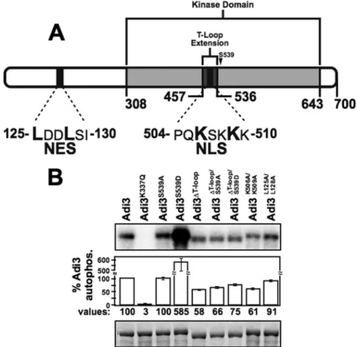
Adi3 Contains a Nuclear Localization Signal in the T-loop Extension and an N-terminal Nuclear Export Signal—A search of the Adi3 protein sequence for cellular localization signals using the WoLF PSORT protein localization predictor (36) revealed a monopartite basic nuclear localization signal (NLS) in the Adi3 T-loop extension from amino acids 504 –510 (PQKSKKK, Fig. 1A). Additionally, the NESnet nuclear export signal (NES) search engine, identified a leucine-rich NES in the N-terminal region of Adi3 from amino acids 125–130 (LDDLSI, Fig. 1A).
FIGURE 1.T-loop extension deletion effects on autophosphorylation activity of Adi3.A, Adi3 protein domains showing location of the T-loop extension and NLS and NES signals.B, kinase activity of Adi3 mutants tested byin vitro
autophosphorylation assays. MBP-tagged proteins were expressed enE. coli, purified, and 5g of each protein incubated with [␥-32P]ATP in anin vitrokinase assay. Quantification software was used to normalize the
Because the Adi3 NLS is located in the T-loop extension, two different mutants to analyze contributions of this sequence to Adi3 function were generated as well as a mutant of the NES sequence. The Adi3 T-loop extension was deleted by overlap-ping PCR to generate the Adi3⌬T-loopmutant, and the first (Lys-506) and third (Lys-509) Lys residue of the NLS were mutated to Ala creating the Adi3K506A/K509Amutant. Mutation of
equiv-alent Lys residues in NLS sequences similar to that in Adi3 is sufficient to prevent protein nuclear localization (37, 38). Sim-ilarly, in NES sequences analogous to that in Adi3, mutation of one or more Leu residues is sufficient to show nuclear accumu-lation of the protein (39). Thus, Leu-125 and Leu-128 were mutated to Ala to generate the Adi3L125A/L128Amutant.
To determine if deletion of the Adi3 T-loop extension affects Adi3 kinase activity, autophosphorylation activity of the Adi3⌬T-loopprotein was tested and compared with known Adi3
autophosphorylation mutants. As seen previously, the S539D mutation produces a constitutively active Adi3 with greatly enhanced kinase activity (13) (Fig. 1B). Deletion of the T-loop extension (Adi3⌬T-loop) reduced Adi3 autophosphorylation
activity⬃42%, which may be a cause of deleting the 79 amino acids of the T-loop extension within the catalytic kinase domain. Introduction of the S539D mutation into Adi3⌬T-loop
(Adi3⌬T-loop/S539D) increased Adi3⌬T-loopautophosphorylation
by⬃19% (Fig. 1B) indicating that mimicking Pdk1 phosphory-lation of Adi3⌬T-loop (Adi3⌬T-loop/S539D) does not increase
Adi3⌬T-loopautophosphorylation activity to the extent of wild
type. Interestingly, the NLS mutant (Adi3K506A/K509A) had
autophosphorylation activity close to that of Adi3⌬T-loop(Fig. 1B) suggesting these Lys residues offer important structural features to Adi3 required for autophosphorylation activity. The NES mutant, Adi3L125A/L128Adid not drastically affect Adi3
autophosphorylation activity (Fig. 1B). The K337Q mutation (Adi3K337Q) eliminates ATP binding and kinase activity in
wild-type protein (13). Because the T-loop extension deletion and the NLS mutation affected Adi3 autophosphorylation activity, we next tested if these mutations had effects on Adi3 CDS activ-ity and cellular localization.
In Vivo Expression Levels of GFP-Adi3 Expressed from the 35S Promoter and Functionality of the GFP-Adi3 Protein—Our Adi3 functional and localization studies use a GFP-Adi3 pro-tein expressed from the cauliflower mosaic virus 35S promoter (35Spro). Use of the 35Spro can lead to overexpression and accumulation of large amounts of protein, possibly leading to mislocalization. Thus, we analyzed the GFP-Adi3 protein levels in tomato protoplasts expressing35Spro:GFP-Adi3and com-pared this to the protein levels of two other proteins expressed from the35Spro:AvrPto-BFP, a Psteffector protein fused to BFP, and GFP alone. Western blot analysis indicated that the detection levels of GFP-Adi3 were very low and required 8 times and 40 times as much total protein to be detected com-pared with AvrPto-BFP or GFP, respectively (Fig. 2A). This sug-gests that GFP-Adi3 proteins do not reach levels that may cause problems in cellular localization analysis. In addition, we found one nonspecific band in our Western blot that could be detected only when high concentrations of total protein were used (asteriskin Fig. 2Aandsupplemental Fig. S1). This protein is larger than that of GFP alone suggesting it is not GFP and
indicating the presence of intact GFP-Adi3 as expressed in pro-toplasts. Additionally, the GFP-Adi3 protein used for subse-quent localization and functional analyses was also fully kinase active as expected (Fig. 2B).
Ser-539 Phosphorylation and an Intact NLS Are Important for Adi3 CDS Activity—We used our Adi3 expressing proto-plast system (13) to test different Adi3 kinase activity mutants for compromised cell death control (Fig. 2C). These assays make two premises. First, PCD is a process that is always “on” and there are proteins such as PKB and Adi3 that continually suppress PCD to prevent cell death (13–15). Loss of these pro-teins relieves suppression of PCD and initiates cell death as has been seen for PKB (40, 41) and we have seen for Adi3 (13). Second, overexpression of functionally compromised or consti-tutively active forms of PCD suppressor kinases will outcom-pete the endogenous protein, acting in a dominant negative or dominant manner, to induce or suppress cell death, respec-tively. This has been shown for PKB (42– 46) and Adi3 (13). In our current assays, GFP-Adi3 proteins were expressed in pro-toplasts for 24 h and cell viability measured by Evans blue stain-ing as we have previously done (13). These assays used proto-plasts from PtoR tomato plants, which contain a functionalPto
gene and show strong induction of HR cell death in response to thePsteffector protein AvrPto (10). For this reason, we used AvrPto expression as a positive control for strong cell death induction. Expression of wild-type Adi3 showed a moderate increase in cell viability over the vector alone (Fig. 2C) support-ing its role in CDS (13). The kinase-inactive Adi3K337Qshowed
a reduction in CDS and the constitutively active Adi3S539Dhad increased CDS (Fig. 2C) further indicating that Adi3 kinase activity and Pdk1 phosphorylation (via the Pdk1 phosphoryla-tion mimic) is required for Adi3 PCD control. Expression of Adi3⌬T-loopshowed a reduction in CDS similar to Adi3K337Q
(Fig. 2C). However, introduction of the phosphomimic S539D mutation (Adi3⌬T-loop/S539D) was able to restore CDS activity
for Adi3⌬T-loopto near wild-type levels (Fig. 2C). Introduction
of the nonphosphorylatable S539A mutation into Adi3⌬T-loop (Adi3⌬T-loop/S539A) showed a strong loss of CDS (Fig. 2C).
The NLS mutant Adi3K506A/K509Aalso showed loss of CDS
equal to that of Adi3⌬T-loop(Fig. 2C). The Adi3 NES mutant,
Adi3L125A/L128A, had increased CDS comparable to Adi3S539D
(Fig. 2C). These data would suggest that a functional Adi3 NLS is required for proper CDS and that mimicking the Pdk1 phos-phorylation can restore CDS activity to nonfunctional NLS mutants.
Subcellular Distribution of Adi3⌬T-loopProteins—To analyze
the effect on Adi3 subcellular localization due to the loss of the T-loop extension, subcellular fractions from protoplasts expressing GFP-Adi3 proteins for 16 h were analyzed by Western blot (Fig. 2D). The Adi3 and Adi3S539D proteins
showed similar localization in the nucleus and membrane frac-tions, while no protein was detected in the soluble fraction (Fig. 2D). This localization pattern of GFP-Adi3 was confirmed with Adi3 tagged with the small, 9-amino acid HA epitope (supplemental Fig. S2A) indicating the large GFP protein did not affect localization of the Adi3 protein. The Adi3⌬T-loop
indicating a lack of a functional NLS in the Adi3⌬T-loopprotein.
Introduction of the S539D mutation (Adi3⌬T-loop/S539D) caused
a substantial shift of the Adi3⌬T-loopprotein to the nuclear
frac-tion and reducfrac-tion in membrane localizafrac-tion (Fig. 2D). The Adi3⌬T-loop/S539Aprotein was not found in the nuclear fraction but was detectable in the membrane fraction only as a high molecular weight smear, suggesting post-translational modifi-cation (Fig. 2D). These data indicate that the Adi3 NLS in the T-loop extension directs nuclear localization and that mimick-ing Pdk1 phosphorylation of Adi3 without the T-loop exten-sion (Adi3⌬T-loop/S539D) can redirect the protein to the nucleus.
Taken together with the CDS assays (Fig. 2C), these data sug-gest that loss of Adi3 nuclear localization (deletion of T-loop
extension or NLS mutant) impairs the ability of Adi3 to suppress cell death. Restoring nuclear localiza-tion (S539D introduclocaliza-tion into Adi3⌬T-loop) is capable of restoring Adi3 CDS. Additionally, our data support our earlier findings that overexpression of non-functional mutant forms of Adi3 act in a dom-inant negative manner with regard to the endogenous Adi3 protein (13).
Confirmation of Adi3 Nuclear Localization—To confirm Adi3 nuclear localization, GFP-Adi3 expressing protoplasts were treated with the nuclear export inhibitor leptomycin B, which restricts nucle-ar/cytoplasmic shuttling proteins to the nucleus (47, 48). The protoplasts were also treated with the DNA stain Hoechst 33342 to identify nuclei (49) followed by confocal microscopy imaging of the GFP and Hoechst 33342 signals. The results showed that leptomycin B treat-ment reduced cytoplasmic GFP-Adi3 and restricted the protein to the nucleus (Fig. 3A). We also cloned the nuclear export protein Exportin-1 (Xpo1), which binds leucine-rich NESs (34) and showed that Adi3 interacts with Xpo1 in a yeast two-hybrid assay (Fig. 3B). The functionality of the Adi3 NLS was also confirmed by fusing the NLS to the C terminus of GFP (GFP-NLS) and analyzing for nuclear accumulation of GFP. The protein was expressed in tomato protoplasts treated with hoechst 33342 for nucleus identification and cellular localization viewed by confocal microscopy. Protein expression was confirmed by␣-GFP Western blot (Fig. 3D). GFP-NLS protein localization was reduced in the cytoplasm, but retained in the nucleus suggesting the Adi3 NLS is a functional nuclear localization signal (Fig. 3C). A similar analysis was carried out with the Adi3 NES sequence, which was fused to the N terminus of GFP (NES-GFP). If the NES is functional, NES-GFP protein should accumulate in the cyto-plasm. However, the Adi3 NES was incapable of reducing GFP nuclear localization (Fig. 3C) indicating the Adi3 NES may be a weak export signal unable to maintain nuclear exclusion of the small GFP protein, which can freely enter the nucleus (50, 51). Adi3 Nuclear Localization Is Required for PCD Suppression in Tomato and Tobacco Leaves—Because Adi3 NLS loss, either by deletion of the T-loop extension or NLS mutation, causes FIGURE 2.Cellular localization of GFP-Adi3 proteins regulates cell death suppression (CDS) activity.
A, GFP-Adi3 protein expression levels under the 35S promoter. Tomato protoplasts were transformed with the indicated DNA constructs and protein expressed for 16 h followed by total protein extraction and␣-GFP Western blot analysis.B, GFP-Adi3 autophosphorylation activity. Recombinant GFP-Adi3 protein was immu-noprecipitated using␣-GFP-agarose and tested in anin vitrokinase assay as in Fig. 1B. Values are reported as a percentage of wild-type Adi3 autophosphorylation and are average of three independent experiments.Error barsare standard error.Top panel: kinase assay (phosphorimage).Bottom panel: assay input (Coomassie Blue-stained gel).C, cell viability of protoplasts expressing GFP-Adi3 plus NLS and NES mutants. Tomato protoplasts expressing the indicated proteins for 24 h were analyzed for cell viability by Evans Blue staining to identify dead cells. Data represent the average of five independent experiments.Error barsare standard error.One asterisk
Adi3 to have compromised PCD control in protoplasts, func-tionality of GFP-Adi3 and GFP-Adi3⌬T-loopproteins in leaf
tis-sue was analyzed utilizingAgrobacterium-mediated transient expression in tomato (Fig. 4A) andN. benthamiana(Fig. 4B). As with our protoplast functionality assays (Fig. 2C), we used PtoR tomato plants, which produce strong HR cell death in response to thePsteffector AvrPto (10).N. benthamianaplants also produce HR cell death in response to AvrPto (10). Thus, expression of AvrPto-FLAG was used as a strong cell death inducer positive control (Fig. 4,AandB). In the assays, leaves (tomato) or leaf disks (N. benthamiana) were taken at 12, 24, and 48 h afterAgrobacteriuminfiltration, stained with Evans blue to identify dead cells, depigmented, and photographed. Protein expression in leaf tissue was confirmed by␣-GFP and ␣-FLAG Western blot (Fig. 4C). In both tomato and N. benthamiana, expression of Adi3 showed no loss of CDS (Fig. 4, A and B). The Adi3⌬T-loopprotein showed a
loss of CDS by 24 h after infiltration in tomato (Fig. 4A)
and 48 h inN. benthamiana(Fig. 4B). Expression of Adi3⌬T-loop/S539D
was capable of restoring CDS to wild-type levels in both tomato and N. benthamiana, while the expres-sion of Adi3⌬T-loop/S539A caused a
very strong loss of CDS nearly equivalent to that of AvrPto (Fig. 4, AandB). This would suggest that Adi3 nuclear localization driven by mimicking Pdk1 phosphorylation of Ser-539 is required for CDS.
A reliable assessment of plant cell death is to measure cell ion leakage from leaf disks by mea-suring conductivity of a surround-ing solution (52). Cell death con-trol in leaf disks from Fig. 4B was analyzed by conductivity (Fig. 4D). The results matched the visu-alization of cell death seen with Evans blue staining; Adi3⌬T-loop
and Adi3⌬T-loop/S539Awere unable
to suppress cell death, with Adi3⌬T-loop/S539A showing the
strongest loss (Fig. 4D). The Adi3⌬T-loop/S539D protein had
restored ability to suppress cell death (Fig. 4D). These CDS assays using intact leaves confirm the results seen in our protoplast sys-tem (Fig. 1C) and confirms that the protoplast system is a reliable plat-form for assessing Adi3 function.
As a further confirmation that nuclear Adi3 is required for its CDS activity, we coexpressed GFP-Adi3 in PtoR tomato protoplasts with a GFP-binding protein (GBP) for 16 h and analyzed for loss of Adi3 CDS by Evans blue-stained dead cells. This GBP protein has been shown in plants to effectively bind GFP fusion proteinsin vivo and disrupt the function of GFP fusions of nuclear-localized cell death-regulating proteins by restricting them to the cytoplasm (53). Expression of GBP alone did not alter cell viability (Fig. 4E). However, coexpression of GBP with Adi3, Adi3S539D, or
Adi3L125A/L128A/S539Dshowed a strong loss of Adi3 CDS
activ-ity (Fig. 4E) further indicating a nuclear-localized Adi3 is required for full CDS. A reduction in the nuclear localization of the GFP-Adi3 proteins expressed with GBP was confirmed by ␣-GFP Western blot (supplemental Fig. S2B).
Deletion of the T-loop Extension Causes Localization of Adi3 to Punctate Cellular Structures—To confirm subcellular frac-tionation results (Fig. 2D), confocal microscopy was used to visualize the localization of GFP-Adi3 and GFP-Adi3 NLS mutants inAgrobacterium-infiltrated intact tomato leaf meso-phyll cells (Fig. 5A) and tomato mesophyll protoplasts (Fig. 5B). Protein expression of GFP-Adi3 proteins were confirmed by
FIGURE 3.Adi3 contains a functional NLS.A, leptomycin B treatment. Tomato protoplasts expressing GFP-Adi3 for 14 h were incubated with 20 nMleptomycin B for 90 min followed by incubation with 10MHoechst
33342 for 2 h and visualized by confocal microscopy.Merge, overlay of GFP, Hoechst 33342, and autofluores-cence images.Bar, 20m;arrowhead, nucleus.B, Adi3 interaction with Xpo1. Xpo1 from tomato was cloned and tested for yeast two-hybrid interaction with Adi3 in the LexA system.C, functional analysis of Adi3 NES and NLS. The indicated GFP fusion proteins were expressed in tomato protoplasts for 14 h, treated with 10M
␣-GFP Western blot analysis (Fig. 5C). Both systems showed similar results. Adi3 wild type appeared to be localized throughout the entire cell, while GFP-Adi3⌬T-loop protein
was localized to punctate structures throughout the cell (Fig. 5, A and B). Localization of the GFP-Adi3⌬T-loop protein appears to be regulated by the mimicking phosphorylation status of the Pdk1 phosphorylation site, Ser-539. Introduction of the S539D mutation into the Adi3⌬T-loopprotein decreased
punctate structure localization and directed some protein to the nucleus (Fig. 5,AandB), which was also seen in the subcel-lular fractionation (Fig. 2D). Introduction of the S539A muta-tion caused an increase in the number of Adi3⌬T-looppunctate
structures with no nuclear localization (Fig. 5B). Expression of the Adi3K506A/K509Aprotein also showed localization to
punc-tate structures similar to the Adi3⌬T-loopprotein (Fig. 5,Aand
B). The number of GFP-Adi3⌬T-looppunctate structures seen in
protoplasts were quantified for each mutant and showed that expression of GFP-Adi3⌬T-loop/S539A is located to the most
punctate structures (supplemental Fig. S3). Because the Adi3⌬T-loop/S539Aprotein has very strong loss of CDS activity
and a high amount of localization to punctate cellular struc-tures, there appears to be a strong correlation between the number of punctate structures and induction of PCD. Several attempts to identify these punctate structures have not yielded positive results. However, the Adi3⌬T-loopproteins do appear to
be enriched in the membrane fraction (Fig. 2D) suggesting the punctate structures are an intracellular membrane system.
Nuclear-localized Adi3 Can Suppress AvrPto-induced Cell Death—To test our findings that nuclear-localized Adi3 can suppress cell death, we tested the ability of constitutively active and nuclear forms of Adi3 to suppress the induction of cell death caused by AvrPto. PtoR tomato protoplasts expressing GFP alone (vector) or wild-type and mutant GFP-Adi3 proteins for 14 h were transformed with BFP-AvrPto or a water control and cell death measured with Evans Blue over a 4-h time period. Expression of Adi3 and AvrPto were confirmed by ␣-GFP Western blot (supplemental Fig. S4). Cell death was strongly induced by expression of AvrPto alone as compared with GFP expression only (Fig. 6A, compare first and last column of each time point). Wild-type Adi3 was capable of suppressing this cell death induced by AvrPto at 0.5 h and to a small level at 1.5 h after AvrPto expression (Fig. 6A, compare second and last column of each time point). The constitutively active Adi3S539Dand the constitutively nuclear-localized and active
Adi3L125A/L128A/S539Dwere both able to very strongly suppress AvrPto-induced cell death up to 4 h after AvrPto expression
(Fig. 6A, compare fourth and fifth column with last column of each time point). Interestingly, kinase-inactive Adi3K337Qwas
able to suppress AvrPto-induced cell death at 0.5 h after AvrPto expression (Fig. 6A, compare third and last column of each time point). These assays were also carried out in protoplasts from
prf-3tomato plants which have a mutation in thePrfgene (54, 55). This gene is required for Pto-mediated cell death in response to AvrPto, and cell death is not induced inprf-3plants when treated with AvrPto (10, 54, 55). As expected, cell viability was maintained at or near the GFP alone control in all combi-nations of Adi3 and AvrPto inprf-3protoplasts (Fig. 6B).
We also tested the ability of Adi3 to suppress non-pathogen induced forms of cell death in yeast, such as the cell death induced by H2O2(56). Expression of GFP-Adi3 in yeast was
capable of reducing the cell death induced by H2O2 ( sup-plemental Fig. S5) suggesting the Adi3 CDS mechanism is highly conserved in eukaryotes. In these assays thePsteffector protein AvrPtoB was used as a positive control for H2O2CDS
(57).
Taken together, these data confirm that nuclear-localized Adi3 is capable of suppressing cell death and that the function of overexpressed, active forms of Adi3 can be analyzedin vivo
because they outcompete endogenous Adi3 protein as we have seen previously (13). Ourin vivofunctional studies, which use GFP-Adi3 proteins, also suggest that the localization of the GFP-Adi3 proteins is correct since the CDS activity of these GFP-Adi3 proteins matches predicted Adi3 function and that previously seen for constitutively active Adi3S539D(13).
DISCUSSION
Identification of plant proteins that regulate PCD has been difficult and yielded few results as compared with mammalian systems (1, 4, 5, 58). However, several MAP kinases and tran-scription factors have been identified that regulate plant PCD (11, 59 – 64). Additionally, a few homologues of mammalian PCD regulators have been found in plant systems (65). Given the important roles of PCD in plant development and pathogen interactions, identification, and characterization of such pro-teins is critical for understanding how plant PCD functions. We identified the protein kinases Adi3 and Pdk1 as negative regu-lators of plant PCD (13). Our studies here firmly place Adi3 as a suppressor of PCD in tomato and provide insights into the role of cellular localization in controlling Adi3 CDS activity that are strikingly similar to the apoptosis suppressing function of PKB in mammalian systems. Because Adi3-like sequences are found
FIGURE 4.Nuclear-localized Adi3 is required for CDS.A, CDS loss in tomato leaves expressing Adi3⌬T-loopproteins.Agrobacterium tumefaciens-mediated
transient expression of GFP-Adi3 and GFP-Adi3⌬T-loopproteins was carried out in PtoR tomato leaves. After indicated times, whole leaves were Evans Blue
stained, depigmented, and photographed. AvrPto-FLAG was used as positive control for cell death.B, CDS loss inN. benthamianaleaves expressing Adi3⌬T-loop
proteins.Agrobacterium-mediated transient expression of GFP-Adi3 and GFP-Adi3⌬T-loopproteins was carried out inN. benthamianaleaves. After indicated
times, 1 cm in diameter leaf disks were treated as inAfor visualization of induced cell death.C, Western blot of proteins expressed in tomato leaves. Total protein extract was obtained from leaves transiently expressing GFP-Adi3 and GFP-Adi3⌬T-loopproteins and Western analysis carried out using␣-GFP (top panel) and␣-FLAG (bottom panel) antibodies.Filled triangle, GFP-Adi3;open triangle, GFP-Adi3⌬T-loopproteins;gray triangle, AvrPto-Flag;open diamond,
RuBisCo loading control.D, ion leakage as indication of cell death induction. Conductivity tests were carried out on three 1-cm diameter disks from tomato leaves transiently expressing GFP-Adi3 and GFP-Adi3⌬T-loopproteins at indicated times. Values are the average of three independent experiments.Error barsare
in many plants (Arabidopsis, rice, corn), it is possible that PCD is regulated in a similar manner in most plants.
The NLS in the Adi3 T-loop Extension Directs Nuclear Localization—Our data show that the Adi3 T-loop extension contains an NLS that is required for nuclear localization. Other studies have suggested that this T-loop extension of plant AGC kinases also functions in determining cellular localization. The
T-loop extension of the plant AGC kinases PID and AGC1–7 directs these proteins to the plasma mem-brane and cytoplasm, respectively (21). Nuclear localization of two other plant VIIIa AGC kinases, KIPK and WAG1, has been shown, but the requirement of the T-loop extension has not been reported (21). The Ndr family of mammalian AGC kinases also contains T-loop exten-sions similar to plant VIIIa AGC kinases. The extension encodes an NLS for Ndr1, but it appears to inhibit autophosphorylation in other Ndr kinases (29 –32). The Adi3 T-loop extension does not appear to be an inhibitor of autophosphorylation. Sequence identity and amino acid length of both mammalian and plant AGC kinase T-loop exten-sions are quite variable. However, they all contain a stretch of basic amino acids near the C-terminal end of the extension that appears to give functionality to the extension (21, 29). In Ndr1 (31) and Adi3, these basic amino acids encode the NLS and in other Ndr kinases they are responsible for autophosphory-lation inhibition (32). Examination of other plant VIIIa AGC kinase T-loop extensions is required to determine if these basic amino acids are required for cellular localization or kinase activity.
Pdk1 Phosphorylation of Adi3 Is Required for Nuclear Localization and CDS—The presented results indicate that in addition to phos-phorylation of Adi3 at Ser-539 for activation of kinase activity, mim-icking Pdk1-mediated phosphoryla-tion at Ser-539 is sufficient for Adi3 nuclear localization (Figs. 2Dand 5, AandB). Interestingly, the S539D mutation can alter localization of the Adi3⌬T-loopprotein from
punc-tate structures to the nucleus even though this protein lacks the identi-fied NLS (Fig. 5,AandB), suggest-ing the S539D phosphorylation mimic may produce a confor-mational change sufficient for interaction with nuclear import machinery or exposure of a nonconsensus NLS. Pdk1-depen-dent nuclear localization of plant AGC kinases has not been reported. However, the mammalian AGC kinase PKB is phos-phorylated at the plasma membrane on Thr-308 by Pdk1, which mobilizes PKB to the nucleus (14, 66, 67).
FIGURE 5.Adi3 NLS mutants are located in punctate cellular structures.A, localization of GFP-Adi3 proteins in intact PtoR tomato leaf mesophyll cells. AfterAgrobacterium-mediated transient expression for 48 h (GFP-Adi3), 24 h (GFP-Adi3⌬T-loop; GFP-Adi3⌬T-loop/S539D), or 12 h (GFP-Adi3⌬T-loop/S539A; GFP-Adi3K506A/K509A), leaf
sections were cut from 3 different leaves and visualized by confocal microscopy. Images are representative of three independent experiments.Merge, overlay of GFP and autofluorescence images.Bar, 20m;arrowhead, nucleus.B, localization of GFP-Adi3 proteins in PtoR tomato protoplasts. The indicated GFP-Adi3 proteins were expressed in protoplasts for 16 h and visualized by confocal microscopy. Labeling as inA.C, Western blot using ␣-GFP antibody on total protein from expression of the indicated GFP-Adi3 proteins.Filled triangle, GFP-Adi3;
open triangle, GFP-Adi3⌬T-loop proteins; gray triangle, RuBisCo loading control (Coomassie Blue-stained
Mimicking Pdk1 phosphorylation of Adi3 Ser-539 is also required for CDS as indicated by restoration of CDS to the Adi3⌬T-loop/S539D protein and strong loss of CDS for
Adi3⌬T-loop/S539A (Figs. 2C and 4, A, B, D) as well as the
ability of Adi3S539D and Adi3L125A/L128A/S539D to suppress AvrPto-induced cell death (Fig. 6A). This is similar to Pdk1-phosphorylated, activated, and nuclear-localized PKB, which can phosphorylate nuclear substrates to suppress cell death (67–71). Interestingly, the S539D mutation does not increase Adi3⌬T-loopkinase activity to the level of wild-type Adi3 (Fig.
1B) even though Adi3⌬T-loop/S539Dis capable of CDS,
suggest-ing full autophosphorylation activity is not required for com-plete Adi3 CDS and that one mechanism by which Adi3 sup-presses cell death may be protein interaction for functional inhibition independent of full kinase activity. This is supported by the finding that the kinase-inactive Adi3K337Qis capable of limited CDS (Fig. 6A). From our results a picture is evolving of a Pdk1/Adi3 kinase signaling cascade for suppressing cell death that is similar to mammalian Pdk1/PKB; Pdk1 phosphorylation of Ser-539 activates Adi3 kinase activity, which is sufficient to direct nuclear localization and is required for CDS activity.
A Model for Adi3 Function in Cell Death Control—Based on our results presented here and previous studies (13) we suggest a model of Adi3 function in PCD regulation. We have shown that the Pdk1-Adi3 interaction is constitutive (13) and most likely takes place at the plasma membrane because that is where Pdk1 is known to activate substrates (72). Mimicking phosphor-ylation of Adi3 Ser-539 by Pdk1 drives Adi3 nuclear localization where it functions to suppress PCD. Loss of Adi3 nuclear
local-ization through deletion of the T-loop extension or NLS muta-tion directs Adi3 to intracellular punctate structures where it no longer has CDS activity. These punctate structures appear to be an intracellular membrane system. This model, and the fact that Adi3 interacts with Pto and AvrPto (12, 13), raises the possibility that the induction of HR cell death during the patho-gen resistance response is mediated by redirecting Adi3 from the nucleus to the punctate membrane structures seen here. This would inactivate Adi3 and induce PCD. In the future, it will be important to determine exactly how Adi3 is being mobi-lized to the nucleus and how this localization is regulated under pathogen-challenged situations. Additionally, it will be essen-tial to identify the punctate membrane structures to which non-nuclear Adi3 is located, as well as to identify non-nuclear Adi3 sub-strates for CDS.
Acknowledgments—We thank Dr. Stan Vitha of the Texas A&M Microscopy and Imaging Center for assistance and critical advice with confocal imaging and members of the Devarenne Laboratory for comments and constructive discussions. The eBFP2 construct was kindly provided by Michael W. Davidson, Florida State University. The GFP-binding protein construct was kindly provided by Sophien Kamoun, The Sainsbury Laboratory.
REFERENCES
1. Lam, E. (2004)Nat. Rev. Mol. Cell Biol.5,305–315
2. Greenberg, J. T., and Yao, N. (2004)Cell Microbiol.6,201–211 3. Mur, L. A., Kenton, P., Lloyd, A. J., Ougham, H., and Prats, E. (2008)J. Exp.
Bot.59,501–520
4. Hoeberichts, F. A., and Woltering, E. J. (2003)Bioessays25,47–57 5. Lam, E. (2008)Crit. Rev. Plant Sci.27,413– 423
6. Jirage, D., Tootle, T. L., Reuber, T. L., Frost, L. N., Feys, B. J., Parker, J. E., Ausubel, F. M., and Glazebrook, J. (1999)Proc. Natl. Acad. Sci. U.S.A.96,
13583–13588
7. Falk, A., Feys, B. J., Frost, L. N., Jones, J. D. G., Daniels, M. J., and Parker, J. E. (1999)Proc. Natl. Acad. Sci. U.S.A.96,3292–3297
8. Kachroo, P., Shanklin, J., Shah, J., Whittle, E. J., and Klessig, D. F. (2001)
Proc. Natl. Acad. Sci. U.S.A.98,9448 –9453
9. Liang, H., Yao, N., Song, J. T., Luo, S., Lu, H., and Greenberg, J. T. (2003)
Genes Dev.17,2636 –2641
10. Pedley, K. F., and Martin, G. B. (2003) Annu. Rev. Phytopathol. 41,
215–243
11. del Pozo, O., Pedley, K. F., and Martin, G. B. (2004) EMBO J. 23,
3072–3082
12. Bogdanove, A. J., and Martin, G. B. (2000)Proc. Natl. Acad. Sci. U.S.A.97,
8836 – 8840
13. Devarenne, T. P., Ekengren, S. K., Pedley, K. F., and Martin, G. B. (2006)
EMBO J.25,255–265
14. Vivanco, I., and Sawyers, C. L. (2002)Nat. Rev. Cancer2,489 –501 15. Vara, J. A. F., Casado, E., de Castro, J., Cejas, P., Belda-Iniesta, C., and
Gonzales-Baron, M. (2004)Cancer Trt. Reviews30,193–204
16. Bo¨gre, L., Okre´sz, L., Henriques, R., and Anthony, R. G. (2003)TRENDS Plant Sci.8,424 – 431
17. Sobko, A. (2006)Sci. STKE2006, re9
18. Bivona, T. G., Quatela, S. E., Bodemann, B. O., Ahearn, I. M., Soskis, M. J., Mor, A., Miura, J., Wiener, H. H., Wright, L., Saba, S. G., Yim, D., Fein, A., Pe´rez de Castro, I., Li, C., Thompson, C. B., Cox, A. D., and Philips, M. R. (2006)Mol. Cell21,481– 493
19. Cha, T. L., Zhou, B. P., Xia, W., Wu, Y., Yang, C. C., Chen, C. T., Ping, B., Otte, A. P., and Hung, M. C. (2005)Science310,306 –310
20. Zegzouti, H., Anthony, R. G., Jahchan, N., Bo¨gre, L., and Christensen, S. K. (2006)Proc. Natl. Acad. Sci. U.S.A.103,6404 – 6409
21. Zegzouti, H., Li, W., Lorenz, T. C., Xie, M., Payne, C. T., Smith, K., Glenny, FIGURE 6.Nuclear-localized Adi3 protects against AvrPto-induced cell
death.A, PtoR tomato protoplasts orB,pfr-3tomato protoplasts were trans-formed with indicated GFP-Adi3 constructs, proteins were expressed for 14 h and transformed with anavrPtoconstruct, samples were taken at the indi-cated times, and cell was viability determined by Evans Blue staining. Graphs represent the average of three independent experiments.Error bars are standard error.One asteriskindicates significant increase in cell viability com-pared with AvrPto only expression within each time point (Student’sttest,
S., Payne, G. S., and Christensen, S. K. (2006) J. Biol. Chem. 281,
35520 –35530
22. Anthony, R. G., Henriques, R., Helfer, A., Me´sza´ros, T., Rios, G., Testerink, C., Munnik, T., Dea´k, M., Koncz, C., and Bo¨gre, L. (2004)EMBO J.23,
572–581
23. Anthony, R. G., Khan, S., Costa, J., Pais, M. S., and Bo¨gre, L. (2006)J. Biol. Chem.281,37536 –37546
24. Galva´n-Ampudia, C. S., and Offringa, R. (2007)Trends Plant Sci.12,
541–547
25. Briggs, W. R., and Christie, J. M. (2002)TRENDS Plant Sci.7,204 –210 26. Oyama, T., Shimura, Y., and Okada, K. (2002)Plant J.30,289 –299 27. Rentel, M. C., Lecourieux, D., Ouaked, F., Usher, S. L., Petersen, L.,
Oka-moto, H., Knight, H., Peck, S. C., Grierson, C. S., Hirt, H., and Knight, M. R. (2004)Nature427,858 – 861
28. Robert, H. S., and Offringa, R. (2008)Curr. Opin. Plant Biol.11,495–502 29. Hergovich, A., Cornils, H., and Hemmings, B. A. (2008)Biochim. Biophys.
Acta1784,3–15
30. Tamaskovic, R., Bichsel, S. J., and Hemmings, B. A. (2003)FEBS Lett.546,
73– 80
31. Millward, T., Cron, P., and Hemmings, B. A. (1995)Proc. Natl. Acad. Sci. U.S.A.92,5022–5026
32. Bichsel, S. J., Tamaskovic, R., Stegert, M. R., and Hemmings, B. A. (2004)
J. Biol. Chem.279,35228 –35235
33. Higuchi, R., Krummel, B., and Saiki, R. K. (1988)Nucleic Acids Res.16,
7351–7367
34. Haasen, D., Ko¨hler, C., Neuhaus, G., and Merkle, T. (1999)Plant J.20,
695–705
35. Nelson, B. K., Cai, X., and Nebenfu¨hr, A. (2007)Plant J.51,1126 –1136 36. Horton, P., Park, K. J., Obayashi, T., Fujita, N., Harada, H., Adams-Collier,
C. J., and Nakai, K. (2007)Nucleic Acids Res.35,W585–587
37. Lee, C., Hodgins, D., Calvert, J. G., Welch, S. K., Jolie, R., and Yoo, D. (2006)
Virology346,238 –250
38. Meares, G. P., and Jope, R. S. (2007)J. Biol. Chem.282,16989 –17001 39. Stommel, J. M., Marchenko, N. D., Jimenez, G. S., Moll, U. M., Hope, T. J.,
and Wahl, G. M. (1999)EMBO J.18,1660 –1672
40. Chen, W. S., Xu, P. Z., Gottlob, K., Chen, M. L., Sokol, K., Shiyanova, T., Roninson, I., Weng, W., Suzuki, R., Tobe, K., Kadowaki, T., and Hay, N. (2001)Genes Dev.15,2203–2208
41. Lawlor, M. A., and Alessi, D. R. (2001)J. Cell Sci.114,2903–2910 42. Luo, H. R., Hattori, H., Hossain, M. A., Hester, L., Huang, Y., Lee-Kwon,
W., Donowitz, M., Nagata, E., and Snyder, S. H. (2003)Proc. Natl. Acad. Sci. U.S.A.100,11712–11717
43. Dudek, H., Datta, S. R., Franke, T. F., Birnbaum, M. J., Yao, R., Cooper, G. M., Segal, R. A., Kaplan, D. R., and Greenberg, M. E. (1997)Science275,
661– 665
44. Arico, S., Pattingre, S., Bauvy, C., Gane, P., Barbat, A., Codogno, P., and Ogier-Denis, E. (2002)J. Biol. Chem.277,27613–27621
45. Kulp, S. K., Yang, Y. T., Hung, C. C., Chen, K. F., Lai, J. P., Tseng, P. H., Fowble, J. W., Ward, P. J., and Chen, C. S. (2004) Cancer Res. 64,
1444 –1451
46. Zhu, J., Huang, J. W., Tseng, P. H., Yang, Y. T., Fowble, J., Shiau, C. W., Shaw, Y. J., Kulp, S. K., and Chen, C. S. (2004)Cancer Res.64,4309 – 4318 47. Kudo, N., Khochbin, S., Nishi, K., Kitano, K., Yanagida, M., Yoshida, M.,
and Horinouchi, S. (1997)J. Biol. Chem.272,29742–29751
48. Kudo, N., Matsumori, N., Taoka, H., Fujiwara, D., Schreiner, E. P., Wolff, B., Yoshida, M., and Horinouchi, S. (1999)Proc. Natl. Acad. Sci. U.S.A.96,
9112–9117
49. Rodin, S. A., Pis’menskii, V. F., Nikitin, A. M., Surovaya, A. N., and Gursky, G. V. (2001)Dokl. Biochem. Biophys.379,235–238
50. Bo¨hm, C., Seibel, N. M., Henkel, B., Steiner, H., Haass, C., and Hampe, W. (2006)J. Biol. Chem.281,14547–14553
51. Seibel, N. M., Eljouni, J., Nalaskowski, M. M., and Hampe, W. (2007)Anal. Biochem.368,95–99
52. Rizhsky, L., Shulaev, V., and Mittler, R. (2004) inMeasuring Programmed Cell Death in Plants,Apoptosis Methods and Protocols(Brady, H. J. M., ed) pp. 179 –180, Humana Press, Totowa
53. Schornack, S., Fuchs, R., Huitema, E., Rothbauer, U., Lipka, V., and Ka-moun, S. (2009)Plant J.60,744 –754
54. Salmeron, J. M., Barker, S. J., Carland, F. M., Mehta, A. Y., and Staskawicz, B. J. (1994)Plant Cell6,511–520
55. Salmeron, J. M., Oldroyd, G. E., Rommens, C. M., Scofield, S. R., Kim, H. S., Lavelle, D. T., Dahlbeck, D., and Staskawicz, B. J. (1996)Cell86,123–133 56. Madeo, F., Fro¨hlich, E., Ligr, M., Grey, M., Sigrist, S. J., Wolf, D. H., and
Fro¨hlich, K. U. (1999)J. Cell Biol.145,757–767
57. Abramovitch, R. B., Kim, Y. J., Chen, S., Dickman, M. B., and Martin, G. B. (2003)EMBO J.22,60 – 69
58. Lam, E., Kato, N., and Lawton, M. (2001)Nature411,848 – 853 59. Ligterink, W., Kroj, T., zur Nieden, U., Hirt, H., and Scheel, D. (1997)
Science276,2054 –2057
60. Yang, K. Y., Liu, Y., and Zhang, S. (2001)Proc. Natl. Acad. Sci. U.S.A.98,
741–746
61. Zhang, S., and Klessig, D. F. (2001)Trends Plant Sci.6,520 –527 62. Ren, D., Yang, H., and Zhang, S. (2002)J. Biol. Chem.277,559 –565 63. Li, S., Samaj, J., and Franklin-Tong, V. E. (2007)Plant Physiol. 145,
236 –245
64. Kaneda, T., Taga, Y., Takai, R., Iwano, M., Matsui, H., Takayama, S., Isogai, A., and Che, F. S. (2009)EMBO J.28,926 –936
65. Doukhanina, E. V., Chen, S., van der Zalm, E., Godzik, A., Reed, J., and Dickman, M. B. (2006)J. Biol. Chem.281,18793–18801
66. Scheid, M. P., Parsons, M., and Woodgett, J. R. (2005)Mol. Cell Biol.25,
2347–2363
67. Miyamoto, S., Rubio, M., and Sussman, M. A. (2009)Cardiovasc. Res.82,
272–285
68. Shiraishi, I., Melendez, J., Ahn, Y., Skavdahl, M., Murphy, E., Welch, S., Schaefer, E., Walsh, K., Rosenzweig, A., Torella, D., Nurzynska, D., Kajs-tura, J., Leri, A., Anversa, P., and Sussman, M. A. (2004)Circ. Res.94,
884 – 891
69. Adini, I., Rabinovitz, I., Sun, J. F., Prendergast, G. C., and Benjamin, L. E. (2003)Genes Dev.17,2721–2732
70. Lee, S. B., Xuan Nguyen, T. L., Choi, J. W., Lee, K. H., Cho, S. W., Liu, Z., Ye, K., Bae, S. S., and Ahn, J. Y. (2008)Proc. Natl. Acad. Sci. U.S.A.105,
16584 –16589
71. Xuan Nguyen, T. L., Choi, J. W., Lee, S. B., Ye, K., Woo, S. D., Lee, K. H., and Ahn, J. Y. (2006)Biochem. Biophys. Res. Commun.349,789 –798 72. Biondi, R. M. (2004)Trends Biochem. Sci.29,136 –142