Persistent immune activation in HIV 1 infected individuals|btrigering mechanisms = Hiperactivación inmune en pacientes infectados por VIH 1 : origen multifactorial
253
0
0
Texto completo
(2) A Carmen y Julio, por su cariño e incondicional apoyo.
(3) AGRADECIMIENTOS. A mis compañeros del piramidín y piramidón, con quienes compartimos largas jornadas, incluyendo clínicos, investigadores y personal del hospital. Destaco la labor de mi director de Tesis, Alex, por la formación y apoyo que recibí, del Jefe de servicio, Santi, de quien aprecio sus valiosos consejos, y de las amistades que entablé, especialmente con mis "anatómicas". A profesores e investigadores que me transmitieron su entusiasmo por la Ciencia. Gracias. A mi familia en general, con la que mantengo una estrecha relación y de la que me siento muy orgullosa. Destaco a mi abuela, por su jovialidad y sentido del humor que tanto nos divierte. Gracias. A mi cuadrilla de Huesconsin, por las bonitas y buenas amistades. A pesar de la distancia y paso del tiempo, continuamos siendo las de siempre, con entrañables encuentros casuales o tradicionales en nuestro infalible 9 de agosto. Gracias. A mi pequeña "familia madrileña", que hicieron de palabras típicas en Aragón, un diccionario, de Alcalá de Henares y Madrid, mis ciudades, y de su preciada amistad, un periodo en mi vida único e irrepetible. A mis compañeras de piso, por aprender juntas a sacar lo positivo de nosotras en convivencia. Gracias. El valor y calidad de las estancias reside en las personas que te rodean, con quienes descubrir otras lenguas y culturas integrándome en sus círculos de amigos y familiares, conocer el entorno acumulando anécdotas, expandir mis conocimientos mediante colaboración y compañerismo y entablar amistades a lo largo y ancho de este mundo. A todos los que hicieron distinguible mi paso por Verona y Salt Lake, consiguiendo incluso que me olvidara de la gélida nieve de las Rocosas y del extravío de mi maleta a esas temperaturas. Gracias. A mi portugués, por ser como es, y hacer que todo parezca fácil y sencillo. Un buen concierto finaliza con una buena pieza musical, dejo así en esta posición a dos personas a las que admiro profundamente, a mis padres, Carmen y Julio, que además de ser financiadores de excelencia, recibí de ellos las mayores lecciones en esta escuela de vida. La tenacidad de herencia maternal y el optimismo de herencia paternal, fue la mejor combinación para mi incorporación en la investigación. Muchas gracias..
(4) Abbreviations. TABLE OF CONTENTS. ABSTRACT. 1. RESUMEN. 3. Introduction. 5. ANTIRETROVIRAL THERAPY: SUCCESSES AND CHALLENGES. 6. 1.1 From lethal disease to chronic pathology: 1996. 6. 1.2 Limitations of antiretroviral therapy. 7. PERSISTENT INMUNE ACTIVATION 1.3 Immunological non-response to cART 1.4 Immunosenescence PERSISTENT IMMUNE ACTIVATION: CAUSES RESIDUAL VIREMIA: ONGOING VIRAL REPLICATION. 8 8 10 11 11. 1.5 Generation of 2-LTR circles: recent infection events. 12. 1.6 Anatomical sites: HIV-1 reservoirs. 12. 1.7 Antiretroviral treatment intensification: maraviroc and raltegravir. 13. MICROBIAL TRANSLOCATION. 14. 1.8 Gut-associated lymphoid: anatomical viral reservoir. 14. 1.9 Intestinal epithelium: HIV-1 associated CD4+ T cell depletion. 15. 1.10 Microbial translocation. 16. 1.11 Possible driver mechanism of HIV-1 associated immune activation. 17. 1.12 Recovery of gut mucosa integrity. 18. HIV-1 COINFECTIONS. 18. 1.13 Visceral leishmaniasis: Leishmania infantum. 19. 1.14 Hepatitis C virus (HCV). 20. 1.15 Human T-lymphotropic virus (HTLV). 22. AIMS OF THE STUDY. 25.
(5) Abbreviations. Material and Methods. 27. FLOW CYTOMETRY STAINING. 28. CELL STAINING AND FLOW CYTOMETRY. 28. MICROBIAL TRANSLOCATION. 31. LIPOPOLYSACCHARIDE (LPS). 31. SOLUBLE CD14 (SCD14). 31. LIPOPOLYSACCHARIDE-BINDING PROTEIN (LBP). 32. BACTERIAL 16S RIBOSOMAL DNA (16S rDNA). 32. ANTIRETROVIRAL TREATMENT INTENSIFICATION. 35. 2.1 Study design and ethic statement. 35. 2.2 Patients and specimen collection. 36. 2.3 Immune activation. 36. 2.4 T cell subsets. 37. 2.5 Microbial translocation. 37. 2.6 Statistical analysis. 37. COINFECTION WITH LEISHMANIA COMPARED TO IMMUNOLOGICAL DISCORDANT RESPONSE TO ANTIRETROVIRAL THERAPY. 38. 2.7 Study design, ethic statement and sample collection. 38. 2.8 Immune activation and Immune senescence. 39. 2.9 Inflammation. 39. 2.10 Microbial translocation. 39. 2.11 Statistical analysis. 40. COINFECTION WITH HTLV-2 IN BOTH HIV-1/HCV COINFECTED MONOINFECTED INDIVIDUALS. AND HIV-1 40. 2.12 Screening for HTLV infection among intravenous drug users infected by HIV-1 41 2.13 Patients`characteristics. 43. 2.14 HTLV-2 proviral load. 44. 2.15 Immune activation. 49.
(6) Abbreviations. 2.16 Statistical analysis SUBSTUDY 1. 49 50. 2.18 Microbial translocation. 50. 2.19 Cytokine profile. 50. 2.20 Epidemiological characterization of the HTLV-2 isolates. 50. SUBSTUDY 2 3.21 Patients`s characteristics. Results ANTIRETROVIRAL TREATMENT INTENSIFICATION. 54 54 55 56. 3.1 Patients characteristics. 56. 3.2 Effect of treatment intensification on T-cell count. 58. 3.3 Effect of treatment intensification on T-cell immune activation. 59. 3.4 Effect of treatment intensification on T cell subsets. 61. 3.5 Effect of treatment intensification on microbial translocation. 62. 3.6 Effect of treatment intensification on the expression of gut homing β7 receptor on activated T cells. 63. 3.7 Good correlation between microbial translocation measurements except for 16S rDNA quantification 64 3.8 16S rDNA levels correlated with activated CD4+ T cells, while no correlation with CD8+ T-cell activation was evident 65 3.9 Association of microbial translocation and immune activation with T-cell subsets. 66. 3.10 Association between microbial translocation and the expression of gut homing β7 receptor on activated CD4+ and CD8+ T cells. 67. COINFECTION WITH LEISHMANIA COMPARED TO IMMUNOLOGICAL DISCORDANT RESPONSE TO ANTIRETROVIRAL THERAPY. 68. 3.11 Patients´ characteristics. 68. 3.12 Immune activation. 69. 3.13 Inflammation and microbial translocation. 70. 3.14 Immune senescence. 71.
(7) Abbreviations. 3.15 Factors independently associated with immune activation. 72. 3.16 Correlation between immune activation, inflammation and microbial translocation. 73. COINFECTION WITH HTLV-2 IN BOTH HIV-1/HCV COINFECTED AND HIV-1 MONOINFECTED INDIVIDUALS. 74. 3.17 Screening for patients infected by HTLV-2. 74. 3.18 HTLV-2 infected patients. 75. 3.19 HTLV-2 proviral load. 75. 3.20 HTLV-2 non-infected control groups. 77. 3.21 Characteristics associated to HIV-1 infection modified by HTLV-2 coinfection. 78. 3.22 Immune activation. 78. 3.23 Influence of HTLV-2 over HCV infection. 79. SUB-STUDY 1. 81. 3.24 Patients´ characteristics. 81. 3.25 Microbial translocation. 82. 3.26 Cytokine profile. 82. SUB-STUDY 2. 84. 3.27 Patients´ characteristics. 84. 3.28 T cell count. 84. 3.29 HTLV-2 proviral load. 84. Discussion. 86. COINFECTION WITH LEISHMANIA COMPARED TO IMMUNOLOGICAL DISCORDANT RESPONSE TO ART. 89. COINFECTION WITH HTLV-2, HEPATITIS C VIRUS (HCV) AND HIV-1. 90. CONCLUSIONS. 94. Discusión. 96. INTENSIFICACIÓN DEL TRATAMIENTO ANTIRETROVIRAL. 96. INDIVIDUOS COINFECTADOS CON LEISHMANIA EN COMPARACIÓN CON INDIVIDUOS CON RESPUESTA DISCORDANTE A TRATAMIENTO. 97.
(8) Abbreviations. COINFECCIÓN POR HTLV-2 Y HCV EN INDIVIDUOS INFECTADOS VIH-1 CONCLUSIONES. 98 100. References. 102. APPENDIX 1: List of Publications. 122. APPENDIX 2: Articles submitted. 205.
(9) Abbreviations. ABBREVIATIONS AEMPS: Spanish Agency for Medications and Health Products AIDS: Acquired Immunodefiency Syndrome ALT: Alanine transaminase APC: Allophycocyanin APC700: Allophycocyanin 700 APC-Cy7: Allophycocyanin-Cyanin 7 ART: Antiretroviral therapy AST: Aspastate transaminase bp: base pair cART: Combination antiretroviral therapy CCL3: CC-Chemokine ligand 3 CCR5: C-C chemokine receptor type 5 CD: Cluster of Differentiation CDC: Centers for Disease Control and Prevention cDNA: copy DNA CR: Concordant patients CTL: Cytotoxic T cells CTLA-4: Cytotoxic T-Lymphocyte Antigen 4 DS: Discordant patients EDTA: Ethylenediaminetetraacetic acid eF450: eFluorTM 450 ELISA: Enzyme-linked immunosorbent assays env: Envelope FITC: Fluorescein Isothiacyanate Flc: Fluorescein FRET: Fluorescence Resonance Energy Transfer FSC: Forward scatter gag: Group antigens GALT: Gut-associated lymphoid GAPDH: Glyceraldehyde 3-phosphate dehydrogenase GI: Gastrointestinal Tract GM-CSF: Granulocyte-macrophage colony stimulating factor GOT: Glutamic oxoloacetic transaminase gp120: Glycoprotein GPT: Glutamic pyruvic transaminase. HAART: Highly active antiretroviral therapy HCV: Hepatitis C Virus HERN: HTLV European Research Network HIV-1 : Hunam Immunodeficiency Virus type 1 HLA: Human leukocyte antigen HSx: Unprotected heterosexual contact HTLV: Human T-lymphotropic Virus IDU: Injecting drug user IFN: Interferon Ig: Immunoglobulin IL: Interleucin ILC: Idiopathic CD4 lymphopenia INR: Immunological non-responders IQR: Interquartile range KO: KromeTM Orange LB: Luria Broth LBP: Lipopolysaccharide binding protein LFA-1: Lymphocyte functionassociated molecule 1 LPS: Lipopolysaccharide LTNP: Long-term non-progressors LTR: Long terminal repeat MIP-1: Macrophage Inflammatory Protein mRNA: messenger RNA MSM: Men who have sex with men MVC: Maraviroc NK: Natural killer NNRTI: Non-nucleoside reverse transcriptase nPCR: Nested PCR NRTI: Nucleoside reverse transcriptase inhibitor OD: Optical density PBMCs: Peripheral Blood Mononuclear cells PBS: Phosphate Buffered Saline PCR: Polimerase chain reaction PD-1: Programmed cell death 1 PE: Phycoerythrin PE-Cy7: Phycoerythrin-Cyanin7.
(10) Abbreviations. PerCP: Peridinin Chlorophyll Protein Complex PI: Protease inhibitor pVL: Proviral load RAL: Raltegravir RANTES: Regulated upon activation, normal T cell expressed and Secreted rDNA: Ribosomal DNA RM: Rhesus macaques RT-PCR: Real-time polimerase chain reaction sCD14: Soluble CD14 SIV: Simian Immunodeficiency Virus SM: Sooty mangabeys SSC: Side scatter STLV: Simian T-lymphotropic virus TCM: T central memory TEM: T effector memory Th: T helper cells TLR: Toll-like receptor TNF: Tumor necrosis factor VL: Visceral Leishmaniasis WB: Western Blot.
(11) Abstract. ABSTRACT. The use of combination antiretroviral therapy (cART) in individuals infected by human immunodeficiency virus type 1 (HIV-1) resulted in substantial reductions in morbidity and mortality in many countries worldwide. Successful treatment increases levels of CD4+ T cells and suppresses virus replication. However, persistent immune activation and inflammation remained significantly elevated in these individuals. Several studies associated chronic immune activation with an increased risk of non-AIDS associated morbidity and mortality during treated HIV infection, such as cardiovascular disease, metabolic disorders, neurocognitive abnormalities, liver and renal diseases, osteoporosis and neoplasias. The exact mechanisms responsible for persistent immune activation in HIV-1 infected individuals are not entirely understood. Some studies proposed ongoing viral replication in anatomical sites such as the gastrointestinal tract, where antiretroviral therapy may not reach inhibitory levels. Others, however, suggested that microbial translocation through damaged gut mucosa was the main driver mechanism of inflammation and immune activation via secretion of pro-inflammatory cytokines. While others highlighted the influence of frequent HIV-1 coinfections, such as with hepatitis C virus (HCV), as key potential determinants of persistent immune activation. The aim of the study was to further investigate which factors could contribute to chronic immune activation and disease progression in HIV-1 infected individuals. Microbial translocation, inflammation, immune activation, coinfection with Leishmania and the influence of HTLV-2 coinfection frequently found in HIV-1/HCV coinfected and HIV-1-monoinfected patients, were analyzed. HIV-1 infected patients with regular medical appointments in the Hospital Ramón y Cajal, in Madrid, Spain, were analyzed in this study. All participants were grouped based on immunological and virological characteristics into three well-differentiated scenarios. Within the first scenario, the effect of treatment intensification was evaluated on HIV-1 infected individuals with good immune recovery. The second scenario included patients with leishmaniasis compared to two control groups that encompassed both immunological discordant responders and concordant patients to cART. Finally,. 1.
(12) Abstract. the third scenario included the influence of HTLV-2 infection on HIV-1/ HCV coinfected and HIV-1 monoinfected individuals. Results showed reduced immune activation in HIV-1 infected individuals ongoing treatment intensification. However, no parallel decrease was found on microbial translocation, which in fact increased during the maraviroc intensification follow-up period. There was good correlation between the techniques employed to measure microbial translocation, including levels of LPS, LBP and sCD14, though no correlation was found between these measurements and immune activation in long term suppressed patients. HIV-1 infected individuals coinfected with Leishmania showed higher levels of activated CD8+ T cells, IL-6, sCD14 and activated CD8+ T cell senescence when compared to immunological non-responders to antiretroviral treatment. HTLV-2 infection in HIV-1 infected individuals showed lower plasma HIV RNA levels before starting antiretroviral therapy, and higher levels of CD8+ T cell count when compared to individuals non infected by HTLV-2. Moreover, HIV-1/HCV coinfected individuals also infected by HTLV-2 showed decreased levels of activated CD8+ T cells, and also lower levels of both alanine transaminase and liver fibrosis. In conclusion, this study demonstrated that microbial translocation is associated with inflammation and immune activation in HIV-1 infected patients. However, this association was not so evident in long-term suppressed individuals with relatively good immune recovery on treatment intensification. Besides, coinfection with Leishmania aggravated the HIV-1-associated immunosuppression with immune characteristics worse than those found in immunological non-responders to antiretroviral treatment. And finally, we corroborated the protective role of HTLV-2 infection on HIV-1 infection and disease progression, and evidenced further beneficial role of HTLV-2 infection in HIV-1/HCV coinfected individuals, reducing immune activation and decreasing risk of HCV-associated liver diseases.. 2.
(13) Abstract. RESUMEN. La implantación de tratamiento antirretroviral de alta eficacia (TARGA) en pacientes infectados por el virus de la inmunodeficiencia humana tipo 1 (VIH-1) ha reducido considerablemente la morbilidad y mortalidad de estos pacientes a nivel mundial. La terapia antirretroviral controla la replicación viral y contribuye a la recuperación inmunológica del individuo. No obstante, a pesar de disminuir los niveles de inflamación y activación inmunológica, estos niveles permanecen significativamente elevados. Varios estudios han relacionado la expresión de marcadores de inflamación con un peor pronóstico clínico y eventos no-SIDA como enfermedades cardiovasculares, neurocognitivas, metabólicas, osteoporosis, enfermedades hepáticas y renales, y cáncer. Los mecanismos responsables de la activación inmune en individuos infectados por VIH-1 continúa siendo tema de controversia. Algunos estudios señalan continuos ciclos de replicación viral como posible factor desencadenante de una activación crónica generalizada. Esta replicación viral, a niveles indetectables por métodos convencionales, podría estar sucediendo en lugares anatómicos como el tejido linfoide asociado al intestino, donde el tratamiento antirretroviral podría no alcanzar los niveles inhibitorios óptimos. Otros estudios, en cambio, proponen la translocación bacteriana como un proceso desencadenante de inflamación local y activación inmune mediante la secreción de citoquinas proinflamatorias. Estos fenómenos se agravarían en presencia de coinfecciones, frecuentemente producidas en individuos infectados por VIH-1, entre las que destaca el virus de la hepatitis C (VHC). La finalidad de este estudio es contribuir a clarificar los efectos que pueden tener varios factores biológicos en la activación inmune persistente asociada a la infección por VIH-1, en particular, la translocación bacteriana y subsecuente expresión de citoquinas proinflamatorias, y la influencia de algunas coinfecciones frecuentes en este tipo de pacientes, ya sea parasitaria, como la infección por Leishmania., o víricas como la infección por VHC y el virus linfotrópico de células T humanas (HTLV). En este estudio se incluyeron individuos infectados por VIH-1 que acuden periódicamente a revisiones médicas en el Hospital Ramón y Cajal, en Madrid. Los pacientes. se analizaron. de manera separada. atendiendo. a características. inmunológicas y virológicas en un total de tres grupos claramente diferenciados. El 3.
(14) Abstract. primero de ellos incluía individuos con una buena recuperación inmunológica que comenzaron intensificación con tratamiento como estrategia para reducir la replicación viral residual. El segundo grupo lo componen individuos con leishmania visceral cuyos parámetros analizados los comparamos con dos grupos control formados por individuos con respuesta concordante o discordante a tratamiento. Finalmente, el último grupo de estudio se compone de una gran cohorte de individuos infectados por VIH-1 coinfectados por HTLV-2 y VHC. Los resultados muestran una reducción en la activación inmune de individuos infectados que se someten a intensificación de tratamiento con maraviroc o raltegravir. Sin embargo, sólo en aquellos pacientes que recibieron maraviroc se observó un incremento en los niveles de translocación bacteriana. Las técnicas utilizadas para medir el grado de translocación bacteriana correlacionan entre sí, sin embargo no se observa correlación entre la translocación bacteriana y la activación inmune. Los individuos coinfectados por Leishmania muestran niveles de activación inmune, inflamación, translocación bacteriana y senescencia celular superiores a los individuos inmunológicamente discordantes a tratamiento. Por otro lado, la coinfección por HIV-1/HTLV-2 muestra menores niveles de carga viral por VIH-1 previo inicio de tratamiento, además de mayores niveles de células T CD8+ en comparación con individuos negativos para HTLV-2. En individuos coinfectados por HIV-1/HCV, la infección por HTLV-2 muestra menores niveles de activación celular, alanina transaminasa y fibrosis hepáticas que aquellos individuos no infectados por HTLV-2. En conclusión, este estudio demuestra que los niveles de translocación bacteriana se asocian con la inflamación y activación inmune en individuos infectados por VIH-1. Sin embargo, esta asociación no se evidencia en individuos virológicamente suprimidos en tratamiento bajo un largo periodo de tiempo. Por otro lado, la coinfección por Leishmania acelera el nivel de inmunosupresión relacionado con la infección por VIH-1 con características inmunológicas peores que individuos inmunológicamente discordantes a tratamiento. Por último, la infección por HTLV-2 ejerce una función protectora en la infección por VIH-1 ralentizando la progresión a SIDA, y se evidencia por primera vez que esta función protectora es extensible a la infección por VHC, disminuyendo la hepatotoxicidad asociada.. 4.
(15) Introduction. 5.
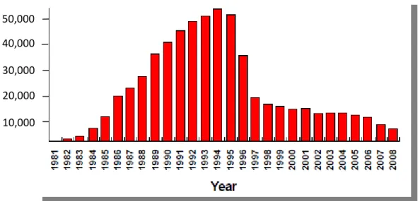
(16) Introduction. ANTIRETROVIRAL THERAPY: SUCCESSES AND CHALLENGES. Thirty decades after the recognition of acquired immunodeficiency syndrome (AIDS) in United States, HIV-1 has spread throughout the world, with growing heterogeneity. In 2010, the estimated number of people with HIV-1 world-wide was over 34 million people1, more than 95% living in low-and middle-income countries. The number of people infected by HIV-1 is expected to increase, mostly because of inadequate access to prevention and treatment2.. 1.1 From lethal disease to chronic pathology: 1996. The AIDS mortality in 1993 was the leading cause of death among persons ages 25 to 44 years3. However, after 1996, the number of deaths among people with AIDS drops every year since then. According to the Centers for Disease Control and Prevention (CDC), AIDS mortality fell from more than 51,000 in 1995 to about 16,000 in 2002 (Figure 1.1). These dramatic declines in AIDS mortality rate were due largely to the availability of combination antiretroviral therapy4-5, although with differences among demographic areas6. In this way, the development of antiretroviral therapy (ART) has been one of the greatest accomplishments of basic and translational research of the late 20th century. Combination ART (cART) with at least three drugs has resulted in substantial reductions in morbidity and mortality in many countries worldwide7. Successful cART increases levels of CD4+ T cells and reduces HIV-1 viral load indefinitely to undetectable levels (below 40 HIV-1 RNA copies/ml)8. Besides, cART has proven efficacious in HIV prevention, reducing the risk of HIV-1 viral transmission from mother-to-child and serving as post-exposure prophylaxis for individuals exposed to HIV-19-10.. 6.
(17) Introduction. 50,000 40,000 30,000 20,000 10,000. Figure 1.1 The estimated AIDS mortality from 1981 until 2008 in United States (www:cdc.gov). 1.2 Limitations of antiretroviral therapy. Although lifelong suppression of HIV-1 replication with cART should be possible in adherent patients, as soon as treatment is stopped, HIV-1 viremia rapidly rebounds11-12. Additionally, despite the inherent potency of cART to suppress virus replication, full life expectancy for patients living with HIV-1 has not been restored, remaining significantly less than population controls. Although ongoing refinements to improve the increased risk of morbidity and mortality, HIV-1-associated complications persist, including increased risk of cardiovascular disease, metabolic disorders, neurocognitive abnormalities, liver and renal disease, osteoporosis, and cancer 13-21. The exact mechanisms responsible for this increased risk are not entirely understood, however, toxic effects of long-term antiretroviral treatment22 or persistence of immune activation and inflammation emerged as key potential determinants of nonAIDS associated morbidity and mortality during treated HIV-1 infection23.. 7.
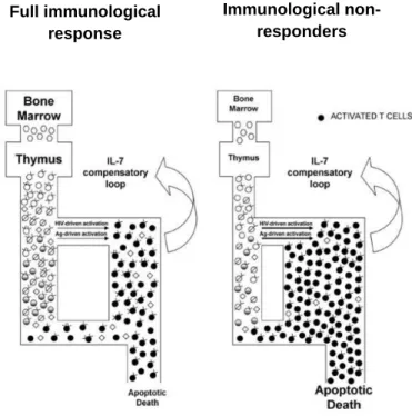
(18) Introduction. PERSISTENT INMUNE ACTIVATION. Chronic immune activation is a characteristic feature of progressive HIV-1 disease24-25. Generalized immune activation contribute to the inefficiency of the HIV-1 immune response and to the impairment of the regenerative potential of the entire immune system, including deleterious effects on. generation of effective immune. responses and on T-cell homeostasis and changes in the composition of lymphoid tissues that impede immune function26. The increase in immune activation and inflammation is also accompanied by HIV-1 specific exhaustion of CD4+ and CD8+ T cells, defined by decreased proliferative and effector functions, alterations in homing receptor expression patterns consistent with migration to sites of inflammation and increased expression of inhibitory receptors such as PD-1, or CTLA-4 on T cells27-28. The most widely accepted evidence that immune activation plays a major role in HIV-1 immunopathogenesis is the nonpathogenic outcome of SIV infection in its natural host. SIV-infected sooty mangabeys (SM) have strikingly low levels of immune activation in the chronic stage of infection and almost never develop immunodeficiency despite high levels of SIV viremia29. Conversely, SIV infection of rhesus macaques (RM) and other non-natural hosts results in high levels of systemic immune activation despite having lower or similar levels of SIV viremia, CD4+ T cell depletion, and rapid progression to clinical manifestations similar to AIDS30-31. It remains unclear, however, how the natural hosts of SIV infection are able to maintain low levels of immune activation despite high levels of viral replication.. 1.3 Immunological non-response to cART. Abnormally high levels of T cell activation persist despite years of ART-mediated viral suppression compared to uninfected individuals32. Control of HIV-1 replication reduces CD4+ T cell loss from direct cytolysis33-34, and partially restores T cell homeostasis35, increasing the number of CD4+ T cell count. Although many patients continue to have CD4+ T cell recovery for several years after receiving ART36, from 6% to 30% patients known as immunological non-responders (INR) or immunologic discordant patients (ID) failed to increase their absolute counts of CD4+ T cells37-39, 8.
(19) Introduction. having increased risk of non-AIDS-related complications40-41 and detrimental clinical consequences42-43. Elevated immune activation has been established as a marker of disease progression in untreated patients44-45. Even though its role during viral suppression may be less significant46, compelling evidences for decreased thymic production47 and increased CD4+ T cell hyperactivation that undergo rapid proliferation and subsequent cellular apoptosis seems to have a significant effect on CD4+ T cell recovery during ART48-49 (Figure 1.2). These findings were consistent with other studies in which elevated CD4+ T cell activation was observed in patients with idiopathic CD4 lymphopenia (ILC) who have low CD4+ T cells counts in the absence of HIV-145, suggesting that CD4+ T cell activation may be at least partly related to lymphopenia-driven T cell proliferation50-51. Although highlighting the possible pathogenic role of enhanced CD4+ T cell activation in well suppressed HIV-1-infected individuals, these findings also raise the questions on the driver mechanisms of persistently elevated CD4+ T cell activation52.. Immunological nonresponders. Full immunological response. +. Figure 1.2 Activated CD4 T cells undergo proliferation and + apoptosis having a significant effect on CD4 T cell recovery 52 during HAART. (Modified from Gazzola et al, 2008 ).. 9.
(20) Introduction. 1.4 Immunosenescence. Ongoing HIV-1-related immune dysfunction and inflammation during ART underlies premature aging in HIV-1-infected individuals53-55. Several physiological alterations. and. comorbidities. related. to. old. ages,. such. as. osteoporosis,. 56-59. atherosclerosis, and neurocognitive decline, occurs in HIV-1 disease. .. During aging, a reduction in T cell renewal together with progressive enrichment of terminally differentiated T cells occurs. Remarkably similar, HIV-1 reduces the capacity of the thymus to produce new cells60-62. Continuous stimulation of the immune system in HIV-1 disease with loss of CD4+ T cells by activation-induced cell death coupled with poor T-cell restoration due to lower thymic function results in an impaired T-cell homeostasis. The naïve T-cell pool cannot be replenished efficiently, and therefore old senescent CD8+ T cell clones or depleted CD4+ T cells cannot either be replaced, leading to an imbalance in T-cell phenotypes, similar to that observed in the elderly63-65. HIV-1 infection induces premature aging of naïve CD4+ T cells and both memory CD4+ and CD8+ T cells. Levels of cellular activation (CD38+ HLA-DR+)66-68 are the major driving factor of proliferation and T-cell differentiation, resulting in the generation of antigen-experienced cells with limited proliferative potential that eventually lose expression of CD28 and increase the expression of CD57, which is a key predictor of immune incompetence in the elderly and HIV-1-infected individuals69-71. The association of HIV-1-related immune dysfunction and non-AIDS defining comorbidities and premature aging is represented in the figure 1.3. The accelerated aging model in HIV-1 infection would start with ongoing HIV-1-related immune activation despite cART as the central event in the senescent pathway. Activated T cells undergo clonal expansion due to circulating antigen, resulting in differentiation and accumulation of nonfunctional end stage senescent cells that cannot be replenished due to the loss of thymic function altering T cell homeostasis increasing the risk of developing nonAIDS. morbidities. associated. to. HIV-1-associated. inflammation72.. 10. immune. activation. and.
(21) Introduction. 65. Figure 1.3 Accelerated aging model in HIV-1 infection (Modified from Desai et al, 2010 ).. PERSISTENT IMMUNE ACTIVATION: CAUSES. The underlying causes of persistent immune activation during cART-mediated viral suppression are likely multifactorial but remain incompletely defined. Several potential causes have been proposed, highlighting HIV-1 ongoing replication, microbial translocation and co-infections, as continuous trigger mechanisms of immune activation73.. RESIDUAL VIREMIA: ONGOING VIRAL REPLICATION. First, the effectiveness of current therapeutic regimens has been monitored by using HIV-1 RNA assays which have a sensitivity of 40 to 50 copies/ml. Suppression of plasma viremia below detectable levels can be sustained in HIV-1 infected individuals by antiretroviral regimens currently in use74-75. Nevertheless, replication-competent cell-. 11.
(22) Introduction. free virions can be found for many years in the plasma of most patients on cART, and viremia rapidly rebounds when antiretroviral suppression is interrupted76-79. Still infected individuals remain clinically stable on cART, persistent virus expression may contribute to long-term complications53. Using recently developed realtime HIV-1 RNA assay, with sensitivity below one HIV-1 RNA copy per ml, allows a more detailed analysis of the viral decay kinetics in patients on suppressive cART80,81. Several studies revealed persistent low-level viremia of around 3-5 copies/ml in plasma of most patients on cART and persists in patients even after years of therapy82. The source and dynamics of this residual viremia are currently unknown, representing a challenge to future attempts to induce a drug-free remission of HIV-1 disease and eradication of the infection83. Viremia could arise from ongoing cycles of viral replication in a sanctuary site where there is suboptimal drug penetration, from long-lived productively infected cells, or from activation of virus expression from latently infected cell reservoirs84-87.. 1.5 Generation of 2-LTR circles: recent infection events. Efforts to characterize the extent of ongoing viral replication in suppressed individuals have been hampered by a lack of convenient surrogates of ongoing viral replication88. Viral episomes containing one or two long terminal repeats (1-LTR or 2LTR-containing episomes) are formed after completion of viral cDNA synthesis and translocation to the host cell nucleus, where recombination and direct ligation lead to the formation of episomes containing one and two LTRs, respectively. These episomal cDNA forms are labile and, as such, their presence is indicative of recent infection events89-91.. 1.6 Anatomical sites: HIV-1 reservoirs. Persistent. ongoing. viral. replication. may. remain. in. anatomical. sites. immunologically sheltered from the blood and lymphoid systems with low antiretroviral 12.
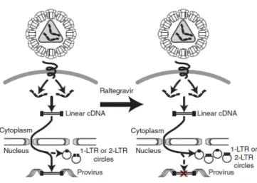
(23) Introduction. drug penetration92-93. These anatomical sites, including the lymphoid tissue, central nervous system, respiratory, gastrointestinal and genitourinary tracts, may act as reservoirs of HIV-1 replication94-96.. Figure 1.4 Generation of 2-LTR and 1-LTR circles after intensifying 110 suppressive regimen with raltegravir (Buzon et al, 2010 ).. 1.7 Antiretroviral treatment intensification: maraviroc and raltegravir. Several studies have looked at the intensification treatment with an additional antiretroviral drug, not previously used by the patients, as a possible strategy to reduce low-level viremia and subsequent immune activation whether ongoing new cycles of viral replication were contributing to residual viremia97. Additionally, the emergence of new antiretroviral drugs with different mechanisms of action, such as CCR5 antagonists and integrase inhibitors, provides new opportunities to assess the viral reservoirs and residual viremia that persist in suppressed individuals99-100. Particularly, these new antiretroviral drugs referred are; maraviroc (MVC), a potent new antiretroviral agent approved for the treatment of HIV-1 infection that blocks interaction between the virus and the CCR5 co-receptor, targeting HIV-1 entry process, a crucial step in the HIV-1 life cycle101-102, and raltegravir (RAL), the first-in-class integrase inhibitor approved for the treatment of HIV-1 infection. It blocks integration of linear viral cDNA that is subsequently circularized by host DNA repair enzymes to form 13.
(24) Introduction. episomes containing two copies of 2-LTR circles or undergoes recombination to form a 1-LTR circle103-104, as represented above (Figure 1.4). Disappointingly, the vast majority of treatment intensification trials to date have failed to show any decline in low-level viremia or cell-associated HIV-1 DNA105-107, highlighting two studies that showed no change in the gastrointestinal tract108 or in the cerebrospinal fluid109. Nevertheless, the addition of raltegravir lead to an increase in 2LTR circles within two weeks in one-third of the suppressed HIV-1-infected individuals, consistent with evidence of residual viral replication, and a significant reduction on T cell activation after 48 weeks intensifying with RAL, although there was still no change in persistent low-level HIV-1 RNA or cell-associated DNA, following intensification110.. MICROBIAL TRANSLOCATION. Secondly, microbial translocation has been pointed as a possible mechanism underlying immune activation and non-AIDS-associated morbidity in HIV-1 disease during suppressive regimen111-112.. 1.8 Gut-associated lymphoid: anatomical viral reservoir. The gastrointestinal (GI) tract continues to serve as an important viral reservoir of HIV-1-infected cells even during long-term cART and contributes to viral persistence113114. . The GI tract harbors most of the total lymphoid tissue in the body and an. abundance of activated effector memory CD4+ T cells that are CCR5+ and their predominantly activated state make them highly sensitive to HIV-1 infection115-117. Of these CD4+ CCR5+ T cells, Th17 cells, which also expressed the gut homing marker CCR6, are even more susceptible to HIV-1 infection and subsequent cellular depletion118-119.. 14.
(25) Introduction. 1.9 Intestinal epithelium: HIV-1 associated CD4+ T cell depletion. HIV-1 infection cause massive depletion of CD4+ T cells from the gut-associated lymphoid (GALT) tissue in the first few weeks of the infection, attributable to the cytopathic effects of the viral infection or the activation-induced uninfected CD4+ T cell death120-121. Insufficient production and tissue delivery of CD4+ memory T cells may contribute to sustained CD4+ T cell depletion during disease progression122. The loss of CD4+ T cells was coincident with increased prevalence of CD8+ T cells in GALT and the subsequent marked changes in the T-cell subset distribution in GALT, that were not adequately reflected in peripheral blood123. This dysregulation in T-cell homeostasis leads to increased levels of pro-inflammatory cytokine124-125, increased apoptosis of epithelial cells, and altered tight junction protein composition126 resulting in a functional degradation of the intestinal barrier127. The mucosa damage leads to a process known as microbial translocation, whereby microbial products from the intestinal lumen cross the disrupted mucosal barrier into the systemic circulation in the absence of overt bacteraemia128-129. Additionally, the cytokines IL-17 and IL-22 produced by Th17 cells enhance epithelial regeneration and defend against microbial translocation by recruiting neutrophils to the GALT to clear microbial products and by stimulating epithelial cell proliferation and antibacterial defensin expression130-132. Therefore, loss of Th17 cells may cause impaired mucosal healing increasing intestinal permeability and the subsequent microbial translocation133-134. Hence, both HIV-1-infected individuals and SIV-infected rhesus macaques have preferential significant depletion of gut Th17 CD4+ T cells with a sequential increase in Th1 CD4+ T cells, contributing to epithelial injury and enhance local inflammation and immune activation135. However, SIV-infected sooty mangabeys and African green monkeys, the natural hosts for SIV infection that do not have increased microbial translocation and do not develop AIDS, maintain, strikingly, Th17 cells in the gut despite mucosal CD4+ T cell losses136 -137. Taking together all data, intestinal epithelium in HIV-1 infected individuals is characterized by massive CD4+ T cell depletion, particularly Th17 cells138-140, abnormal enterocyte differentiation141, enterocyte apoptosis and destruction of the tight junctions142 caused by increased production of interferon-γ (IFN-γ) and tumor necrosis factor (TNF), contributing to increased microbial translocation143 (Figure 1.5). 15.
(26) Introduction. Figure 1.5. Intestinal epithelium in HIV-1-infected individuals (Sandler et al, 2012).. 1.10 Microbial translocation. Increased levels of circulating microbial products in HIV-1-infected individuals, such as lipopolysaccharide (LPS), peptidoglycan, bacterial CpG DNA, flagellae, and viral genomes, molecules that can directly stimulate the innate immune system through Toll-like receptors (TLRs), are strongly associated with increased immune activation144146. (Figure 1.6). Several parameters have been indistinctly used to determine bacterial. translocation147-149, though LPS and soluble CD14, have been the most commonly used150. Lipopolysaccharides (LPS) are the major component of gram-negative bacteria cell walls and a potent immunostimulatory product151. Circulating LPS promotes hepatic synthesis of LBP, a plasma protein that increases the binding of LPS to CD14/TLR4 receptor complex on monocyte/macrophages152-153, triggering monocyte 16.
(27) Introduction. activation and resulting in the release of soluble CD14 (sCD14) and the production of inflammatory cytokines, such as interleukin-6 (IL-6), interleukin-1β (IL-1β), Tumor Necrosis Factor (TNF) and type I Interferons (IFN-I)154-155, contributing to HIV-1associated immune activation (Figure 1.6).. Figure 1.6. Intestinal epithelium in HIV-1-infected individuals (Sandler et al, 2012).. 1.11 Possible driver mechanism of HIV-1 associated immune activation. The association between microbial translocation and immune activation were reported in several studies in the first years of chronic HIV-1 infection or in the most advance stages of HIV-1/AIDS156-160. Due to the ongoing viral replication in GALT and elevated chemokine production, there is a recruitment of CD4+ T cells from the periphery into the gut via α4β7, an integrin that mediates migration of lymphocytes to the gut161. Engagement of α4β7 on 17.
(28) Introduction. CD4+ T cells by gp120 viral envelope protein leads to activation of lymphocyte functionassociated molecule 1 (LFA-1) or αLβ2, facilitating formation of virologic synapses and increasing the efficiency of HIV-1 infection providing new targets for the virus162.. 1.12 Recovery of gut mucosa integrity. On the other hand, studies utilizing the SIV model showed that the major mechanism of early and near complete CD4+ T cell restoration in GALT, involved trafficking of CD4+ T cells from the periphery to gut mucosa during primary SIV infection163-165. Moreover, longitudinal follow-up studies revealed that the kinetics of restoration of the gut mucosal immune system in patients with HIV-1 infection during cART was substantially delayed and incomplete compared with that observed in the peripheral blood compartment and was independent of the time of initiation of cART166. This delay in mucosal immune restoration was attributed to incomplete suppression of viral replication in GALT during therapy and increased levels of local inflammation and immune activation167. Noteworthy, viral replication was detected in GALT of HIV-1-infected patients receiving cART despite undetectable viral loads in the peripheral blood168. The lack of complete viral suppression with the subsequent loss of CD4+ T cells in the mucosa may contribute to discordant CD4+ T cell restoration and viral suppression between GALT and peripheral blood169.. HIV-1 COINFECTIONS. Thirdly, and finally, the vast majority of HIV-1-infected individuals are coinfected with other chronic viral infections or parasitic diseases, many of which get worse as a consequence of immunodeficiency during untreated HIV-1 disease, and may continue to contribute to systemic immune activation during treated HIV-1 infection73.. 18.
(29) Introduction. 1.13 Visceral leishmaniasis: Leishmania infantum. Visceral leishmaniasis (VL) caused by Leishmania infantum is a frequent disease among HIV-1 infected patients in countries of the Mediterranean basin170-171 (Leishmania lifecycle in Figure 1.7). Despite cART, which indeed led to a reduction of its incidence, this disease still constitutes the third most frequent parasitic opportunistic infection in Europe172. Even in treated patients VL has case-fatality rates of 10-20%173, especially in HIV-1-co-infected patients174. Leishmania and HIV-1 share some of the same target cells, namely macrophages and dendritic cells175-176, and similar immune compromising mechanisms, most notably pro-inflammatory responses177-178 and CD4+ T cell lymphocyte depletion making immunosuppression as the hallmark of both Leishmania-HIV-1 pathogens179. The parasite infection in HIV-1-infected individuals is thought to induce chronic immune activation, viral replication and HIV-1 disease progression180-181, diminishing their life expectancy175; whereas immunological disturbances caused by HIV-1, despite cART, are particularly favorable for the uncontrolled multiplication of the parasite182, with increased parasite burden, drug resistance and frequent relapses183-184. Studies addressing microbial translocation in infectious diseases are scarce. Considering the involvement of microbial translocation in activation mechanisms in HIV-1-infected individuals115, and the similar pathogenic features shared between VL and HIV/AIDS185, it was also expected that gut parasitation by Leishmania amastigotes lead to mucosal barrier breach predisposing to microbial translocation in these patients. This phenomenon was, in fact, detected in VL186-187, and may potentiated in co-infected patients contributing to the activation status by enhancing the plasma cytokine storm, aggravating. the. disease´s. clinical. outcome. in. Leishmania/HIV-1. co-infected. individuals188. The contribution of microbial translocation to the pathogenesis of different infectious diseases with persistent immune activation is likely to vary. Thus, microbial translocation per se has been proposed as a common pathway causing disease progression that is shared by different pathogens, such as HIV-1, Leishmania, and HCV189.. 19.
(30) Introduction. Figure 1.7 Leishmania life cycle (Centers for Disease Control and Prevention). Considering human stages, the female phlebotomies sandflies inject the promastigote form of Leishmania phagocytized by macrophages that reach the puncture wound. Promastigotes transform into amastigotes form within these cells, where they multiply and infect other mononuclear phagocytic cells and affect different tissues causing clinical manifestations of visceral leishmaniasis.. 1.14 Hepatitis C virus (HCV). The overall burden of HIV-1/HCV coinfection is estimated at 4 or 5 million people worldwide190, finding the highest prevalence of HCV infection in HIV-1 infected individuals with a history of intravenous drug use, with a rate reported to be 82% to 93%191-193. In Europe, liver disease has emerged as a leading cause of death among HIV-1-infected individuals, mostly due to chronic viral hepatitis194, increasing mortality from liver in coinfected individuals with the consumption of alcohol195-196. The increased mortality of coinfected individuals over that of HCV-monoinfected ones reflects accelerated. progression. of. chronic. immunosuppression197-198. 20. liver. disease. with. HIV-1-related.
(31) Introduction. The determinants of progression to cirrhosis and liver failure are poorly defined. One contributor to chronic inflammation and fibrosis in chronic HCV monoinfection is microbial translocation199-202. Microbial translocation may promote liver fibrosis either by direct interaction with Kupffer cells and hepatic stellate cells upregulating proinflammatory and pro-fibrogenic cytokines such as tumor necrosis factor TNF-α, IL-1, IL-6, and IL-12 or indirectly via induction of systemic immune activation and activationinduced apoptotic cell death203-204.. +. Figure 1.8 Microbial translocation due to HIV-1-related CD4 lymphocyte depletion contributes to both immune activation and progression of liver disease in HIV/ HCV coinfected individuals 209 (Balagopal 2008 ).. In addition, HIV-1/HCV seems to augment the local and systemic effects of microbial translocation caused by CD4+ T lymphocyte depletion and immune activation, accelerating liver disease consistent with the chronic hepatic inflammation observed in chronic HCV infection205-209 (See model below, Figure 1.8).. 21.
(32) Introduction. 1.15 Human T-lymphotropic virus (HTLV). During the 1980s and 1990s, Spain had one of the highest prevalence of drug injection in Europe210. Sharing needles practice led to the spread of different viruses among injection drug users (IDUs) and their sex partners, such as HIV-1, HCV and HTLV211. While the prevalence of HTLV-1 infection in Spain is low, there is a high prevalence of HTLV-2 infection specially in injecting drug users, reaching prevalence rates around 18% in prisoners, and around 5% outside the prison212-214. The molecular characterization of HTLV-2 isolates from North America and Europe demonstrated that HTLV-2a subtype was the predominant circulating variant in North America215 and northern Europe216, while HTLV-2b subtype was almost exclusive in southern Europe, particularly Spain and Italy217-218, where co-infection with HCV and HIV-1 is frequent. Up today, there is still no clear evidence that HTLV-2 causes any human disease, although it has been occasionally linked to neurological and lymphoproliferative disorders219-221. Several studies analyzing the effects of HTLV-2 on HIV-1 pathogenesis in dually infected HIV-1/ HTLV-2 individuals, revealed delayed progression of HIV-1 to AIDS222-223 linked, in some cases, to a long-term nonprogressor phenotype224-225. This protective role is thought to be due to maintenance of CD4+ T cells, lowering levels of HIV-1 replication and immune activation in dually infected HTLV-2/HIV-1 individuals compared to patients infected with HIV-1 only226-228. Additionally, several IDU long-term non-progressors (LTNPs) with stable CD4 counts, in the absence of ART, have been reported to be infected with HTLV-2229, and the median value of HTLV-2 proviral loads (pVL) tended to be higher in the LTNP coinfected group compared to those who required antiretroviral treatment230. The possible mechanism underlying the milder HIV-1 disease course has been attributed to the modulation of cytokine and chemokine networks by HTLV-2 that would modify innate host immune responses. In this way, HTLV-2 induces the expression of three CC-chemokines, natural ligands for CCR5 from PBMCs and monocytes; Macrophage inflammatory protein 1-alpha (MIP-1α)/CC-Chemokine ligand-3 (CCL3); MIP-1β/ CCL4; and RANTES (Regulated upon activation normal T-cell expressed and secreted (RANTES)/CCL5 inhibiting HIV-1 entry by binding to CCR5231-233, and the concomitant downregulation of the CCR5 receptor on lymphocytes234-235, interfering with HIV-1 infection process (Figure 1.9). 22.
(33) Introduction. Notably, different viruses have specific cytokine and chemokine requirements for maintenance in vivo. What appears to be optimal for one strain may be detrimental for another236-238. Therefore, the reported upregulation of granulocyte-macrophage colonystimulating factor (GM-CSF) and IFN-γ secretion239, which can also inhibit CCR5 expression contributing to slow HIV-1 disease progression240. In fact, it has been demonstrated in a clinical trial conducted on AIDS patients that GM-CSF decreases viral replication and increases the number of circulating CD4+ T cells, the primary target of HIV-1241. In addition, IFN-γ is the pivotal cytokine triggering a phagocyte-dependent Th1 response leading to CTL response against invading pathogens242. In this regard, a poor Th1 response and a dominant Th2 response have been implicated in the pathogenesis and progression of HIV-1 infection243-244. Thus, HTLV-2 infection may exert protective effect on HIV-1 disease progression inducing Th1 response against invading pathogens via up-regulating IFN-γ expression239,242.. Figure 1.9 Scheme illustrating how HTLV-2 may interfere on + HIV-1 pathogenesis. The HTLV-2 infected CD8 T cells and their expansion induces the secretion of CC-chemokines, which can + reinforce the cytotoxic T lymphocyte response against CD4 HIV227 1 infected cells (Casoli 2007 ).. 23.
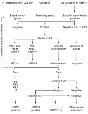
(34) Introduction. On the other hand, clinical features of coinfected individuals have been overlooked, despite its high frequency among injecting drug users245. Only higher frequencies of severe fibrosis has been found among HCV/HTLV-2 individuals compared to HCV-monoinfected group, although no significant246. Consistent with the fact that hepatic measures were reported to be worse in HTLV-1/ HCV coinfected individuals with more severe immunosuppression247, cellular immune response should be analyzed in HCV/HTLV coinfected individuals to elucidate the effects on HTLV-2 on HCV infection and progression of liver disease248. From public health perspective, prevention of HTLV-1/2 is crucial as there is no current treatment for this infection. Several studies analyzed the effects of antiretroviral agents widely used for treating HIV-1 infection on HTLV virus expression, showing no effects on HTLV proviral burden or HTLV mRNA expression249-251. Raltegravir was an integrase inhibitor approved for HIV-1 infection treatment. Given the similarities between HIV-1 and HTLV integrases, raltegravir was thought to be effective against HTLV-1. Furthermore, two studies reported a decrease HTLV-1 pVL in vitro and also ex vivo252-253. Despite these promising in vitro results, no decrease on HTLV-1 pVL was reported in the unique in vivo study254, attributed to the lack of new rounds of viral replication that is mainly maintained through cell division255.. 24.
(35) AIMS OF THE STUDY. Despite viral suppression and partially immune recovery, persistent increased immune activation continues in HIV-1-infected individuals. Microbial translocation and the subsequent pro-inflammatory cytokines expression has been proposed as the major driver mechanisms of HIV-1-associated immune activation and inflammation, also influenced by frequent HIV-1-coinfections. The aim of the study was to analyze microbial translocation, cytokines profile, and immune activation in different scenarios, involving, HIV-1-infected individuals on treatment intensification, HIV-1-infected individuals with different immune response to antiretroviral therapy, and HIV-1-infected individuals with other coinfections, such as Leishmania, HCV and HTLV.. Objective 1. To evaluate in a longitudinal study the effects of treatment intensification with maraviroc or raltegravir and further intensifying drug discontinuation on immune activation, microbial translocation and dynamics of T cell subsets including expression of gut homing β7 receptor on T cells.. Objective 2. To determine correlations between different techniques employed to measure microbial translocation, including levels of LPS, sCD14, LBP and 16S rDNA, and between all these techniques and immune activation in HIV-1 infected patients on treatment intensification.. Objective 3. To investigate in a cross-sectional study how visceral leishmaniasis impacts immune activation compared to two control groups consisted of HIV-1 infected both immunological concordant and non-responders to antiretroviral therapy, and which factors could impair immune response in these individuals, including inflammation, microbial translocation and immunosenescence.. 25.
(36) Objective 4. To investigate in a cross-sectional study how HTLV-2 infection influences both HIV-1 disease progression and liver disease associated to HCV infection. Immune activation, microbial translocation, cytokine profile and HTLV-2 proviral load are determined for this purpose.. Objective 5. Evaluation of HTLV-2 proviral load in coinfected HIV-1/HTLV-2 individuals ongoing treatment intensification with raltegravir.. 26.
(37) Material and Methods. 27.
(38) Results. FLOW CYTOMETRY STAINING. Many immunologically important cells can be defined based on proteins present on their membrane. Human clusters of differentiation (CD) are used to classify many epitopes on the cell surface of leukocytes. The study of cellular characteristics using flow cytometry involves the use of fluorescent molecules such as fluorophore-labeled antibodies that binds to a specific molecule on the cell surface or inside the cell. In further analysis through flow cytometer, when laser light of the right wavelength strikes the fluorophore, a fluorescent signal is emitted and detected by the flow cytometer, demonstrating the presence of both superficial and intracellular proteins. The combination of several immunological markers is used to define cellular immune functions or properties. The commercial antibodies used to study cellular characteristics using flow cytometry are shown in Table 2.6. These antibodies had the following fluorochromes FITC (Fluorescein Isothiacyanate), PE (Phycoerythrin), PerCP (Peridinin Chlorophyll Protein Complex), PE-Cy7 (Phycoerythrin-Cyanin7), APC (Allophycocyanin), APC700 (Allophycocyanin 700), APC-Cy7 (Allophycocyanin-Cyanin 7), eF450 (eFluorTM 450) and KO (KromeTM Orange) directly conjugated to the antibodies used as described above. Fluorocrhome peack excitation and emission wavelengths are also shown in Table 2.1. All antibodies against different CDs were purchased from Becton Dickinson (Becton Dickinson, NJ, USA).. CELL STAINING AND FLOW CYTOMETRY. Briefly, 100 µl of blood were lysed with 200 µl of FACS lysing solution (Becton Dickinson, NJ, USA) for 30 min at room temperature, incubated with the corresponding superficial antibodies during 20 min at 4ºC, washed twice with phosphate-buffered saline (PBS) containing 1% azida, fixed with BD CytofixTM (Becton Dickinson, NJ, USA) for 10 min at 4ºC and finally washed and resuspended in PBS containing 1% azida. An unstained control was performed for all samples.. 28.
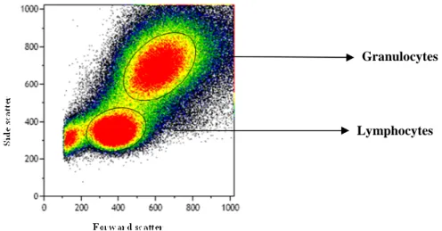
(39) Results. Table 2.1 Fluorochrome-conjugated antibodies used for flow cytometry staining.. Excitation Laser 638 nm Red. Antibody. Fluorocrhome. Excitation Max (nm). Emission Max (nm). HLA-DR. APC-Cy7. 650. 774. CD38. APC700. 696. 719. β7. 488 nm Blue. 405 nm Violet. APC. 645. 660. CCR7. PE-Cy7. 496, 565. 774. CD8. PerCP. 482. 675. CD45RA. PE. 496, 565. 575. CD57. FITC. 493. 525. CD3. eF450. 410. 455. CD4. KO. 398. 528. Cells stained with fluorochrome-conjugated antibodies were visualized using flow cytometry using a Gallios flow cytometer (Beckman-Coulter, California, US). Flow cytometry allows the analysis of multiple parameters of individual cells within hererogeneous populations. The flow cytometer analysis is performed by passing thousands of cells through a laser beam and capturing the light that emerges from each cell as it passes through one at a time. As a cell passes through the laser, it will refract or scatter light at all angles. Forward scatter (FSC), or low-angle light scatter, is the amount of light that is scattered in the forward direction as the laser light strikes the cell. The magnitude of forward scatter is roughly proportional to the size of the cell. Light scattering at larger angles, to the side (SSC), is caused by granularity and structural complexity inside the cell. The generation of two-dimensional dot or scatter plots, using forward and side scatter, allow us to distinguish between cell populations. The scatter plot from a peripheral blood cell run is shown below (Figure 2.1). The populations that emerge include lymphocytes which are small cells possessing low internal complexity; monocytes which are medium-sized cells with slightly more internal. 29.
(40) Results. complexity, and neutrophils and other granulocytes which are large cells that have a lot of internal complexity. A threshold was set to avoid information coming from a very large number of minute particles, like platelets and debris. Thereby, the majority of events that the cytometer collects are the cells of interest, despite small particles are still passing through the instrument. Gallios flow cytometer can analyze 12 parameters, FSC, SSC and 10 different channels simultaneously, allowing to gather statistical data on large number of cells and to use that information to correlate multiple parameters within a cell population.. Granulocytes. Lymphocytes. Figure 2.1 Populations emerged from a peripheral blood run defined by Forward and Side scatter.. At least 105 CD3+ T cells were collected for each sample and analyzed up to nine different parameters with Kaluza software v.1.1 (Beckman coulter) by initially gating lymphocytes according to morphological parameters. Gating was always the same between the different time points. At least 20000 events were gated for the T cell subsets. 30.
(41) Results. MICROBIAL TRANSLOCATION. CD14 is a glycoprotein that mediates the interaction of lipopolysaccharide (LPS, endotoxin) with cells, thereby signaling the presence of gram-negative bacteria. The binding of LPS to CD14 requires an acute phase protein, LPS-binding protein (LBP). These three factors appear to participate in a complex feedback mechanism of immune regulation involving both up-regulation and down-regulation of the inflammatory process triggered by LPS. The 16S ribosomal RNA, however is a component of the 30S small subunit of prokaryotic ribosomes, and thus, presented in all bacteria, gramnegative and gram-positive.. LIPOPOLYSACCHARIDE (LPS). Lipopolysaccharides (LPS) are found in the outer membrane of Gram-negative bacteria. Levels of LPS are determined in duplicated 50 µl of plasma samples using a quantitative test QCL-1000 Limulus Amebocyte Lysate (Lonza®, Basel, Switzerland). This method utilizes lyophilized lysated prepared from the circulating amebocytes of the horseshoe crab Limulus polyphemus which was an extremely sensitive indicator of the presence of endotoxin. This technique requires to be used with appropriate precautions to avoid microbiological or endotoxin contamination.. SOLUBLE CD14 (SCD14). CD14 is expressed mainly by macrophages, neutrophils and dendritic cells. CD14 acts as a co-receptor with Toll-like receptor-4 (TLR4) for the detection of bacterial. lipopolysaccharide. (LPS),. to. which. it. binds. in. the. presence. of. lipopolysaccharide-binding protein (LBP). The soluble form of the receptor (sCD14) is secreted mainly by monocytes. Levels of soluble CD14 were measured in duplicated 200-fold diluted plasma samples using the Quantikine® Human sCD14 Immunoassay (R&D Systems, 31.
(42) Results. Minneapolis, Minnesota, US). This assay employs the quantitative sandwich enzyme immunoassay technique (ELISA), in which a monoclonal antibody specific for sCD14 has been pre-coated onto a microplate.. LIPOPOLYSACCHARIDE-BINDING PROTEIN (LBP). Lipopolysaccharide-binding protein (LBP) is a soluble protein that binds to bacterial lipopolysaccharide (LPS) to elicit immune responses by presenting the LPS to the receptor CD14 and the pattern recognition receptor TLR4. Levels of LBP were determined in 800-fold diluted plasma samples, run in duplicate, using the Enzyme Immunoassay for Quantification of free human LBP kit (Enzo Life Siences, Farmingdale, NY). However, LBP levels were only determined at baseline, at week 48 after treatment intensification, and at weeks 12 and 24 after intensifying drug discontinuation.. BACTERIAL 16S RIBOSOMAL DNA (16S rDNA). Because LPS is a component of cell wall of gram-negative bacteria, present on a proportion of enteric bacterial microbiota, the well-conserved 16S ribosomal DNA (16S rDNA) subunit common to most bacteria was also quantified during the follow-up 48 weeks of treatment intensification. The bacterial 16S rDNA was quantified from the DNA extracted from 200 µl of plasma using QIAamp DNA kit (Qiage, Hilden, Germany). DNA concentrations were determined using ND-1000 spectrophotometer, according to manufacturer´s protocol. DNA was kept at -20ºC until further use. Standard plasmids were generated through the amplification of partial 171bp 16S rDNA gene. Reaction mixtures were performed in a total volume of 25 µl containing PCR Buffer II (AccuPrime Taq High Fidelity, Bioline, London, UK), 0,5 U Taq 32.
(43) Results. polymerase (AccuPrime Taq High Fidelity, Bioline, London, UK), 200 ng of genomic DNA from the plasma of diagnosed HIV-1-infected individual, and 20 pmol of the following primer pairs were used, (16S-1/ 16S-2) for partial bacterial 16S rDNA gene, in the Table below (Table 2.2). Thirty-five cycles of denaturation at 95ºC for 15s, annealing at 56ºC for 10s, and extension at 68ºC for 20s, followed by a final extension at 68ºC for 2min were performed.. Table 2.2 List of oligonucleotides used for generating the 16S rDNA standard plasmids.. Primer. Sequence. 16S-1. 5´-TAGCGATTCCGACTTCATGGA-3´. 16S-2. 5´-ACACACGTGCTACAATGGC-3´. Length (bp) 171bp. Standard plasmids were generated by cloning PCR amplicon products in a pUC19 plasmid DNA following the instructions provided by TOPO® Cloning reactions (TOPO. TA. Cloning®. kits,. Invitrogen,. Life Technologies,. US). according. to. ®. manufacturer´s protocol. Subsequently, competent One Shot Escherichia coli were transformed and diluted bacteria were spread plated with Ampicillin (100 µg/ml) and incubated at 37ºC overnight (See Molecular Cloning 2.6.2). The presence of the cloned amplicon was verified in some colonies by colony PCR using the same amplification conditions for 171 bp 16S rDNA gene explained above. The selected colonies were grown in 2 ml of LB liquid medium overnight, and plasmid DNA was extracted from transformed bacteria using the QIAprep Spin Miniprep kit (Qiagen GmbH, Hilden, Germany). A standard curve for copy number quantification were generated in each run using six 5-fold serial dilutions of each plasmid specific amplicon template. The bacterial 16S rDNA levels in plasma of HIV-1-infected patients were determined by real time PCR assay, with newly designed primers and probe are described in Table 2.3. A TaqMan® Probe (Sigma-Aldrich, Missouri, US) was used with 6-FAMTM and TAMRA as a fluorophore and a quencher, respectively. 33.
(44) Results. Since any contaminating DNA is likely to be co-amplified and could interfere with quantification, we considered to perform a DNase treatment of commercial amplification FastSart Buffer kit prior to proceed with the real-time quantification. Thus, LightCycler FastStart DNA Master PLUS HybProbe 5X (Roche Diagnostics) was treated with DNase I (Invitrogen, Life Technologies, US) at 37ºC for 10 min followed by 10 min at 95ºC to inhibit the enzyme activity, according to manufacturer´s indications.. Table 2.3 Primers and probes used for partial 16S rDNA gene quantification by RT- PCR.. Primer. Sequence. 16S-3. 5´-TTGCAGACTCCAATCCGGACT-3´. 16S-4. 5´-GCATAGAAAGAGAAGCGACCT-3´. Length (bp) 122bp. 16S Taqman Probe. 5´-CAGCGTCAAAGGTGAGGAGTGGGTGTCG-3´. Since any contaminating DNA is likely to be co-amplified and could interfere with quantification, we considered to perform a DNase treatment of commercial amplification FastSart Buffer kit prior to proceed with the real-time quantification. Thus, LightCycler FastStart DNA Master PLUS HybProbe 5X (Roche) was treated with DNase I (Invitrogen, Life Technologies, US) at 37ºC for 10 min followed by 10 min at 95ºC to inhibit the enzyme activity, according to manufacturer´s indications. The amplification reaction was carried out in triplicate using the capillary-based LightCycler® 2.0 (Roche). Standards curves were preparared for each amplification and analyzed at each PCR-run, including negative and positive controls. Reactions were carried out in triplicate in a total volume of 20 µl containing LightCycler FastStart DNA Master PLUS HybProbe 5X (Roche), 200 ng of DNA, 50 pmol of each primer, 16S-3/ 16S-4 along with 2 pmol of the following 16S Taqman probe. The cycling parameters included a hot start at 95ºC 10 min, and continued with 40 cycles of denaturation at 95ºC for 10s, annealing at 55ºC for 10s and extension at 72ºC for 15s.. 34.
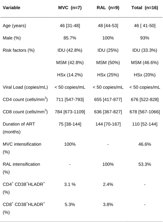
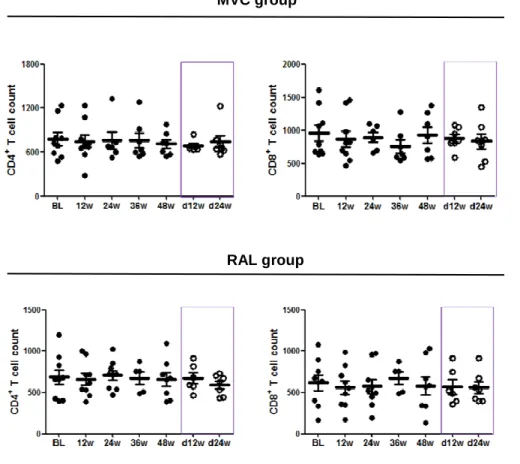
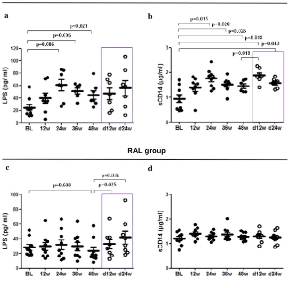
(45) Results. The Ct values were converted into template quantity using the standard curve method, with PCR efficiency >90% and R2 correlation superior to 0.9, avoiding replicates that have more than 0.5 Ct of difference. Levels of 16S rDNA were calculated and the final results were expressed as 16S rDNA DNA copies per microliter of plasma sample.. ANTIRETROVIRAL TREATMENT INTENSIFICATION. In the frame of two pilot open-label phase II clinical trials, we analyzed the effects and dynamics of treatment intensification and intensified treatment discontinuation on immune activation, T cell subsets, expression of gut homing β7 receptor on T cells and microbial translocation in 16 HIV-1-infected patients of whom seven intensified with maraviroc, a CCR5 antagonist, and the other nine patients with raltegravir, an integrase inhibitor. We also analyzed the correlations between different measurements of microbial translocation including lipopolysaccharides (LPS), soluble CD14 (sCD14), lipopolysaccharide-binding proteins (LBP) and quantification of 16S ribosomal DNA (16S rDNA), and also the association between microbial translocation and either immune activation, T cell subsets or expression of gut homing β7 receptor on T cells.. 2.1 Study design and ethic statement. Two independent pilot prospective open-label clinical trials were performed to evaluate the effect of maraviroc (MVC), a CCR5 antagonist (developed and provided by Pfizer, Inc., US) or raltegravir (RAL), an integrase inhibitor (developed and provided by Merck, New Jersey, US) on the HIV-1 latent reservoir in 16 HIV-1 infected patients. Both clinical trials (NCT00795444 and NCT00807443) were conducted at the Hospital Universitario Ramón y Cajal, in Madrid, Spain, between 2008 and 2011 with an intensification period of 48 weeks and a follow-up of 24 weeks after drug discontinuation. Biochemical, immunological and virological parameters were assessed at baseline and at weeks 12, 24, 36 and 48 of treatment intensification, and at weeks 12 and 24 weeks after intensifying drug discontinuation. 35.
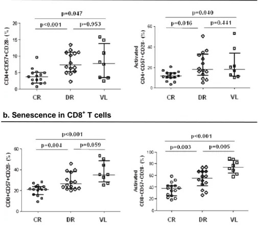
(46) Results. The study was carried out according to the recommendations of the Declaration of Helsinki and current Spanish legislation on clinical trials. It was approved by the AEMPS (Spanish Agency for Medications and Health Products) and our local Independent Ethics Committee (Hospital Ramón y Cajal, Madrid, Spain). All patients provided written informed consent for participation, sample collection, and laboratory determinations.. 2.2 Patients and specimen collection. Sixteen patients (seven from MVC trial and 9 from RAL trial) completed the intensification phase of the study; however one individual from RAL trial declined to participate in the follow-up 24 weeks after intensifying drug discontinuation, so finally fifteen patients completed the follow-up period of 24 weeks. Plasma viral load was measured by quantitative RT-PCR with a detection limit of 40 copies/ml (Roche Taqman HIV-1 test; Roche Molecular Systems), and Tlymphocyte counts were determined by flow cytometry. The main inclusion criteria were as follows: HIV-1 infected adults receiving antiretroviral treatment for at least 2 years; undetectable plasma viral load (below 40 copies HIV-1 RNA/ml) for at least two years; CD4+ T-cell count above 350 cells/mm3; and no previous experience with MVC or RAL. Patients were excluded if they had experienced virological failure, received any immunosuppressive or immunomodulatory therapy, were or planned to become pregnant, or planned to interrupt treatment for any reason. A total of 50 ml of whole blood with ethylenediaminetetraacetic acid (EDTA) was drawn for plasma and isolation of peripheral blood mononuclear cells (PBMCs). Plasma samples were stored at -80ºC and PBMCs in liquid nitrogen until further use.. 2.3 Immune activation. Fresh EDTA anticoagulated whole blood was used to analyze CD4+ and CD8+ T cell activation with the following antibody combination: CD3-eFluorTM 450 (eF450), CD4-KromeTM Orange (KO), CD8-peridinin chlorophyll protein complex (PerCP), CD3836.
(47) Results. Allophycocyanin-700 (APC-700), and HLA-DR-allophycocyanin-Cy7 (APC-Cy7). Also staining with β7-allophycocyanin (APC) to analyze the expression of gut homing β7 on both T cells or activated T cells, respectively. All the antibodies (Becton Dickinson, NJ, USA) are shown in Table 2.1.. 2.4 T cell subsets. Lymphocyte subpopulations were defined as naïve (CD45RA+CCR7+), T central memory (CD45RA+CCR7+, TCM), T effector memory (CD45RA+CCR7+, TEM), and T effector memory RA+ (CD45RA+CCR7+, TemRA). To analyze lymphocyte subsets this antibody combination was used: CD3-eFluorTM 450 (eF450), CD4-KromeTM Orange (KO), CD8-peridinin chlorophyll protein complex (PerCP), CD45RA-phycoerythrin (PE) and CCR7-allophycocyanin (APC). Additionally, staining with β7-allophycocyanin (APC) was used to analyze the expression of gut homing β7 on both T cells or activated T cells, respectively. All the antibodies (Becton Dickinson, NJ, USA) are shown in Table 2.1.. 2.5 Microbial translocation. Microbial trnalocation was measured in plasma by two commercial kit assays according to the manufacturer´s protocol. Plasma bacterial LPS was measured using QCL-1000 Limulus Amebocyte Lysate (Lonza, Basel, Switzerland), plasma sCD14 was quantified using the Quantikine Human sCD14 Immunoassay (R&D Systems, Minneapolis, MN) and plasma LBP was measured by LBP soluble ELISA kit (Enzo Life Sciences, Farmingdale, NY). Real-time quantification was used to perform the bacterial 16SrDNA.. 2.6 Statistical analysis. Continuous variables were expressed as median and interquartile range (IQR) and discrete variables as percentages. The t test for independent samples was used to 37.
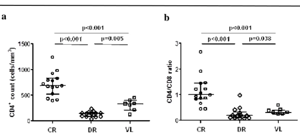
(48) Results. compare normally distributed continuous variables and the Mann-Whitney U test to compare non-normally distributed continuous variables. Categorical variables were described as proportions. The association between categorical variables was evaluated using the chi-square test. The Spearman correlation coefficient was used to analyze the correlations between continuous variables. Statistical analysis was performed using IBM SPSS software 21.0 (IBM, Chicago, Illinois, USA).Graphs were generated using GraphPad Prism 5.01 (La Jolla, California, USA).. COINFECTION WITH LEISHMANIA COMPARED TO IMMUNOLOGICAL DISCORDANT RESPONSE TO ANTIRETROVIRAL THERAPY. In this cross-sectional comparative study, we analyzed levels of microbial translocation, immune activation, inflammation through IL-6 levels, and immune senescence in HIV-1 infected patients with concordant immunological response to antiretroviral therapy, compared to infected individuals with discordant immunological response to treatment. We also studied correlations between microbial translocation measured by LPS and soluble CD14 levels, and either markers of inflammation or immune activation.. 2.7 Study design, ethic statement and sample collection. A cross-sectional, descriptive and comparative study was performed in HIV-1 infected patients at the Hospital Universitario Ramón y Cajal, in Madrid, Spain. A total of 29 HIV-1-infected adults with undetectable viral load (lower than 40 HIV-1 copies/ml) and receiving antiretroviral therapy for at least one year were analyzed. 15 of these HIV-1 infected adults showed concordant immunological response to the therapy, with CD4+ T cell count above 400 cells/mm3, while the other 14 HIV-1-infected patients showed discordant immunological response with CD4+ T cell count below 200 38.
(49) Results. cells/mm3. Whole blood was collected in Vacutainer-EDTA tubes (Becton-Dickinson, Madrid, Spain) for plasma and PBMCs isolation. Plasma samples were stored at -80ºC while the PBMCs were stored in liquid nitrogen until further use. The study was carried out according to the recommendations of the Declaration of Helsinki and current Spanish legislation on clinical trials. It was approved by the Institution Review Board and Ethics Committee (Hospital Ramón y Cajal, Madrid, Spain). All patients provided written informed consent for participation, sample collection, and laboratory determinations.. 2.8 Immune activation and Immune senescence. Fresh EDTA anticoagulated whole blood was used to analyze CD4+ and CD8+ T cell activation with the following antibody combination: CD3-eFluorTM 450 (eF450), CD4-KromeTM Orange (KO), CD8-peridinin chlorophyll protein complex (PerCP), CD38Allophycocyanin-700. (APC-700). and. HLA-DR-allophycocyanin-Cy7. (APC-Cy7).. Immunosenescence was defined as the expression of CD57-fluorescein isothiacyanate (FITC), and the absence of CD28-allophycocyanin-H7. All the antibodies (Becton Dickinson, NJ, USA) are shown in Table 2.1.. 2.9 Inflammation. Inflammation was measured by plasma interleukin 6 (IL-6) quantification using hsIL-6 (Human IL-6 Quantikine High-Sensitive ELISA kit, R&D Systems). IL-6, along with TNF-α and IL-1, drives the acute inflammatory response and is important in the transition from acute inflammation to either acquired immunity or chronic inflammatory disease.. 2.10 Microbial translocation. Microbial translocation was measured in plasma by the quantification of both lipopolysaccharide (LPS) using QCL-1000 Limulus Amebocyte Lysate (Lonza®, Basel, 39.
Figure
+7
Documento similar
