ENDOCRINOLOGY
VOLUME 61 JULY, 1957 NUMBER 1
Copyright © 1957 by the Endocrine Society
CHANGES IN DOGS DEVOID OF A CELLS
1SERGIO A. BENCOSME, S. MARIZ AND J. FREI
Department of Pathology, Queen's University, Kingston, Ontario, Canada
T
HE recent and excellent reviews of Cavallero (1), Foa (2) and Korp and Le Compte (3), have indicated that the inability to produce com-plete and permanent destruction of the A cells of the pancreas has greatly hindered our understanding of their function, particularly as a possible site of origin of glucagon. Recently, several substances have been used in an attempt to destroy the A cells, but there is as yet no general agreement on the exact nature of the A cell lesion thus obtained, nor on the effective-ness of these drugs in various animal species (4, 5, 6, 7, 8). The discovery in our laboratory that the uncinate process of the dog pancreas is nor-mally devoid of A cells, whereas the B and D cells are present (9), and than glucagon is probably absent in this portion of pancreas but abundant in the rest of the organ (10), prompted us to investigate the changes oc-curring in dogs deprived of their A cells. This was accomplished by means of the selective surgical removal of the A cell-containing portion of the pancreas. It was hoped that this procedure would allow the dogs to pro-duce enough endogenous insulin to prevent the development of diabetes, while at the same time permitting the establishment of a non-complicated A cell deficiency syndrome. In addition, it was hoped that this procedure would yield information on whether the uncinate process could generate A cells once the A cell-containing portion of the pancreas was removed.MATERIAL AND METHOD
Eighteen healthy mongrel dogs of both sexes, weighing between 25 and 45 pounds, were kept in individual metabolic cages until the completion of the experiment. Animals were weighed about once a week, had free access to water, drank daily about \ pint of homogenized milk and ate raw tripe. Pancreatectomized dogs received tripe cleaned of fat to facilitate digestion in the absence of pancreatic juice. Blood samples were obtained from the leg veins about 1-2- hours after the morning meal and glucose determined by a micro modification (11) of the Folin-Wu method. Twenty-four hour urine specimens
Received August IS, 1956. 1
BENCOSME, MARIZ AND FREI Volume 61
were collected in clean vessels to which toluol had been added, and glucose was mined by the quantitative Benedict method (12). Blood and urine sugar were deter-mined approximately twice a week. Glucose tolerance tests were done as follows: glucose (0.3 gm./kg.; in a 20% solution) was injected intravenously in the fasted anaesthetized (Nembutal) dog within 20 to 30 seconds. Blood samples were taken prior to and at 30, 60 and 90 minutes after glucose injection.
For qualitative estimation of the glucagon content of pancreatic tissue, extracts obtained by the method of Best et al. (13), were tested according to the method of Staub and Behrens (14). The details of these procedures as performed in this laboratory have been given elsewhere (10). Unless otherwise stated, extracts from 0.5 gm. instead of 1 gm. of pancreas were tested in triplicate whenever sufficient material was available. At the completion of the experiment, dogs were killed by an over-dose of Nembutal. The time at which each dog was killed is indicated in Figure 1. Samples of the following-organs were taken at autopsy for histological examination: pituitary, lacrimal gland, thyroid, parathyroid, salivary glands, oesophagus, lung, heart, abdominal aorta, stom-ach, duodenum, liver, pancreas, spleen, adrenal, gonads, kidney, muscle, sciatic nerve, abdominal skin, abdominal lymph node and sternal bone marrow. Careful search was
N G E S I N D O G S D E V O I D
O F A - C E L L S .
4 0 30 2 0 •40 30 20 4 0 30 20 4 0 30 20 4 0 30 20 80 40 0 8 0 4 0 0 80 4 0 0 80 4 0 0 80 4 0 0
6 0 0 T D M P - _
200 0 600 400 200 0 600 400 200 0 600 400 200 0 600 400 200 0
ID4I _^
...". '
URINE GLUCOSE BLOOD SUGAR WEIGHT ACETONURIA B BIOPSY OF PANCREAS P PANCREATECTOMY
40 80 120 160
DAYS AFTER PANCREATECTOMY
July, 195r< CHANGES IN DOGS DEVOID OF A CELLS
P A N C R E A T I C G L U C A G O N C O N T E N T . O F NORMAL DOGS P A R T I A L L Y P A N C R E A T E C T O M I Z E 0 DOGS
B I O P S Y B A U T O P S Y
25 0 5 15 25 0 5 • 15 25 0 5 15 25 0 5 15
MINUTES AFTER INJECTION
IN EACH CASE EXTRACT OF .5 GM OF PANCREAS WAS ASSAYED
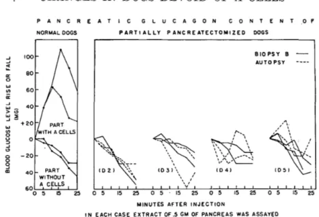
FIG. 2. Shows the lack of hyperglycemic effect of the extract from the normal and the transplanted uncinate process.
made at the site of operation for an}' residual pancreatic fragments, and all suspect tissue was removed and examined histologically. Tissues were fixed in Zenker formol and formol 10% and processed according to methods previously described; paraffin sections of 2.5 n were stained with Masson's haemalum-phloxin saffron trichrome method, (15). In addition, pancreas and pituitary were stained with aldehyde fuchsin (16), and with a modification of the chrome-alum-hematoxylin and Masson's trichrome methods (17). Glycogen was stained in the pancreas, lung, liver and kidneys with the Best-Carmine and the Periodic Acid Schiff controlled by diastase digestion.
The animals used in this experiment were divided into two groups. Group A.
{Con-trol): Eight untreated dogs served as morphologic controls. In addition extracts for
glucagon assay were made from the pancreas of three animals of this group. Extracts from the A cell-free uncinate process and from the A cell-containing parts were pooled. Separately each pooled extract was tested twice, using amounts equivalent to 0.5 gm. (Fig. 2) and 1 gm. of pancreas (Table 1). Group B {Partial pancreatectomy): All surgical procedures were performed under Nembutal anaesthesia. In 10 dogs, body and tail of the pancreas were removed and the uncinate process transplanted subcutaneously. A pancreatic fistula was created through the skin in order to prevent atrophy of the islet tissue which otherwise would occur in the transplant (19). To make sure that the trans-planted portion of the pancreas was devoid of A cells, a complete transverse section of the uncinate process proximal to the body of the pancreas was taken (biopsy A) for histological examination at the time of transplantation. Previous work performed in this laboratory has shown that this procedure will tell invariabl}'- whether A cells are present in the region distal to the biopsy (9, 10). In 2 dogs, the two-stage pancreatic transplant procedure of Ivy et al. (20) was followed in full. In one of these animals (Dl) the pancreatic transplant became completely dependent for its blood supply on the vascular connections established with the subcutaneous vessels. In the other 8 animals, the operative procedure was modified allowing the original vascular pedicle of the transplanted uncinate process to remain intact. Five of the operated dogs (1 female, Dl, and 4 males, D2, D3, D4 and D5) survived the experimental period; the others died of various post-operative complications, (pneumonia, eventration, etc.).
BENCOSME, MARIZ AND FREI Volume 61
TABLE 1. BLOOD SUGAR RESPONSE OF PASTING ANAESTHETIZED NORMAL CATS TO INTRAVENOUS INJECTION OF DOG PANCREATIC TISSUE
Material injected
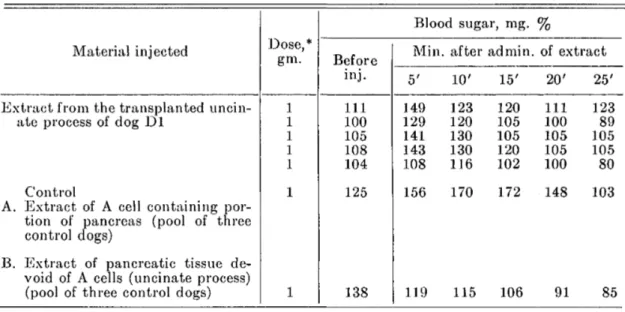
Extract from the transplanted uncin-ate process of dog Dl
Control
A. Extract of A cell containing por-tion of pancreas (pool of three
control dogs)
B. Extract of pancreatic tissue de-void of A cells (uncinate process) (pool of three control dogs)
Dose,* gm. 1 1 1 1 1 1 1 Before inj.
I l l 100 105 108 104 125 138
Blood sugar, rag.
Min 5' 149 129 141 143 108 156 119
. after
10' 123 120 130 130 116 170 115 admin. 15' 120 105 105 120 102 172 106 %
of extract
20' 111 100 105 105 100 148 91 25' 123 89 105 105 80 103 85
* Calculated in term of \vt. of fresh pancreas.
order to preserve the pancreatic fistula. Thus the blood supply of the remaining part of the transplant became dependent on the newly established vascular connection with the skin vessels, becoming comparable, in this respect, to the pancreatic transplant of dog Dl. The amount of tissue resected for biopsy B in each dog is given in Table 2. A complete transverse section was used for microscopic studies, while the remainder was used for glucagon extraction in all dogs, but Dl. At the end of the experiment, immediately after killing the animals, identical studies were made on the remaining part of the pancreatic transplant.
In dogs D2, D3 and D4, one glucose tolerance test was done before partial pancreatec-tomy and another one week before the second pancreatic biopsy. In dog D5, the second glucose tolerance test was not done.
KESULTS
Group A (Controls): In agreement with our previous findings (9, 10), the uncinate process contained B, D and X cells, but not A cells and its extracts were devoid of hyperglycemic activity (Fig. 2 and Table 1). In one dog, at the junction between the body and the uncinate process, there was gross evidence of incomplete embryological fusion and microscopic examination revealed a sharp demarcation line (Figs. 3, 4 and 5).
Unex-TABLE 2. MORPHOLOGICAL CHANGES IN THE TRANSPLANTED UNCINATE PROCESS OF DOGS DEPRIVED OF A CELLS BY SELECTIVE PARTIAL PANCREATECTOMY
Dog D l D2 D3 D4 D5 B-cell degran. Biop. A n 0 0 0 0 B 1 1 3 1 Autop. 4 3 2 4 3 Glycogen infiltration B-cells Biop. A 0 0 0 0 0 B 0 0 1 0 Autop. 4 1 0 4 1 Duct cells Biop. A 2 1 1 2 2 B 2 1 2 2 Autop. 4 3 2 4 3 ^ancreatic Biop. A 0 0 0 0 0 B 0 ±3 1 Autop. 1 1 1 0 1 Pancreas used for glue, ext
Gm. Biop. B 1.7 2 1.5 4 Autop. 5.8 5 5 2 2 Total trans. Gm.* 5.8 6.7 7 3.5 6
July, 1957 CHANGES IN DOGS DEVOID OF A CELLS 5
pected but interesting was the finding of glycogen in the small and medium-sized pancreatic ducts of all control dogs (Figs. 6, 7 and 8). It was also noticed that many of the tubular cells of the kidney contained large amounts of histochemically demonstrable glycogen.
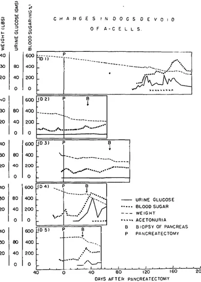
Group B (Partial pancreatectomy): Little changes were noted in blood and urine glucose of dogs D2, D3, and D5 after partial pancreatectomy, while in D4 a mild diabetes appeared (Fig. 1). On the other hand the post-operative glucose tolerance test was invariably diabetic in type (Fig. 9), except for dog D5, in which the test was not done. Variable glycosuria and hyperglycemia appeared in all dogs after biopsy B, and were most severs in dogs D4 and D5 which had the smallest transplant. Dog D4, which showed the greatest weight loss and highest hyperglycemia and glycosuria, developed a permanent ketonuria (trace) about one week after biopsy B (Table 2 and Fig. 1).
The subcutaneous pancreatic fistula was functioning in all dogs until the time of death. However, it seems that mild pancreatic duct obstruction was present in most instances, as suggested by minimal to moderate fibrosis and diffuse duct hyperplasia found in biopsy B and at autopsy (Fig. 10). The intensity of these changes is given for each case in Table 2 under the heading of "pancreatic fibrosis." Only in dog D4, a microscopic fragment of pancreas was found attached to the duodenal wall, probably remaining from the time of pancreatectomy. This showed well preserved acinar tissue, ducts cells markedly infiltrated with glycogen, and two small accumula-tions of 10-15 cells which were probably islet cells. However, no A cells were seen in the numerous serial sections obtained from this area.
In all dogs, biopsy A confirmed the fact that the transplant was devoid of A cells. Variable amounts of glycogen were found in biopsy A in all dogs (Table 2 and Figs. 6, 7 and 8). In no case were A cells found in biopsy B, or in the remnant of the pancreatic transplant obtained at autopsy, nor was the structure of the D and X cells altered in these two sections of the pancreas. Some degranulation of the B cell and variable amounts of glyco-gen infiltration of both duct and B cells were observed in the operated dogs (Figs. 10, 11 and 12). The intensity of these changes in both biopsy B and in the pancreatic tissue removed at autopsy is given for each animal in Table 2. The degree of glycogen infiltration of the B cells could be roughly correlated with the severity of hyperglycemia and glycosuria present in these operated dogs. Of interest was the presence in the pancreas of these dogs of neuro-insular complexes containing X cells, and showing glycogen infiltration in the B cells (Fig. 13).
BENCOSME, MARLZ AND FREI Volume 61
July, 1957 CHANGES IN DOGS DEVOID OF A CELLS
GLUCOSE TOLERANCE CURVES OF PARTIALLY PANCREATECTOMIZED DOGS
60 90 0 30 MINUTES AFTER INJECTION
60 90
FIG. 9. Shows that all partially pancreatectomized dogs had a diabetic type of glucose tolerance.
of protamine zinc insulin (Conaught Laboratories). Traces of acetone, how-ever, continued for a few weeks, then cleared spontaneously (Fig. 1). Until the time of death, the subcutaneous pancreatic fistula remained functional. At the time of autopsy, the pancreatic tissue of dog Dl showed severe de-granulation of B cells and glycogen infiltration of B cells and ductular epithelium. No changes were found in the X or in the D cells. Because of
EXPLANATION OF FIGURES 3-8
FIG. 3. General view at the site of junction of the ventral and dorsal pancreas in one dog which presented the incomplete fusion of these two portions of the pancreas. On the left side is the portion of pancreas with large islets containing A cells, whereas on the right side, no islet can be distinguished with this magnification. Aldehyde fuchsin.
X20.
FIG. 4. High power from Figure 3 showing the zone containing A cells. Note the large size of islets when compared with those of Figure 5. X100.
FIG. 5. High power from Figure 3 showing the zone devoid of A cells. X100.
FIG. 6. Uncinate process of dog Dl at the time of the partial pancreatectomy (biopsy A). Glycogen in duct cells. PAS. X580.
FIG. 7. Body of the pancreas of dog Dl at the time of the partial pancreatectomy (biopsy A). Glycogen in duct cells. PAS. X580.
BENCOSME, MARIZ AND FREI Volume 61
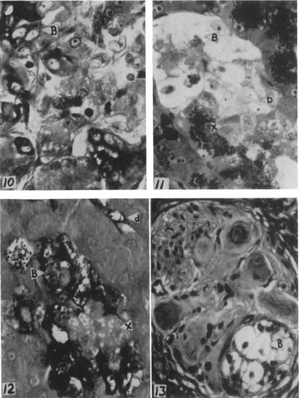
FIG. 10. Biopsy B of dog D4 showing disruption of the normal structure of acini and the presence of a few islet cells with marked glycogen infiltration. PAS. X580. FIG. 11. Uncinate process of dog Dl obtained at autopsy, showing marked hydropic degeneration of the B cells, normal appearing non-granular D cells and coarsely granu-lated X cells. Masson's trichrome. X580.
FIG. 12. Uncinate process of dog D4 obtained at autopsy, showing marked glycogen infiltration of B cells and intact X cells. The small vacuolization of these cells is of normal occurrence in them. There is also marked glycogen infiltration of a small duct (d). PAS. X580.
July, 1957 CHANGES IN DOGS DEVOID OF A CELLS 9
the results of the glucagon assay in this pancreas, 200 serial sections were examined and only two cells were observed which stained like A cells.
Bio-assay of pancreatic extract: As seen in Figure 2, no significant hyper-glycemic effect was induced with extracts obtained from biopsy B, or from the remaining portion of the transplanted uncinate process removed at autopsy, from dogs D2, D3, D4 and D5 (Fig. 2). Pancreatic extracts from these dogs elicited similar hypoglycemic effects in the cat, regardless of whether the B cells were hydropic, or slightly or severely degranulated (Fig. 2). The pancreatic extracts from the transplanted pancreas of dog Dl elicited a moderate initial hyperglycemia (Table 1) which was followed by almost no hypoglycemia, despite the fact that the pancreas obtained at the time of autopsy from dog D4 was histologically indistinguishable from the pancreas of dog Dl.
In general, the extrapancreatic changes observed in all treated dogs were similar to those found in other types of experimental diabetes: glyco-gen infiltration (hydropic change) was considerably greater in the kidney tubules of dogs Dl, D4 and D5 than in the controls; liver glycogen was normal in Dogs D2 and D3, decreased in the other animals. The livers of dogs Dl and D4, which had the most severe diabetes, showed moderate and minimal centrolobular fatty change respectively. There was marked reduction of spermatogenesis in dogs D3, D4 and D5. In the seminiferous tubes of D4 and D5, one could see a few giant cells, similar to those de-scribed in animals with some nutritional deficiencies (21).
DISCUSSION
Despite the absence of A cells in the experimental animal no A cell developed in the transplanted uncinate process during the period of 28 to 187 days. Since during this time, neither the X nor the D cell changed in structure or in their relative number, one may conclude that the A, B, D and X cells are histogenetically and functionally independent cells, that they are not capable of differentiating from one to another and finally, that the uncinate process has no precursors for the A cells. This is in agreement with previous work on the histogenesis of islet cell in the rabbit (6, 7). It is possible, however, that the procedure used did not provide the proper stimulus for the formation of A cells in the uncinate process or that perhaps residual fragments of pancreas containing A cells became hyperactive and compensated for the A cells removed in these operated animals. The latter explanation does not seem tenable since careful exploration at autopsy failed to reveal any fragment save for a minute amount in one case.
10 BENCOSME, MARIZ AND FREI Volume 61
the work of Allen (22), suggesting that the changes in the B cells did not require the presence of A cells nor of glucagon in order to develop.
The presence in normal dogs of histologically demonstrable glycogen in the epithelium of small and medium-sized pancreatic ducts is of consider-able interest. Perhaps the greater degree of glycogen infiltration seen in the duct cells of diabetic animals might represent some sort of exaggera-tion of a normal funcexaggera-tion (i.e., post-prandial hyperglycemia?), rather than a degenerative phenomenon. Glycogen was never seen in the B cells of the control dogs.
The type of pancreatectomy carried out in this group of animals is such that there is a complete elimination of A cells, and a diminution in B cell content. The conventional pancreatectomy reported by others results in a porportional diminution of both A and B cell elements. Since the type and severity of diabetes in the two experimental procedures proved to be sim-ilar, one may reasonably cast doubt on the diabetogenic influence of the A cells in the dog. In view of the supposed origin of glucagon from the A cells (10, 23, 24) and the known diabetogenic activity of this agent in rats (25) further studies on the metabolic effects of glucagon in the dog are required.
The lack of alterations in the pituitaries of dogs deprived of A cells is difficult to reconcile with several reports in which a relationship between the anterior pituitary and these cells had been stated to occur (1, 2, 3), unless adaptive mechanisms have occurred to mask this relationship at the morphological level. The presence of peculiar giant cells in the testes of two dogs and of marked diminution of the spermatogenesis in all operated male animals was probably due to nutritional deficiencies related perhaps to the absence of pancreatic juice, rather than to a lack of A cells, since similar lesions have been found in other nutritional deficiencies (21).
SUMMARY
A procedure for the removal of the A cells of the pancreas by means of a selective partial pancreatectomy has been described. This permits meta-bolic studies in dogs devoid of A cells without the accompaniment of frank diabetes.
Despite the removal of the A cell-containing portion of the pancreas, no A cells appeared in the transplanted uncinate process which is normally devoid of A cells, even though the operated dogs were observed for a period as long as 187 days. No changes were found in the D and X cells of the transplanted uncinate process. No hyperglycemic materials were found in the extracts of transplanted uncinate process of all dogs except one, which had a moderate hyperglycemic effect.
July, 1957 CHANGES IN DOGS DEVOID OF A CELLS 11
diabetic condition as revealed by a decreased tolerance to glucose and even-tually by the development of hyperglycemia and glycosuria. With the method used here, the diabetic syndrome in dogs devoid of A cells was essentially similar to that described in dogs partially pancreatectomized by the conventional method where many A cells remained. Morphological alterations outside the pancreas were in general no different from what one finds in other types of experimental diabetes.
In conclusion, there is strong evidence to support the idea that glucagon is produced by A cells and that the A, B, D and X cells are independent cells which do not convert into one another, even when all pancreatic A cells have been removed.
Although no clear-cut A cell deficiency syndrome has been produced, the method described may prove useful for studying the physiological signifi-cance of the A cell.
REFERENCES
1. CAVALLBRO, C : Rev. Canad. de Biol. 12: 189. 1953.
2. FOA, P. P.: Glucagon, the hyperglycemic-glycogenolytic hormone of the pancreas. Advances in Internal Medicine. Year Book Publishers, Inc. 1954.
3. KORP, W. AND P. M. LECOMPTE: Diabetes. 4 : 347. 1955.
4. VOLIV, B. W., S. S. LAZARUS AND M. G. GOLDNER: Arch. Int. Med. 93: 87. 1954. 5. CREUTZFELDT, W. AND W. SCHMIDT: Arch. Exper. Path. U. Pharmakol. 222: 487.
1954.
6. BENCOSME, S. A.: Rev. Canad. de Biol. 14: 175. 1955.
7. BENCOSME, S. A.: Am. J. Anat. 96: 103. 1955.
8. FODDEN, J. H.: Arch. Path. 61: 65. 1956.
9. BKNCOSME, S. A. AND E. LIEPA: Endocrinology. 57: 588. 1955.
10. BKNCOSME, S. A., E. LIEPA AND S. S. LAZARUS: Proc. Soc. Exper. Biol. & Med. 90:
387. 1955.
11. HAWK, P. B., B. L. OSER AND W. H. SOMMERSON: Practical Physiological Chemistry,
13th Edition, Blakiston Co. Inc., Toronto. 1954.
12. TODD, J. C. AND A. H. SANDFORD: Clinical Diagnosis by Laboratory Methods. W. B. Saunders Co. Philadelphia. 1942.
13. BEST, C. H., R. E. HAIST AND J. H. RIDOUT: J. Physiol. 97: 107. 1939.
14. STAUB, S. AND 0 . K. BEHRENS: / . Clin. Invest. 33: 1629. 1954.
15. BENCOSME, S. A.: Am. J. Clin. Path. 24: 1324. 1954. 16. GOMORI, G.: Am. J. Clin. Path. 20: 665. 1950. 17. BENCOSME, S. A.: Arch. Path. 53: 87. 1952.
18. TORESON, W. E.: Am. J. Path. 27: 327. 1951.
19. SIMARD, L. CH.: Rev. Canad. de Biol. 1: 264. 1945.
20. IVY, A. C. AND J. I. FARRELL: Am. J. Physiol. 77: 474. 1926.
21. KAUFMAN, N., J. V. KLAVINS AND T. D. KINSEY: Am. J. Path. 32: 103. 1956. 22. ALLEN, F. M.: / . Med. Res. 1: 5. 1922.