Otras secciones de este sitio:
☞ ☞ ☞ ☞
☞ Índice de este número ☞
☞ ☞ ☞
☞ Más revistas ☞
☞ ☞ ☞
☞ Búsqueda
Others sections in this web site:
☞ ☞ ☞ ☞
☞ Contents of this number
☞ ☞ ☞ ☞
☞ More journals
☞ ☞ ☞ ☞ ☞ Search
Artículo:
What do you do when all units are incompatible?
Derechos reservados, Copyright © 2005: Instituto Mexicano del Seguro Social
Revista Médica del IMSS
Suplemento
Supplemento 2 0 0 5
Volumen
edigraphic.com
George Garratty
American Red Cross Blood Services Southern California
Comunication with: George Garratty. Tel.: 909 859 7405. Fax: 909 859 7680.
E-mail: garraty@usa.redcross.or
If all red blood cells (RBCs) (and most or all RBCs of a phenotyped red cell panel) are incompatible when tested with a patient’s serum/plasma, the results may be due to autoantibody(ies) or alloantibody(ies). In a patient who has not been transfused recently, the differentiation is easy by looking at the result with autologous RBCs. If autologous RBCs are not reacting, then the incompatibilities are due to alloantibody(ies). If the autologous RBCs are incompatible, then an autoantibody alone or an autoantibody plus alloantibody may be present. If the patient has been transfused recently, then the results with autologous RBCs may not have much value and further procedures (e.g., separating transfused “older” RBCs from autologous “younger” RBCs) may be necessary. The autoantibody or alloantibody may react optimally at cold (0-5 °C) or warm (37 °C) temperatures. Table I shows some of the speci-ficities that may be involved when all, or most, RBCs tested are reacting.
What do you do when
all units are incompatible?
Incompatibility associated with
autoantibodies
The autoantibody may be a cold autoagglutinin, or reacting at 37 °C [usually by indirect antiglo-bulin test (IAT)]. If the patient has a 37 °C reacting autoantibody, the direct antiglobulin test (DAT) should be positive.
The first question to answer is: Does the patient’s serum contain autoantibody alone, auto-antibody + alloauto-antibody, or alloauto-antibody alone? If the patient has autoimmune hemolytic anemia (AIHA), one should never assume that incom-patibilities are only due to autoantibody.1-6 It is very important to answer the question above as blood incompatible with the autoantibody may not survive any better than the patient’s own RBCs, but will not usually cause an overt hemolytic trans-fusion reaction. In contrast, an alloantibody masked by the autoantibody may cause a severe reaction. We have found that 40 % of our AIHA, Primera versión: 18 de agosto de 2005
Versión definitiva: 19 de agosto de 2005 Aceptado: 23 de agosto de 2005
Palabras clave
inmunohematología
trasfusion de
eritrocitos
Key words
immunohematology
red cell trasfusion
Table I
Specificities of antibodies reacting with most or all RBCs tested
Auto Allo
Cold antibodies I, i, IH, H, Pr, Gd, Sa, Fl, Ju, Ena, Vel, Sda, P, PP1Pk,
Me, Lud, Rx, P, Vel M+Leb, P
1+Leb, M+P1
Warm antibodies “Rh”, LW, Ena, Ge, Kpb, Hr o, Hr, hr
S, hrB, Rh29, 38,
K13, Jk3, U, Sc1, Sc3, Vel, 39; LW; Fy3; k, Kpb Ku, Jsb,
IT, AnWj, Rx KL, K11-16, K18-20, K22;
Jk3; U; Ena; Wrb; Lub;
Lu3-8; Lu11-13; AnWj; Dib;
Yta; Sc1; Sc3; Coa, Co3;
Inb; Vel; Ge; Lan; Ata; Gya,
Hya, Cr; Jra; Cha, Rga; Csa,
George Garratty. Incompatibility in red cells
edigraphic.com
sustraídode-m.e.d.i.g.r.a.p.h.i.ccihpargidemedodabor
:rop odarobale FDP
VC ed AS, cidemihparG
arap
acidémoiB arutaretiL :cihpargideM
sustraídode-m.e.d.i.g.r.a.p.h.i.c associated with 37 °C-reactive autoantibodies, have
alloantibody masked by autoantibody.1,5 To provide blood for transfusion to such patients we suggest that one of the approaches shown in Table II is used before issuing blood for transfusion.
Adsorption with autologous
or allogeneic RBCs
Adsorption with autologous RBCs is the best approach in a patient who has not been transfused recently.1 The patient’s RBCs are cleared of auto-antibody, to expose autoantigens that will adsorb more autoantibody but not alloantibody from the patient’s serum. The antibody can be cleared by 56 °C heat elution, EDTA glycine acid elution, or ZZAP reagent.1,6 If the first two methods are used, the patient’s RBCs are then enzyme-treated; ZZAP contains enzyme so the second step is not necessary. The treated RBCs are then used to adsorb autoantibody from the patient’s serum and the serum tested for alloantibody (e.g., against antibody detection RBCs). As a rough guide: if the original indirect antiglobulin test (IAT) was 1+, 2+, 3-4+, then 1, 2, or ≥ 3 adsorptions respectively are usually necessary.1 If the patient has been transfused within the last 3 months, then the autoadsorption needs to be performed using allogeneic RBCs.1 A single phenotype can be used if it matches the patient’s phenotype. If this is not possible, then we recom-mend three phenotypes, rr, R1R1, and R2R2 that cover Kidd, Kell, and Ss. If ZZAP is used, then only Jka and Jkb need to be covered (i.e., ZZAP destroys all relevant antigens except Kidd –see Table III). We prefer using ZZAP, as it is a
one-step procedure, to elute antibody and destroy many antigens thus stopping adsorption of most alloantibodies except those of the Kidd system.
Approaches using dilutions
of patients’ serum
Because the warm autoantibody is being adsorbed continually by the patient’s RBCs, there is likely to be less autoantibody left in the serum than if an alloantibody is present. If dilutions of the patient’s serum are tested, sometimes the stronger alloantibody becomes obvious.
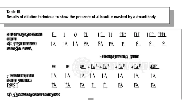
We titrate the patient’s serum against a pool of antibody detection RBCs and select a dilution that is yielding a 1+ reaction.1 This dilution is tested against a panel of red cells to determine specificity. Sometimes a serum that was originally reacting 3-4+ with all panel red cells will now react with only some of the panel [e.g., E+, K+, or Jk(a+) red cells]. Table III shows a result where an alloanti-c was masked by autoantibody. Oyen et al7 described a modification of this using a single dilution (1 in 5) of the patient’s serum. Leger and Garratty2 compared this approach with adsorption using ZZAP-treated allogeneic RBCs and found that 27 % of alloantibodies of potential clinical significance were detected by adsorption but not by the 1 in 5 dilution approach. Nevertheless, the approach is useful if adsorptions cannot be performed or is too time-consuming for a parti-cular patient’s clinical need for transfusion.
Dealing with cold autoantibodies
Most cold autoantibodies are of low titer and thermal amplitude and if interfering with pretrans-fusion testing, can be easily autoadsorbed by incubating the patient’s RBCs and serum plasma at 0-5 °C. The whole blood sample can be incubated, or for more efficiency, the patient’s RBCs should be enzyme-treated before incubating with the patient’s serum/plasma.3
Powerful high titer, high thermal amplitude cold autoantibodies (e.g., those associated with AIHA) are much more difficult to deal with. Autoadsorptions with enzyme-treated RBCs will help but many adsorptions may be needed to adsorb all or even most of the autoantibody.
Table II
Detection of alloantibodies in the presence of warm autoantibodies
Warm autoadsorption
George Garratty. Incompatibility in red cells
edigraphic.com
We find the easiest approach is to develop a systemwhere you can work strictly at 37 °C (see detail in reference 1). A prewarm procedure1,6 is useful, even with its disadvantages.8
Alloantibodies reacting with all,
or most, red cells
When all or most RBCs react, it may be due to a mixture of several alloantibodies rather than a single antibody to a high frequency antigen. This may be obvious from the serology. There may be a variation in strength of reactions with different RBCs on the panel or at different phases (e.g., one antibody specificity by antiglobulin test and another by direct agglutination). There may be differences noted by different test methods (e.g., one specificity with untreated RBCs and another using enzyme-treated RBCs).
Table IV shows some characteristics that are helpful in determining the specificity of an anti-body to a high frequency antigen. Some high frequency antigens are destroyed on RBCs treated with enzymes (e.g., ficin), dithreitol (DTT) and 2-aminoethylisothiouronium (AET). Some anti-gens are depressed or enhanced on cord RBCs. Some show characteristic agglutination appear-ances (e.g., Sda). Some antibodies are associated with certain ethnic groups.9
Obtaining donor blood for
patients with antibodies to high
frequency antigens
Screening random units is not usually productive but can be made more efficient by testing only RBCs from appropriate ethnic groups (see Table V). An effort to obtain siblings for typing is im-portant; there is a much better chance of finding a compatible donor among siblings. Donors may have to be obtained from National or International Blood Donor Registries, but this is time-consuming and expensive. Sometimes blood incompatible with an antibody to high frequency antigens can be transfused with no clinical ill effects. For instance, some anti-Yta, Ge, Lan, Lutheran, Era do not cause hemolytic transfusion reactions.10-13
Determining clinical significance
of antibodies to high frequency
antigens
Thermal amplitude and specificity: The first pieces of useful information are the thermal amplitude and specificity of the antibody. If antibodies are not reactive at 37 °C [by any test (e.g., antiglobulin test)], they have no potential to be clinically significant and incompatible blood can be transfused. Table V shows the relationship of specificity based on published data.
Table III
Results of dilution technique to show the presence of alloanti-e masked by autoantibody
Dilutions of patient’s 2 4 8 16 32 64 128 256 512 1024 serum
IAT on screening 3+ 3+ 3+ 2+ 2+ 1+ 0 0 0 0
cells (pooled)
Panel of group O cells
rr rr r'r R1R1 R1R1 R2R2 R2R2 r''r''
Undiluted serum 3+ 3+ 3+ 3+ 3+ 3+ 3+ 3+
serum diluted
1 in 64 1+ 1+ 1+ 0 0 1+ 1+ 1+
George Garratty. Incompatibility in red cells
edigraphic.com
Red cell survival using 51Cr-labeled red cells:The International Committee for Standardization in Hematology published recommendations for a compatibility test using 51Cr-labeled red cells.14 They suggested using only 0.5 ml of the incom-patible 51Cr-labeled red cells. Their interpretation was as follows: “In cases of urgency or when there is great difficulty in finding completely compati-ble RBCs, donor RBCs may be transfused with minimal hazard when, following a test with 0.5 mL
of donor RBCs the amount of radioactivity in the plasma, both at 10 and 60 minutes, does not correspond to more than 3 % of the radioactivity injected and when the RBC survival at 60 minutes is not less than 70 %.”
Functional cellular assays: As the 51Cr approach is not generally available, some investigators developed an in vitro equivalent using various in vitro
cellular assays, utilizing monocytes/macrophages, to mimic the reticuloendothelial system.10-13,15,16 These were: 1) the antibody-dependent cell-mediated cytotoxicity assay (ADCC)15; 2) the monocyte monolayer assay (MMA10,11); 3) the chemiluminescent test (CLT).16
We have used the MMA successfully for about 20 years to help make decisions on transfusing incompatible RBCs.10,11 The major problem for these assays is that the definition of “clinical significance” varies. To the more conservative, it means that the transfused incompatible RBCs should have normal survival. Some are satisfied if the patient has no clinical or laboratory signs of a reaction, and the less conservative are satisfied if the patient has no clinical signs of a reaction (e.g., they are not concerned with an increased bilirubin, suggesting abnormal red cell survival if the patient has no clinical signs). The latter is
Table IV
Useful results when identifying antibodies reacting with most RBCs
High frequency antigens destroyed by papain/ficin: Inb, Ge, JMH, Ch/Rg, EnaFS
High frequency antigens destroyed by DTT/AET: Kell, Lutheran, LW, Yta, Inb, Yka, McCa, Kna,
JMH, Hy, Gy
Cord RBCs differ from adult RBCs:
– increased on cord: i, LW
– decreased on cord: I, H, Yta, AnWj, Lub, Vel, Sda, HTLA
Appearance of agglutination pattern: Leb, Sda, Sdx, Lub, HTLA
High frequency antigens more frequently missing in certain racial groups:
– Black Hro, Hr, U, Fy5, Jsb, Hy, Ata
– Japanese Dib, Jra
– Mongolian descent/Mexicans Dib
– Polynesian/Filipino Jk3
– Swedish PP1Pk
– Melanesians Ge
– Jewish/Arab/Druse Yta
DTT = dithiothreitol AET = aminoethylisothiouronium bromide
HTLA = high titer low avidity antibody
Table V
Specificity / clinical significance of antibodies reacting at 37C
Usually significant Not significant Sometimes significant
ABO Chido/Rodgers Yta
Rh Knops Lutheran
Kell HTLA Dombrock
Kidd Leb Gerbich
Duffy (Xga) Lan
Colton (Sda) Lea
Ss (JMH) MN
George Garratty. Incompatibility in red cells
edigraphic.com
more acceptable if the patient is hematologicallynormal (e.g., surgery patient) rather than a hematology patient where optimal survival of transfused RBCs is important. An example of a successful transfusion of RBCs incompatible with anti-Dib, published by Leger and Garratty,17 illustrated an example in a surgical patient.
“In vivo” compatibility test
A commonly used method is to give a test dose of 5-10 mL of incompatible RBCs slowly and then draw an anticoagulated sample after 10-60 mins and inspect the plasma for hemolysis. This is a very poor approach and can give physicians a false sense of security. The method will only detect acute complement-mediated destruction of red cells as occurs with ABO incompatible blood. Most antibodies destroy RBCs extravascularly in the spleen and liver and do not cause hemoglo-binemia (especially of small quantities of blood). Thus, most antibodies to high frequency antigens and even powerful Rh, Kidd, Kell, Duffy antibodies would not appear to be clinically significant using this procedure. Only ABO and rare powerful complement activating antibodies such as examples of anti-Vel, -PP1Pk might give a positive result with this approach. The main disadvantage is that physicians may think that a negative result indicates that it is safe to transfuse the incompatible blood and a severe hemolytic transfusion reaction will not could occur; this is not true!
References
1. Petz LD, Garratty G. Immune hemolytic anemias. Second edition. Philadelphia: Churchill Livingstone; 2004.
2. Branch DR, Petz LD. Detecting alloantibodies in patients with autoantibodies. Transfusion 1999;39:6-10.
3. Engelfriet CP, Reesink HW. The detection of alloanti-bodies against red cells in patients with warm-type autoimmune haemolytic anaemia. Vox Sang 2000;78: 200-7.
4. Garratty G, Petz LD. Approaches to selecting blood for transfusion to patients with autoimmune hemo-lytic anemia. Transfusion 2000;42:1390-2. 5. Leger RM, Garratty G. Evaluation of methods for
detecting alloantibodies underlying warm auto-antibodies. Transfusion 1999;39:11-16.
6. Brecher M, ed. Techncial manual. 15th ed. Bethesda, MD: American Association of Blood Banks; 2005. 7. Oyen R, Angeles ML. A simple screening method to evaluate the presence of alloantibodies with concomitant warm autoantibodies. Immunohema-tology 1995;11:85-7.
8. Leger RM, Garratty G. Weakening or loss of antibody reactivity after prewarm technique. Transfusion 2003;23:1611-14.
9. Reid ME, Lomas-Francis C. The blood group antigen facts book. Second edition. New York: Academic Press, 2004.
10. Garratty G. Predicting the clinical significance of red cell antibodies with in vitro cellular assays. Transfus Med Rev 1990;IV:297-312.
11. Arndt PA, Garratty G. A retrospective analysis of the value of monocyte monolayer assay results for predicting the clinical significance of blood group alloantibodies. Transfusion 2004;44:1273-81. 12. Leger RM. In vitro cellular assays and other
appro-aches used to predict the clinical significance of red cell alloantibodies: a review. Immunohematology 2002;18:65-70.
13. Nance ST, Arndt PA. Review: what to dowhen all RBCs are incompatible – serologic aspects. Immuno-hematology 2004;20:147-60.
14. International Committee for Standardization in Haema-tology. Recommended method for radioisotope red-cell survival studies. Br J Haematol 1980;45:659-55. 15. Engelfriet CP, Ouwehand WH. ADCC and other
cellular bioassays for predicting the clinical signifi-cance of red cell alloantibodies. Baillieres Clin Haematol 1990;3:321-37.
16. Hadley A, Wilkes A, Poole J, Arndt P, Garratty G. A chemiluminescence test for predicting the outcome of transfusing incompatible blood. Transfus Med 1999;9:337-42.