Rev Inves Clin. 2017;69:262-269
Prevalence of Human Papillomavirus in
Women from the State of Michoacan,
Mexico, Showed High Frequency
of Unusual Virus Genotypes
Irvin Jácome-Galarza
1,4, María Ayumi Ito-Nakashimada
1, Gloria Figueroa-Aguilar
1,
Ethel García-Latorre
2, M.a. Isabel Salazar
2, Eduardo López-Orduña
3, Alejandro D. Camacho
4,
Juan José Valdez-Alarcón
5, José Manuel Hernández
6and Gloria León-Avila
4*
1Molecular Biology, Laboratorio Estatal de Salud Pública de Michoacán, Morelia, Michoacán; Departments of 2Immunology
and 4Zoology, Escuela Nacional de Ciencias Biológicas, Instituto Politécnico Nacional, Mexico City; 3Amplibio, Mexico City; 5Laboratorio de Epidemiología Molecular y Biotecnología de Enfermedades Infecciosas, Multidisciplinary Center for
Biotechnological Studies, Facultad de Medicina Veterinaria y Zootecnia, Universidad Michoacana de San Nicolás de Hidalgo, Tarímbaro, Mich.; 6Department of Cell Biology, Centro de Investigación y de Estudios Avanzados, Instituto Politécnico
Nacional, Mexico City. Mexico
ABSTRACT
Background: Human papillomaviruses (HPVs), the leading cause of cervical cancer, are distributed worldwide, with high prevalence
in developing countries. Objective: The objective of the study is to know the prevalence and genotypes of HPV in women from the state of Michoacán and the Women’s Hospital in Morelia, Michoacán. Materials and Methods: Cervical smear samples
(159,288) were subjected to HPV detection by hybrid capture 2. A subsample of 484 patients from the Women’s Hospital was studied by Papanicolaou test and linear array HPV genotyping, and when positive, patients were also examined by colposcopy and histopathology. Results: The overall prevalence for HPV in Michoacán State was 7.74%; 7.11% in 2009, 6.46% in 2010, 9.58%
in 2011, and 8.43% in 2012. The highest prevalence was found in the age groups < 25 and 25-34 years. The prevalence at the Women’s Hospital was 8.51%. Cytological examination revealed normal cytology in 64.44% of samples, 26.66 % with low-grade and 8.88 % with high-grade squamous intraepithelial lesion (HSIL). However, by colposcopy, normal tissue appearance was found only in 26.66%; 51% were reclassified as low-grade squamous intraepithelial lesion, 17.77% as HSIL, and in 4.4% atrophy was observed. The most prevalent genotype in single infections was HPV59, followed by HPV51 and HPV45. Double infections occurred with the following genotypes: 52-53, 51-59, 61-67, 66-11, 16-62, 53-62, 59-CP6108, 45-66, and 45-51. Triple infections were identified as: 6-31-39, 51-59-62, 51-62-81, 54-55-59, 16-58-71, and 16-59-62. Conclusions: The prevalent genotype found
among women from Michoacán, HPV59, was different to the rest of the country. The high prevalence of HPV59 could be due to cases imported to Michoacán by agricultural workers migrating to the USA or may be associated to ethnicity differences. Implications of this finding for immunization programs should be explored.
Key words: Human papillomavirus infection. Human papillomavirus prevalence. High-grade squamous intraepithelial lesion. Low-grade squamous intraepithelial lesion.
INTRODUCTION
Human papillomavirus (HPV) is distributed world-wide and is related to more than 99% of all cervical
cancer cases1; it can also infect the vagina, vulva,
urethra, penis, or the perianal area2. The association
of HPV with cancer depends on viral load, genotype, and whether or not the virus episome is integrated. HPV has a double-stranded DNA genome of approxi-mately 8000 bp and three genomic parts: long control
Corresponding author:
*Gloria León-Avila
Departamento de Zoología, Escuela Nacional
de Ciencias Biológicas del Instituto Politécnico Nacional Carpio y Plan de Ayala, s/n
Col. Casco de Santo Tomás
C.P. 11340, Ciudad de México, México E-mail: [email protected]
Received for publication: 03-10-2016 Approved for publication: 24-05-2017 doi: 10.24875/RIC.17002065
region (LCR) and early and late regions. The genome segment contained from the LCR to the E6 and E7 genes encodes oncoproteins described as essential for
HPV-induced carcinogenesis in vivo3. E6 and E7
inter-act with tumor suppressor proteins p534 and pRB5,
respectively, resulting in alteration of the DNA repair process and cell-cycle control.
More than 150 HPV genotypes have been identified and classified into two distinctly different groups, low- or high-risk, based on their association with
pre-malignant and pre-malignant lesions6. The high-risk virus
genotypes are 16, 26, 30, 18, 31, 33, 34, 35, 39, 45, 51, 52, 53, 56, 58, 59, 66, 67, 68, 69, 70, 73, and 82, while the types 6, 11, 32, 40, 42, 44, 54, 55, 61, 62, 64, 71, 72, 74, 81, 83, 84, 87, 89, and 91 are
consid-ered as low-risk7.
In 2011, Jemal et al. reported an estimated prevalence of new cases of cervical cancer close to half a million worldwide, and the calculated percentage of deaths
slightly higher than 50%8. Africa, South-Central Asia,
and South America had the largest incidence rates, while the highest mortality rates were documented in Africa, followed by Melanesia and Central America. In 2007, de Sanjosé et al. reported a crude prevalence of 20.5% in Central America (Costa Rica, Honduras, and
Mexico)9. Bruni et al., in 2010, analyzed eight different
studies and reported a crude prevalence of 20.6% in
Mexico10. The associated risk factors in Mexico were
the lack of formal education, low socioeconomic level, unemployment, poverty, no proper health care, and
the patient’s age11,12.
In 2016, Lazcano-Ponce et al. reported a prevalence of high-risk HPV of 8.6% in 12 states of Mexico, with
a 7% prevalence in Michoacán State.13 In this study,
we determined HPV prevalence in 159,288 women in Michoacán State and HPV genotype in 45 samples from the Women’s Hospital in Morelia, Michoacán.
MATERIALS AND METHODS
Study population
The study population consisted of 159,288 women recruited in eight sanitary districts of Michoacán, Mexico (age 19-82 years) attending regular gyne-cological examination, between January 2009 and December 2012. A woman was considered eligi-ble according to the following inclusion criteria: (i)
resident of Michoacán State, (ii) currently without symptoms of any sexually transmitted infection (STI) or on treatment for any STI, and (iii) not vaccinated against HPV. After patients signed an informed con-sent, cervical samples were obtained following the pro-tocol approved by the Ethics Committee of the State Hospital de la Mujer (Women’s Hospital), Morelia, Michoacán. The women were stratified by age in the following groups: < 25, 25-34, 35-44, 45-54, 55-64, and ≥ 65 years.
Cervical samples collection
Cervical smear samples were obtained and then cer-vical cytology and hybrid capture 2 analysis (HC2) were performed. Patients from the Women’s Hospital additionally were submitted to colposcopy, and their samples were analyzed by histopathology and linear array analysis (Fig. 1).
HPV detection and genotyping
Detection of HPV was performed using the HC2 HPV Test (HC2 High Risk Probe, Digene, USA), which detects 13 cancer-associated HPV types: 16, 18, 31, 33, 35, 39, 45, 51, 52, 56, 58, 59, and 68 and includes
a negative control (Carrier DNA, herring sperm) and calibrators A (1 pg/ml cloned HPV11 DNA) and B (1 pg/ml cloned HPV16 DNA). Plates were read in a DML200 Luminometer (Digene). Samples were con-sidered positive if they were above the threshold of 1 pg HPV DNA/ml. Positive specimens were processed with the Linear Array HPV genotyping test following the manufacturer’s instructions (Roche Diagnostics, Indianapolis, IN) and read manually. The linear array detects 37 high- and low-risk HPV genotypes, includ-ing: 6, 11, 16, 18, 26, 31, 33, 35, 39, 40, 42, 45, 51, 52, 53, 54, 55, 56, 58, 59, 61, 62, 64, 66, 67, 68, 69, 70, 71, 72, 73 (MM9), 81, 82 (MM4), 83 (MM7), 84
(MM8), IS39, and CP6108, and the β-globin
amplifica-tion as a positive control. The array was read manually according to the manufacturer, comparing the blue lines pattern against the reference strip.
Cervical Papanicolaou (PAP) analysis
Cervical smears were processed and cytological abnor-malities were classified according to the Bethesda System diagnostic criteria.
Concurrent infections
The presence of Gardnerella vaginalis, Candida
albi-cans, and Enterococcus spp. was determined by
cul-turing the samples in specific media for their iden-tification. Specific primers were used to detect the pathogens by polymerase chain reaction: Ureaplasma spp. (5’ GAG ATA ATG ATT ATA TGT CAG GAT CA 3’ and 5’ GAT CCA ACT TGG ATA GGA CGG 3’),
Chlamydia trachomatis (5’ TCC GGA GCG AGT TAC
GAGA 3’ and 5’AAT CAT TGC CGG GGA TTG GT 3’), and Mycoplasma hominis (5’ CAA TGG CTA ATG CTG GAT ACG C 3’ and 5’ GGT ACC GTC AGT CTG CAA T
3’). Human β-globin was the positive control and the
following primers were used: 5’ GGT TGG CCA ATC TAC TCC CCG G 3’ and 5’ TGG TCT CCT TAG ACC TGT CTT G 3’.
Statistical analysis
Samples were stratified according to the patient’s age, and results were analyzed using the software SAS. The crude prevalence was calculated and a contingency
table comparison was done. χ2 was calculated to
asso-ciate the incidence between groups and a p < 0.0001 was considered significant. The crude prevalence from
the Women’s Hospital samples was calculated, and a statistical analysis was made as described above.
Diagnostic cytology and colposcopy
validity tests
Results of cytology and colposcopy were compared with histopathology as the gold standard test for detection of lesions. For all tests, lesions were con-sidered positive irrespective of their strength (high- or low-level). Diagnostic validity was performed with
the OpenEpi v3.0, available online14,15. True positives,
false positives, false negatives, and true negatives for cytology and colposcopy were defined against the his-topathological assay.
RESULTS
Study population
During 4 years (2009-2012), 159,288 women were included from the eight sanitary districts of Michoacán State; cervical smear samples were obtained and DNA extraction was performed. The HPV overall preva-lence was 7.74%; prevapreva-lence was 7.11% in 2009, decreased to 6.46% in 2010, rose to 9.58% in 2011, and dropped slightly to 8.43% in 2012. The patients in the age groups < 25 and 25-34 years showed the
highest prevalence (Fig. 2). The χ2 values were very
high and p < 0.0001 indicating that age and locality are not independent, suggesting that in our group of study; there are external influences modifying the
frequencies such as access to medical services, edu-cational level, the number of sexual partners, and the number of pregnancies and abortions.
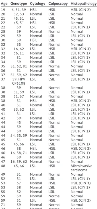
The crude prevalence of HPV in samples from the Women’s Hospital (484 women attending regular gynecological examination) was 9.29% (45/484) with the following age distribution: < 25 years, 1.03%; 25-34 years, 1.65%; 35-44 years, 3.3%; 45-54 years, 2.27%; 55-64 years, 0.61%; ≥ 65 years, 0.413%. Age was significantly associated with HPV infection. Cytological examination of the 45 positive samples revealed normal cytology in 64.44% (29/45), 26.66% (12/45) with low-grade squamous intraepithelial lesion (LSIL), and 8.88% (4/45) with high-grade squa-mous intraepithelial lesion (HSIL) (Table 1). To confirm these results, a colposcopy study was conducted. Only 26.66% of the samples had normal tissue appearance (12/45), while 51% were reclassified as LSIL (23/45), 17.77% as HSIL (8/45), and atrophy was observed in 4.4% (2/45). In women whose samples showed tissue alterations, a tissue biopsy was performed for histo-pathological examination. Within the LSIL samples, 69.56% (16/23) were classified as cervical intraepi-thelial neoplasia 1 (CIN 1). Among the HSIL samples, 25% (2/8) were CIN 2, 50% (4/8) were CIN 3, and one sample showed micro invasive cancer (Table 1). To evaluate the validity of cytology and colposcopy to predict HPV infections that cause lesions, diagnos-tic validity tests were performed. Clearly, colposcopy showed higher values of sensitivity, negative predic-tive value, and diagnostic precision, and correlated better with the histopathological assay. In contrast, cytology had a high level (58.62%) of false negative results (Table 2), thus rendering a low concordance.
Genotyping
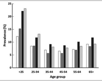
In the linear array HPV genotyping tests, we detected 62% single infections (28/45) and 38% (17/45) had more than one genotype. Stratification by age for sin-gle infections and coinfections, respectively, was as follows: < 25 years, 11.11 and 44.44%; 25-34 years, 19.35 and 6.45%; 35-44 years, 4.91 and 3.82%; and 45-54 years, 4.26 and 2.42%. In the intervals of 55-64 and³ 65 years, only single infections were detected: 4.76 and 5.55%, respectively (Fig. 3).
In single infections, HPV59 was the most prevalent (39.28%), followed by HPV51 (25%); HPV31, HPV45,
Table 1. HPV genotype, cytology and histopathology analysis.
Age Genotype Cytology Colposcopy Histopathology
19 6, 31, 39 HSIL HSIL HSIL (CIN 2)
20 52, 53 Normal LSIL Normal
21 45, 51 LSIL LSIL Normal
22 45, 51 HSIL HSIL Normal
22 59 LSIL LSIL LSIL (CIN 1)
28 59 Normal Normal Normal
29 59 Normal LSIL LSIL (CIN 1)
30 59 HSIL LSIL Normal
32 35 Normal Normal Normal
32 16, 62 LSIL HSIL HSIL (CIN 3) 32 66, 11 Normal LSIL LSIL (CIN 1)
34 51 Normal LSIL LSIL (CIN 1)
34 59 Normal LSIL LSIL (CIN 1)
35 51, 62, 81 Normal Normal Normal
36 51 Normal LSIL LSIL (CIN 1)
37 51, 59, 62 Normal Normal Normal 37 59, HPV
CP6108 LSIL LSIL LSIL (CIN 1)
38 39 Normal Normal Normal
38 51, 59 LSIL LSIL LSIL (CIN 1) 38 61, 67 Normal Normal Normal
38 31 HSIL HSIL HSIL (CIN 3)
40 51 Normal LSIL LSIL (CIN 1)
40 53, 62 LSIL LSIL LSIL (CIN 1)
41 31 LSIL LSIL LSIL (CIN 1)
42 59 Normal LSIL LSIL (CIN 1)
44 45 Normal Normal Normal
44 59 Normal LSIL Normal
44 59 Normal LSIL LSIL (CIN 1)
44 54, 55, 59 Normal Normal Normal
45 51 Normal Normal Normal
45 45, 66 LSIL LSIL LSIL (CIN 1)
46 58 HSIL HSIL HSIL (CIN 3)
46 16, 58, 71 Normal LSIL LSIL (CIN 1)
46 59 Normal LSIL LSIL (CIN 1)
47 16, 59, 62 Normal Normal Normal 48 45, 66 LSIL HSIL Microinvasive
carcinoma
49 51 Normal Normal Normal
52 51 LSIL LSIL LSIL (CIN 1)
53 67 Normal HSIL HSIL (CIN 3)
53 58 Normal LSIL LSIL (CIN 1)
55 52 Normal LSIL Normal
59 59 Normal Normal Normal
59 51 LSIL HSIL HSIL (CIN 2)
71 59 Normal Normal Normal
73 45 Normal Normal Normal
HSIL: high-grade squamous intraepithelial lesion;
LSIL: low-grade squamous intraepithelial lesion; CIN: cervical intraepithelial neoplasia 1; HPV: human papillomavirus
In triple infections, the detected genotypes were 6-31-39, 51-59-62, 51-62-81, 54-55-59, 16-58-71, and 16-59-62 (Fig. 4).
Coinfections were more frequent in the age groups < 25 and 25-34 years, representing 44.44% and 6.45%, respectively. Low-risk HPVs were always detected in association with a high-risk HPV. An inter-esting finding was that among triple infections; the histopathology revealed normal tissue in four cases: LSIL in one, and HSIL in another.
Concurrent infections
Even though we identified in some samples the follow-ing pathogens: G. vaginalis, C. albicans, U. urealyticum,
M. hominis, C. trachomatis, Enterococcus spp., alone or
Table 2. Conc
or
danc
e of r
esults of cyt
olo
gy and c
olposc
op
y f
or HPV det
ection. Method TP FP FN TN
% Sensitivity (95% CI) % Specificity (95% CI)
% PV+ (95% CI)
% PV– (95% CI)
Diagnostic pr
ecision LRP T LRNT Cohen’s k appa Cyt olo gy 13 3 12 17 52 (33.5 -69.97) 85 (63.96 -94.76) 81.25 (56.99 -93.41) 58.62 (40.74 -74.49) 66.67 (52.07 -78.64) 3.467 (1.569 -7.658) 0.564 (0.469 -0.678) 0.3541 (0.08472 -0.6234) Colposc op y 25 6 0 14 100 (86.68 -100.0) 70 (48.1 -85.45) 80.65 (63.72 -90.8) 100 (78.47 -100.0) 86.57 (73.82 -93.74) 3.333 (2.404 -4.621) 0.0 0.7212 (0.441 -1.002) Fn: f alse nega tiv es; F p: f alse positiv
es; LRNT: lik
elihood r
atio of nega
tiv
e t
est; LRP
T: lik
elihood r
atio of positiv
e t
est; PV+: positiv
e pr
edictiv
e v
alue; PV–: nega
tiv
e pr
edictiv
e v
alue; T
n: true nega
tiv
es;
T
p: true positiv
es, HPV: human papilloma
viruses; CI: c
onfidenc
e int
erv
al.
Figure 3. Age-specific prevalence of human papillomavirus mono- or coinfections in 484 women.
some in double infections (G. vaginalis + C. albicans, G.
vaginalis + U. urealyticum, G. vaginalis + C. albicans + U. urealyticum), we found no correlation with severity
of the viral infection.
DISCUSSION
The worldwide prevalence rate of HPV is higher in less-developed regions. Bruni et al. reported a crude prevalence of 20.6% in Central America (Mexico, Honduras, Guatemala, Costa Rica, Belize) in women with normal cytology. However, we found an overall prevalence of 7.74 in the state of Michoacán, Mexico. Cervical cancer is the second most common cancer
among Mexican women16. The traditional routine
method to evaluate cervical tissue is PAP smear since 1941 and it has been very useful to detect cell abnor-malities and reduce mortality rates. However, in our study, we found that 64.44% of the samples analyzed by PAP were classified as without lesions. After colpos-copy study, 51% of samples were reclassified as LSIL.
Granados-García et al.17 found false negative PAPs in
42.6% of samples, while we found 26.66% of false negatives (Table 2). Altogether, these results suggest that colposcopy is a better assay than cytology to predict HPV infections causing lesions in the histopa-thology assay. Cytology was not as good as colpos-copy because of its high level of false negative results as compared to histopathology, thus rendering a low concordance.
Despite these data, it is clear that the PAP coverage in national programs has reduced the mortality of
cervi-cal cancer.18 Moreover, it was reported that PAP
com-bined with HPV test increases the capability to identify
cervical cancer13,19,20.
In 2014, Salcedo et al. analyzed 2,956 samples and found an overall prevalence of HPV of 67.1% and
40 different HPV genotypes21; HPV16 was the most
prevalent (39.4%), followed by HPV18 (7.5 5%), HPV31 (7.1%), HPV59 (4.9%), and HPV58 (3.2%). On the other hand, Aguilar-Lemarroy et al., in an analysis of 822 samples, found HPV16 to be the most preva-lent type in all the studied groups, as in the study by
Salcedo et al.21, but also identified HPV18, HPV45,
HPV52, HPV58, and HPV39 types22.
All the works mentioned above clearly showed a higher prevalence of types 16, 18, 31, 33, 35, 45, 52,
58, and 59. In contrast, we found the highest preva-lence for type 59 (Fig. 4), followed by 51, 45, 31, 58, 35, 39, 52, and 67. The HC2 assay does not detect HPV53, HPV61, or HPV67 although the manufacturer reports that there is cross-reaction with HPV53 and HPV61. In addition, Castle et al. found cross-reaction
with HPV6723. These facts could explain why we
iden-tified positive samples with types not included in the HC2 test and that were later detected by linear arrays. The high prevalence observed for HPV59 could be due to cases imported to Michoacán State since a large number of men have migrated as agricultural workers to the USA. Thus, if poor sex education prevails and migrant men have unprotected intercourse abroad, they may be importing new genotypes on their return. We observed the highest prevalence of HPV infections in young women < 25 and 25-34 years old, which probably corresponds to the more sexually active women. It should be noted that HPV16 was only found in three coinfections and HPV18 was not found at all. Aguilar-Lemarroy et al. also found HPV16 more fre-quently, associated in coinfection with types 18, 39,
and 70 in samples from cervical cancer22. Remarkably,
none of the samples in our study contained any of the high-risk HPV types, including single, double, and triple infections.
In San Luis Potosí, 700 women who underwent col-poscopy presented type 33 as the most prevalent
followed by types 16, 18, and 5124. These results
con-trast with our findings since HPV51 was the second most common genotype.
Flores-Miramontes et al.25 found that types 16 and
51 had the highest prevalence among women without cervical lesions; in contrast, we observed that HPV59 was more frequent in this type of sample. They also detected HPV16 as the most prevalent in women with CIN 1.
González-López et al.26, in a retrospective analysis,
found that CIN 1 was more frequent in women aged 35-44 years old, and CIN 3 in women aged 45-59. We found that CIN 1 was more frequent in women aged 25-34 and 35-44 years, and CIN 2 in women aged 45-64 years.
the major histocompatibility complex (MHC)27-29 and health of the patient, the homeostasis of the immune system, or the historical evolution of the infection, which we did not explore in this study.
On the other hand, there were 51% of LSIL, similar to the proportion previously reported. However, regarding the samples classified as HSIL, the proportion we found was lower than that previously published. This could be due to the fact that those studies were performed in women diagnosed first with intraepithelial lesions or
squa-mous cell carcinoma and CIN or invasive carcinoma30.
The HPV vaccines used initially were designed to pro-tect against types 16-18 or 6-11-16-18. A study car-ried out in Mexican and some foreign women immu-nized with the quadrivalent vaccine showed effective protection against the genotypes included in the vac-cine and also prevented against CIN grade 2/3, adeno-carcinoma in situ, condyloma, and vaginal
intraepithe-lial neoplasia31. However, the efficacy of this vaccine
should be determined in long-term evaluation pro-grams. Skinner et al. in 2016 studied the progression of HPV infection to detectable CIN in 2838 women and followed the persistent infections, finding that women infected with types 16, 31, 33, and 45 had
a higher risk to develop CIN 232. Salmeron et al. are
performing a study in Mexico that will allow a more objective estimate of the efficacy of cervical cancer prevention programs and to establish the number of
doses required for an effective protection33.
In our study, the genotype found to be prevalent in Michoacán was different to the rest of the country. Moreover, none of the HPV genotypes that we iden-tified more frequently are included in the current vaccines. Thus, it is critical to consider in the sero-prevalence studies, the differences according to race,
geographical location, and ethnicity34. In addition, it
seems that the 9vHPV vaccine does not prevent infec-tion or disease caused by genotypes different to those
included in the vaccine35.
In view of the variations in genotypes found in Mexico, there is a need to obtain a complete map of preva-lence in our country and strengthen health education programs and counseling forums for women to pro-vide more information about HPV, its transmission,
and vaccination36,37. In addition to defining the efficacy
of vaccines against HPV genotypes different to those included in the vaccine, the serum antibody response
to vaccine HPV genotypes should be analyzed in future studies to determine antibody neutralization capabilities against genotypes not included in the vac-cine preparations in immunized women. Furthermore, T-cell responses should be evaluated and genotyping of MHC molecules should be performed in patients since the current Mexican population is mostly of Mestizo ethnicity, mixed American Indian, and European descendent.
ACKNOWLEDGMENTS
The authors thank Jimena Hernández-León for cor-rection of the english style. G.L-A. thanks the sup-port from CONACyT proyect 167376 and Instituto Politécnico Nacional (IPN) SIP 2017:0589.
REFERENCES
1. Walboomers JM, Jacobs MV, Manos MM, Bosch FX, Kummer JA, Shah KV, et al. Human papillomavirus is a necessary cause of inva-sive cervical cancer worldwide. J Pathol. 1999;189:12-9. 2. Crosbie EJ, Einstein MH, Franceschi S, Kitchener HC. Human
papil-lomavirus and cervical cancer. Lancet. 2013;382:889-99. 3. Ghittoni R, Accardi R, Hasan U, Gheit T, Sylla B, Tommasino M.
The biological properties of E6 and E7 oncoproteins from human papillomaviruses. Virus Genes. 2010;40:1-13.
4. Fu L, Van Doorslaer K, Chen Z, Ristriani T, Masson M, Travé G, et al. Degradation of p53 by human Alphapapillomavirus E6 pro-teins shows a stronger correlation with phylogeny than oncoge-nicity. PLoS One. 2010;5. pii: e12816.
5. Roman A. The human papillomavirus E7 protein shines a spotlight on the pRB family member, p130. Cell Cycle. 2006;5:567-8. 6. Bernard HU, Burk RD, Chen Z, van Doorslaer K, zur Hausen H, de Villiers
EM. Classification of papillomaviruses (PVs) based on 189 PV types and proposal of taxonomic amendments. Virology. 2010;401:70-9. 7. Doorbar J, Quint W, Banks L, Bravo IG, Stoler M, Broker TR, et al.
The biology and life-cycle of human papillomaviruses. Vaccine. 2012;30 Suppl 5:F55-70.
8. Jemal A, Bray F, Center MM, Ferlay J, Ward E, Forman D. Global cancer statistics. CA Cancer J Clin. 2011;61:69-90.
9. de Sanjosé S, Diaz M, Castellsagué X, Clifford G, Bruni L, Muñoz N, et al. Worldwide prevalence and genotype distribution of cervical human papillomavirus DNA in women with normal cytology: A meta-analysis. Lancet Infect Dis. 2007;7:453-9.
10. Bruni L, Diaz M, Castellsagué X, Ferrer E, Bosch FX, de Sanjosé S. Cervical human papillomavirus prevalence in 5 continents: Meta-analysis of 1 million women with normal cytological findings. J Infect Dis. 2010;202:1789-99.
11. Flores YN, Bishai DM, Shah KV, Lazcano-Ponce E, Lörincz A, Hernández M, et al. Risk factors for cervical cancer among HPV positive women in Mexico. Salud Publica Mex. 2008;50:49-58. 12. Palacio-Mejía LS, Rangel-Gómez G, Hernández-Avila M,
Lazcano-Ponce E. Cervical cancer, a disease of poverty: Mortality differ-ences between urban and rural areas in Mexico. Salud Publica Mex. 2003;45 Suppl 3:S315-25.
13. Lazcano-Ponce E, Lörincz AT, Salmerón J, Fernández I, Cruz A, Hernández P, et al. A pilot study of HPV DNA and cytology testing in 50,159 women in the routine Mexican Social Security Program. Cancer Causes Control. 2010;21:1693-700.
15. Weiss N.S. Clinical epidemiology. In: Rothman KJ, Grennland S, Lash TL, editors. Modern Epidemiology. 3rd ed. Ch. 23. Philadelphia,
PA, USA: Lippincot Williams and Wilkins; 2008. p. 641-51. 16. López-Revilla R, Martínez-Contreras LA, Sánchez-Garza M.
Prevalence of high-risk human papillomavirus types in Mexican women with cervical intraepithelial neoplasia and invasive carci-noma. Infect Agent Cancer. 2008;3:3.
17. Granados-García V, Flores YN, Pérez R, Rudolph SE, Lazcano-Ponce E, Salmerón J. Cost of the cervical cancer screening pro-gram at the mexican social security institute. Salud Publica Mex. 2014;56:502-10.
18. Lazcano-Ponce E, Palacio-Mejia LS, Allen-Leigh B, Yunes-Diaz E, Alonso P, Schiavon R, et al. Decreasing cervical cancer mortality in Mexico: Effect of Papanicolaou coverage, birthrate, and the importance of diagnostic validity of cytology. Cancer Epidemiol Biomarkers Prev. 2008;17:2808-17.
19. Beal CM, Salmerón J, Flores YN, Torres L, Granados-García V, Dugan E, et al. Cost analysis of different cervical cancer screening strategies in Mexico. Salud Publica Mex. 2014;56:429-501. 20. Flores YN, Bishai DM, Lorincz A, Shah KV, Lazcano-Ponce E,
Hernández M, et al. HPV testing for cervical cancer screening appears more cost-effective than Papanicolaou cytology in Mexico. Cancer Causes Control. 2011;22:261-72.
21. Salcedo M, Pina-Sanchez P, Vallejo-Ruiz V, Monroy-Garcia A, Aguilar-Lemarroy A, Cortes-Gutierrez EI, et al. Human papillo-mavirus genotypes among females in Mexico: A study from the Mexican institute for social security. Asian Pac J Cancer Prev. 2014;15:10061-6.
22. Aguilar-Lemarroy A, Vallejo-Ruiz V, Cortés-Gutiérrez EI, Salgado-Bernabé ME, Ramos-González NP, Ortega-Cervantes L, et al. Human papillomavirus infections in Mexican women with normal cytology, precancerous lesions, and cervical cancer: Type-specific prevalence and HPV coinfections. J Med Virol. 2015;87:871-84. 23. Castle PE, Solomon D, Wheeler CM, Gravitt PE, SWacholder PE,
Schiffman M. Human papillomavirus genotype. Specificity of hybrid capture 2. J Clin Microbiol. 2008;6:2595-604.
24. DelaRosa-Martínez R, Sánchez-Garza M, López-Revilla R. HPV genotype distribution and anomalous association of HPV33 to cervical neoplastic lesions in San Luis Potosí, Mexico. Infect Agent Cancer. 2016;11:16.
25. Flores-Miramontes MG, Torres-Reyes LA, Alvarado-Ruíz L, Romero-Martínez SA, Ramírez-Rodríguez V, Balderas-Peña LM, et al. Human papillomavirus genotyping by linear array and next-generation sequencing in cervical samples from Western Mexico. Virol J. 2015;12:161.
26. González-López S, Martínez-Silva MG, Hernández-Hernández DM, Aguilar-Lemarroy A, Jave-Suárez LF. Frequency of
cervical epithelial lesions reported in the Regional Laboratory of Exfoliative Cytology in Jalisco. Rev Med Inst Mex Seguro Soc. 2015;53 Suppl 2:S132-9.
27. Das Ghosh D, Mukhopadhyay I, Bhattacharya A, Roy Chowdhury R, Mandal NR, Roy S, et al. Impact of genetic variations and tran-scriptional alterations of HLA class I genes on cervical cancer pathogenesis. Int J Cancer. 2017;140:2498-508.
28. Chen D, Gaborieau V, Zhao Y, Chabrier A, Wang H, Waterboer T, et al. A systematic investigation of the contribution of genetic variation within the MHC region to HPV seropositivity. Hum Mol Genet. 2015;24:2681-8.
29. Hernández-Hernández DM, Cerda-Flores RM, Juárez-Cedillo T, Granados-Arriola J, Vargas-Alarcón G, Apresa-García T, et al. Human leukocyte antigens I and II haplotypes associated with human papillomavirus 16-positive invasive cervical cancer in Mexican women. Int J Gynecol Cancer. 2009;19:1099-106. 30. Fernández-Tilapa G, Illades-Aguiar B, Martínez-Carrillo DN,
Alarcón-Romero LC, Vences-Velázquez A, Terán-Porcayo MA, et al. Prevalence of human papillomavirus types among Mexican women with intraep-ithelial lesions and cervical cancer: Detection with MY09/MY011 and GP5+/GP6+ primer systems. Am J Infect Dis. 2007;3:62-7. 31. Lazcano-Ponce E, Pérez G, Cruz-Valdez A, Zamilpa L,
Aranda-Flores C, Hernández-Nevarez P, et al. Impact of a quadri-valent HPV6/11/16/18 vaccine in Mexican women: Public health implications for the region. Arch Med Res. 2009;40:514-24. 32. Skinner SR, Wheeler CM, Romanowski B, Castellsagué X,
Lazcano-Ponce E, Del Rosario-Raymundo MR, et al. Progression of HPV infection to detec` cervical lesions or clearance in adult women: Analysis of the control arm of the VIVIANE study. Int J Cancer. 2016;138:2428-38.
33. Salmerón J, Torres-Ibarra L, Bosch FX, Cuzick J, Lörincz A, Wheeler CM, et al. HPV vaccination impact on a cervical cancer screening program: Methods of the FASTER-Tlalpan Study in Mexico. Salud Publica Mex. 2016;58:211-9.
34. Gravitt PE. HPV seroprevalence in the United States: Behavior, biology, and prevention. J Infect Dis. 2016;213:171-2.
35. Joura EA, Giuliano AR, Iversen OE, Bouchard C, Mao C, Mehlsen J, et al. A 9-valent HPV vaccine against infection and intraepithelial neoplasia in women. N Engl J Med. 2015;372:711-23.
36. León-Maldonado L, Allen-Leigh B, Lazcano-Ponce E. Counseling for HPV detection when used to screen for cervical cancer: A qual-itative study on the needs of women from Michoacan, Mexico. Salud Publica Mex. 2014;56:519-27.