Epigenetic Mechanisms in two primary immunodeficiencies: Hyper-IgM Syndrome and Common Variable Immunodeficiency
178
0
0
Texto completo
(2) EPIGENETIC MECHANISMS IN TWO PRIMARY IMMUNODEFICIENCIES: HYPER-IgM SYNDROME AND COMMON VARIABLE IMMUNODEFICIENCY. Tesis Doctoral Virginia Carolina Rodríguez Cortez.
(3)
(4) EPIGENETIC MECHANISMS IN TWO PRIMARY IMMUNODEFICIENCIES: HYPER-IgM SYNDROME AND COMMON VARIABLE IMMUNODEFICIENCY. Memoria presentada por Virginia Carolina Rodríguez Cortez para optar al grado de Doctor en Biomedicina por la Universidad de Barcelona. UNIVERSIDAD DE BARCELONA – FACULTAD DE MEDICINA PROGRAMA DE DOCTORADO EN BIOMEDICINA. Este trabajo ha sido realizado en el Grupo de Cromatina y Enfermedad del Programa de Epigenética y Biología del Cáncer (PEBC) del Institut d' Investigaciò Biomédica de Bellvitge (IDIBELL). Dr. Esteban Ballestar Tarín Director. Virginia Carolina Rodríguez Cortez Doctoranda.
(5)
(6) Todo esfuerzo que va acompañado de sacrificio, perseverancia, disciplina y dedicación tiene una recompensa. Puede que no llegue cuando tú quieras, ni sea lo que esperas, pero sí, definitivamente tiene su recompensa....
(7)
(8) AGRADECIMIENTOS Hace tanto tiempo que deseo estar en ésta etapa, que no me creo que ahora mismo estoy aquí sentada haciendo lo último que dejé por hacer, los agradecimientos. Para mí algo muy especial, porque aprovechas de decir a todas las personas que han formado parte de éste recorrido tan largo que es la tesis, lo importantes que son y han sido para ti y lo mucho que les aprecias. Pues comienzo contigo Esteban, estoy muy agradecida de que me hayas dado la oportunidad de formar parte de tu grupo todos estos años, y de apostar por proyectos que no han sido ideales, ni han dado sus frutos tan rápido como se espera. Pero más que eso, te doy infinitas gracias por tu comprensión y tu flexibilidad en los momentos en que más los necesitaba y tu interés por que estuviera bien. No olvidas que somos personas y que tenemos momentos en los que por diferentes razones no damos nuestro 100%, y creo que es una cualidad importante que todos los jefes deben tener. Eso sí, a veces puedes ser muy duro (muy duro!) pero bueno, ahí nos toca a nosotros tener paciencia. Yo, particularmente me llevo muchas experiencias divertidas contigo y me alegro mucho de que hayas sido mi jefe todos esto años. Javi, para mi has sido un verdadero Maestro, sí, con M mayúscula. Ahora mismo sería incapaz de enumerar todas las cosas que he aprendido de ti. Tienes un don para enseñar! consérvalo siempre porque es un don maravilloso. De todos los "Javi tienes un momentín?" siempre, siempre, salía algo productivo. Ése experimento que creía que había sido una total pérdida de tiempo, siempre se convertía en algo útil. Gracias a ésa motivación, seguí adelante en muchos momentos en que ya estaba decidida a tirar la toalla. Así que miles de gracias para ti!... Henar Henar, mi super compi de ordenador, gracias por todos los momentos que compartimos, eres muy especial para mí, me divertí mucho enseñándote vídeos de Chino y Nacho y de animalitos, compartiendo nuestro día a día y las jornadas de fin de semana poniendo a punto anticuerpos imposibles! Espero que a pesar de la distancia sigamos cosechando la linda amistad que hemos tenido hasta ahora... Lauri! mi queridísima Lauri, mentora de maternidad, compañera y amiga especial. Gracias por todos tus consejos, por compartir tantas cosas conmigo y por brindarme tu amistad en todo momento. Eres una chica super trabajadora y excelente persona, así que solo puedo augurar cosas buenas para ti y por su puesto, espero compartirlas contigo... Roser, mi vecina de poyata, muchísimas gracias también por los momentos amenos de cháchara durante los experimentos, por estar siempre pendiente y ofrecer ayuda en los momentos complicados, y también gracias por las chácharas de los momentos doctorales.
(9) difíciles, porque te das cuenta de que no estás sólo y que es igual de difícil para los demás! te irá muy bien, eres muy trabajadora y muy buena...Loren, gracias por todos los momentos de risas con tus ocurrencias y habilidades de negociación, me divertí mucho!... Biola, Marina, también disfrute mucho de su compañía y aprendí mucho de ustedes, las recuerdo siempre con mucho cariño... Nuestras queridas vecinas, en su momento conocidas como "las apéndices", Lidia, Bruna y Olgui, gracias porque a pesar de que éramos dos grupos diferentes, nos sentíamos como si fuéramos uno. Sobre todo por aquellos intercambios de material (que solían ser unidireccionales) que nos hacían sentir como si fuéramos un solo laboratorio... Lidia, gracias por todos los momentos divertidos que compartimos, fueron geniales!... Bruna, muchas gracias por tu amistad y por estar allí también en momentos difíciles. Eres para mí un ejemplo de tenacidad y de fuerza!... Olgui, mi querida Olgui, que ahora es Olgui, Jordi y Arnau!, qué te puedo decir, estoy encantada de haber compartido con ustedes toda la experiencia de la maternidad. Hemos compartido muchos momentos divertidos y agradables y espero de corazón que sigamos compartiendo muchos momentos más porque los apreciamos mucho! (Julio, Lucía y yo)... Me llevo con especial recuerdo éste PEBC7, las comidas para hablar de todo y de nada, momentos super super divertidos, cumpleaños, fiestas de disfraces, montaña, burritos, baby shower, nacimientos... oh! tantas cosas!, dolores de barriga y lágrimas de tantas risas luego de analizar los beneficios de comer piña, o los curiosos efectos de la primavera y otras cosas que no puedo contar por aquí! han sido y serán momentos muy especiales para mí. Siempre me sentí como en casa... De ésta experiencia forman parte también por su puesto, las allegadas del PEBC7, Vicky, Laura Barberá y Palomita. Chicas, son geniales! les tengo un cariño muy muy especial. Vicky gracias por todos los asesoramientos tesísticos, me ayudaste mucho! muchas gracias también por los momentos que hemos compartido, han sido muy agradables!... Laura, aquí en casa te tenemos especial cariño también, lo pasamos genial los días de patines y todos los otros días que hemos compartido, esperamos que sean muchos más!... Palomita, Palomita, bah! eres una chica genial y super especial, siempre con una sonrisa, siempre con buena vibra, contagiando alegría. Gracias y mil gracias porque tu ayuda ha sido determinante también. He aprendido muchísimas cosas de ti y muchas cosas que he podido hacer durante mi tesis has sido gracias a ti! Deseo de todo corazón que te vaya excelente en cualquier cosa que decidas hacer (que puede ser cualquier cosa, literalmente!). Estoy muy agradecida contigo y con toda la gente del PEBC4, son unos excelentes compañeros! Álex, Anna,.
(10) Laia, Helena, Irene, Nuria, todos, todos, siempre dispuestos a compartir su experiencia y a echar una mano cuando sea. Mil gracias a todos! A los nuevos miembros del PEBC7 también les agradezco mucho! Damiana, Carlos, Antonio, Lluis, Natalia, Alba, Francesc muchas gracias. Lluis, mi nuevo compañero de ordenador, gracias por ser tan listo siempre y ayudarme con mis rollos con la tecnología, siempre una solución a cualquier inconveniente informático! (darle a la tecla de escape y esas cosas)... Damiana, gracias por compartir tus anécdotas con Olivia, son siempre muy divertidas... Carlos, gracias por tu trabajo, seguro te irá muy bien... Nono, gracias por tu excelente disponibilidad y buena vibra con mis ChIPs infernales! (obvia ésta parte Francesc!)... Gracias de antemano a Francesc, porque ha heredado las tareas pendientes del proyecto AID. Aprenderás un montón, resolviendo problemas es cuando más aprendes!... Alba, eres una chica muy simpática, espero que te vaya muy bien en la tesis y que sigamos en contacto! que tenemos un ensayo pendiente eh? no se me olvida... Natalia, muchas muchas gracias, en éste poco tiempo que nos hemos ido conociendo has sido una excelente compañera! espero que te vaya muy bien en la tesis y que sigamos en contacto! hay unos sushis y varias cosas pendiente por allí. Para ti y para todos, estoy disponible para cualquier cosa que necesiten! los aprecio mucho!!! A otras personas del PEBC también les estoy muy agradecida, Fer, Catia, siempre dispuestos a echar una mano... Carmen de microscopía, un ejemplo de fuerza y alegría!!!, todos los compañeros de pasillo que con 2 minutos de conversación y una sonrisa también han formado parte de éste recorrido... Mil Gracias! Ya salgo del PEBC para agradecer a otras personas también, porque aunque no sean del ambiente laboral, su ayuda y su compañia han sido esenciales para mi. Mi querida amiga Patricia, una persona de referencia para mí en todos los sentidos, tus consejos, tu ayuda y tu apoyo para mi han sido vitales. Da igual dónde estemos y si pasamos muchos días sin hablar, la amistad que tenemos va mucho más allá de todo eso... He aprendido muchíiiiiisimo de ti, te quiero mucho y espero que nuestra amistad siga creciendo. Aprovecho para dejar plasmado aquí mi más sincero agradecimiento a tus papás, el Sr. Antonio y la Sra. Rosalía, sin su ayuda hubiese sido imposible venir aquí... eso no lo olvidaré nunca... Gracias por hacerme sentir como en casa, son muy especiales para mí! Al team de Venezuela en Barcelona, María Cecilia, Manuel (los famosos tíos guays), gracias por tanto cariño para mi Lucía y por su apoyo en todo momento, los queremos mucho!. Arnoldo, Irma, Luisda, Sofi, Luis, Yenni, Ainhoa, Yaiza, Carlos,.
(11) Marc, son todos nuestra familia aquí, nuestro pedazo de Venezuela en España! esperamos seguir compartiendo momentos especiales y brindándonos apoyo... los quiero mucho! Mi familia política, mis suegros, mis cuñados y sobrinos, todos siempre pendientes de nosotros, brindándonos amor, apoyo para cualquier cosa que necesitemos. Sra. Carmen, un especial agradecimiento para usted, siempre disponible para ayudarnos en todo, siempre atenta y buscando soluciones a cualquier cosa que se nos presente, la quiero mucho. Los quiero mucho a todos! Mi familia, mi Papá, mi mamá, mi hermana, mis pilares, mi motivación. Lo que soy ahora es gracias a ustedes, gracias por apoyarme siempre, por estar pendientes de mi, por quererme tanto. No ha pasado un día, ni un solo día en que no los extrañe y desee estar cerca de ustedes. Uno de mis deseos más grandes es poder estar para ustedes y poder llegar a ser ése pilar que ustedes han sido para mi, los amo!. Julio, mi kiki (si, kiki, tiene su historia...). Nada de lo que pueda escribir aquí llegará a expresarte todo mi agradecimiento. Han sido tantos años... Tenemos toda una vida juntos. Gracias por todo tu cariño, tu amor, tu apoyo, tu PACIENCIA, tu confianza. Has sacrificado tantas tantas cosas por mi, que lo que más deseo es hacerte feliz y pasar toooda mi vida contigo. Gracias por aguantar tanto y sobre todo por no tirar la toalla cuando te encontrabas en los momentos más dificiles. TE AMO con todo mi corazón y espero que sigamos juntos compartiendo la vida con nuestros retoñitos... Mi Lucía... mi luz, mi inspiración, ahora mi centro y mi mayor motivo para dar lo mejor de mí. Llegaste en un momento clave de mi vida para enseñarme a priorizar, algo que necesitaba muchísimo... Sólo espero darte todo el amor, cariño y paciencia para hacerte una mujer de bien.. Te amo! y tú, mi Noelia (nombre provisional), que aún estás en mi vientre... ya te estoy esperando para igual que a Lucía, darte lo mejor de mí...Te amo también!, perdoname por las sesiones intensas con la tesis y por el estrés de mami que seguro has sentido en algún momento! ya te mimaré mucho para compensarte! Pues termino aquí, me llevo excelentes experiencias, compañeros y amigos y por su puesto, lo que más me ha enseñado la tesis en todo éste tiempo: PACIENCIA y PERSEVERANCIA... creo que son un par de dones vitales para salir airosos de este largo y duro camino... con clínex en mano y al mejor estilo de Gustavo Cerati en concierto, me despido con un: GRACIAS....TOTALES!!!!!.
(12)
(13)
(14) INDEX ABBREVIATIONS ......................................................................................................... I 1. INTRODUCTION ...................................................................................................... 3 1.1. B cell Development ............................................................................................ 3 1.2. B cell Activation .................................................................................................. 5 1.2.1. Class Switch Recombination ........................................................................ 9 1.2.2. Somatic Hypermutation .............................................................................. 11 1.3. Activation Induced Cytidine Deaminase............................................................ 13 1.3.1. AID, the master regulator of antibody maturation ....................................... 13 1.3.2. Functional Domains of AID ........................................................................ 15 1.3.3. AID Expression .......................................................................................... 16 1.3.4. Regulation of AID expression and activity .................................................. 17 1.3.5. Transcriptional Regulation of AID ............................................................... 17 1.3.6. Post transcriptional Regulation of AID ........................................................ 18 1.3.7. AID subcellular localization ........................................................................ 19 1.3.8. Phosporylation of AID ................................................................................ 19 1.3.9. AID interactions.......................................................................................... 20 1.3.10. AID roles beyond CSR and SHM in B cells .............................................. 21 1.4. Epigenetic regulation ........................................................................................ 23 1.4.1 DNA methylation ......................................................................................... 24 1.4.2. DNA demethylation .................................................................................... 25 1.4.3. Histone Modifications ................................................................................. 26 1.4.4. Methylation of histone H4 at lysine 20 ........................................................ 27 1.4.5. Epigenetics mechanisms during antibody diversification ............................ 30 1.4.6. Epigenetics mechanisms in the immune system ........................................ 31 1.5. Primary immunodeficiencies ............................................................................. 33 1.5.1. Hyper-IgM Syndromes ............................................................................... 35 1.5.2. Common Variable Immunodeficiency ......................................................... 37 2. OBJECTIVES ......................................................................................................... 43 3. MATERIALS AND METHODS................................................................................ 47 3.1. Methods used for the epigenetic study on AID and HIGM ................................ 47 3.1.1. Cell culture ................................................................................................. 47 3.1.2. DNA constructs and system for inducible AID expression .......................... 47 3.1.3. Western blot assay .................................................................................... 49 3.1.4. Immunofluorescence and Confocal microscopy ......................................... 49.
(15) 3.1.5. DNAse I digestion-based assay ................................................................. 49 3.1.6. Chromatin immunoprecipitation .................................................................. 50 3.1.7. Amplification of UnMethylated Alu’s ........................................................... 51 3.1.8. Immunoprecipitation ................................................................................... 51 3.1.9. DNA accessibility assay ............................................................................. 52 3.1.10. Expression of μ Germline transcripts ........................................................ 52 3.1.11. List of primers used in the epigenetic study on AID and HIGM ................. 53 3.1.12. List of antibodies used in the epigenetic study on AID and HIGM ............ 55 3.2. Methods used for the study on DNA methylation alterations in CVID................ 55 3.2.1. Patients and ethics statement .................................................................... 55 3.2.2. Sample preparation and isolation of B cell subsets .................................... 55 3.2.3. DNA isolation and Bisulfite modification ..................................................... 56 3.2.4. DNA methylation profiling using universal bead arrays ............................... 56 3.2.5. Bioinformatic analysis and detection of differentially methylated CpGs ...... 57 3.2.6. Gene Ontology Analysis ............................................................................. 57 3.2.7. Bisulfite pyrosequencing ............................................................................ 57 3.2.8. Gene expression data analysis and overlapping between expression and DNA methylation data .......................................................................................... 58 3.2.9. Statistical Analysis ..................................................................................... 58 3.2.10. List of primers used in the CVID epigenetic study .................................... 59 4. RESULTS ............................................................................................................... 63 4.1. AID ability to target epigenetic changes: impact on Hyper-IgM Syndrome ........ 63 4.1.1. AID preferentially associates with heterochromatin .................................... 63 4.1.2. AID expression does not alter the DNA methylation status of IgH locus but results in sequence specific and global increase in H4K20me3 ........................... 65 4.1.3. AID interacts with SUV4-20H family proteins, and modifies its recruitment to the chromatin ....................................................................................................... 69 4.1.4. Hyper-IgM Syndrome AID mutant forms display different effects on nuclear localization and export ......................................................................................... 73 4.1.5. Hyper-IgM Syndrome AID mutants differentially interact with SUV4-20H enzymes .............................................................................................................. 76 4.2. DNA methylation alterations in Common Variable Immunodeficiency ............... 77 4.2.1. Comparison of DNA methylation profiles of monozygotic twins discordant for CVID reveals hypermethylation of a large set of genes ........................................ 78 4.2.2. DNA methylation changes for B cell regulators in CVID are B cell specific . 80 4.2.3. The efficiency of demethylation of B cell regulators is partially impaired in the transition from naïve to memory cells in CVID patients .................................. 84.
(16) 5. DISCUSSION ......................................................................................................... 91 5.1. AID ability to target epigenetic changes: impact on Hyper-IgM Syndrome ........ 91 5.2. DNA methylation alterations in CVID ................................................................ 95 6. CONCLUSIONS ................................................................................................... 103 7. BIBLIOGRAPHY .................................................................................................. 107 8. SUPPLEMENTARY INFORMATION .................................................................... 123.
(17)
(18) ABBREVIATIONS 5hmC. 5-hydroxymethylcytosine. 5hmU. 5-hydroxymethyluracil. AID. Activation-induced Cytidine Deaminase. AUMA. Amplification of UnMethylated Alu’s. BCL-6. B-cell lymphoma 6. BCR. B cell receptor. BER. Base excision repair. BLNK. B-cell linker. BTK. Bruton’s tyrosine kinase. CGI. CpG islands. CLP. Common lymphoid progenitor. C-NHEJ. Non-homologous end joining pathway. CSR. Class Switch Recombination. CVID. Common Variable Immunodeficiency. DDR. DNA damage response. dn. double-null. DNMT. DNA methyltransferase. DSBs. Double strand breaks. EBF1. Early B-cell factor 1. EXO1. Exonuclease 1. GANP. GC associated nuclear protein. GC. Germinal center. GO. Gene Ontology. HA. Haemagglutinin. HBV. Hepatitis B virus. HC. Heavy chain. HDAC. Histone deacetylase. HIGM. Hyper-IgM Syndrome. HIV. Human immunodeficiency virus. HMTase. Histone methyltransferase. HP1. Heterochromatin protein 1. ICF. Centromeric instability and facial anomalies. IGH. Immunoglobulin heavy chain locus. iPS. Induced pluripotent stem cells. KAP-1. Krüppel-associated protein 1 I.
(19) KDM. Lysine demethylase. KMT. Lysine methyltransferase. LC. Light chains. LMB. Leptomycin B. LMPP. Lymphoid-primed multipotent progenitor. MMR. Mismatch repair pathway. MPP. Multipotent Progenitor. NES. Nuclear export signal. NK. Natural Killer. NLS. Nuclear localization signal. PAD. Primary Antibody deficiency. PBMCs. Peripheral blood mononuclear cells. PCH. Pericentromeric heterochromatin. PEI. Polyethylemine. PGCs. Primordial germ cells. PID. Primary Immunodeficiency. PKA. Protein kinase A. PKC. Protein kinase C. Pol η. DNA polymerase η. pre-BCR. pre-B cell receptor. PTBP2. Polypyrimidine-tract binding protein-2. RAG. Recombination-activating gen 1/2. S. Switch repeat regions. SAM. S-adenosyl methionine. SCL. Surrogate light chains. SHM. Somatic Hypermutation. Spt6. Suppressor of Ty6. SSBs. Single strand breaks. ssDNA. Single strand DNA. TCR. T cell receptor. TDG. Thymine DNA glycosylase. TdT. Terminal deoxynucleotidyl Transferase. TET. Ten-eleven translocation. TF. Transcription factor. UTR. Untranslated Region. V(D)J. Variable, Diversity and Joining. II.
(20) WT. Wild type. XLA. X-linked Agammaglobulinemia. μGLT. μ Germline transcripts. III.
(21)
(22)
(23)
(24) 1. INTRODUCTION 1.1. B cell Development The immune response is the result of a highly coordinated action of different cells and molecules that constitute the immune system. It provides protection against internal or external aggressors through two types of defense lines: the innate and the adaptive immunity. Innate immunity, mediated by macrophages, neutrophils, dendritic cells and natural killer lymphocytes; represents the first line of defense against microorganisms through rapid but nonspecific mechanisms. Meanwhile, adaptive immunity, mediated by T and B lymphocytes, confers an extraordinarily specific response, that can be cellular or humoral, with the additional ability to "remember" the recognized aggressors, thus offering stronger and more efficient responses after repetitive exposures to antigens (Abbas and Janeway, 2000). B lymphocytes are considered the main mediators of the humoral response. By its capacity to produce antibodies with high affinity and specificity, B cells lead to the recognition and neutralization of virtually any potential pathogen by the immune system. These cells are developed through a multi-stage process that begins in primary lymphoid tissues (bone marrow and liver in the fetal life and bone marrow in the adult life), and ends with the functional maturation in secondary lymphoid organs, mainly constituted by lymph nodes and spleen (LeBien and Tedder, 2008). The commitment to differentiation towards the B cell lineage is achieved through important changes in gene expression including the acquisition of different cell surface markers and the expression of specific proteins. These changes are tightly regulated, not only by successive networks of transcription factors that ensure the activation of lineage-specific genetic programs and the inactivation of non-required genes, but also by epigenetic mechanisms (Parra, 2009). A central event during B cell differentiation is the assembly of the genes that encode the B cell receptor (BCR), a pivotal molecule in B cell biology. BCR is an immunoglobulin (Ig) receptor formed by the covalent association of two identical heavy chains (HC) and two identical light chains (LC), that additionally requires the heterodimer formed by Igα/Igβ for the signal transduction process (Casola et al., 2004). HC and LC are composed by a variable region, which confers the diversity and specificity against different antigens, and a constant region related to the different effectors functions of the antibodies. V(D)J recombination, is the process of somatic 3.
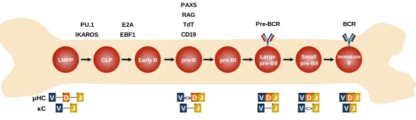
(25) rearrangements that leads to the assembly of variable regions of the HC by combining different V (variable), D (diverse) and J (joining) gene segments. In the LC only V and J segments are recombined. The gene assembly of HC and LC and the expression of the BCR and its accessory molecules is achieved by highly ordered events throughout B cell differentiation. B cells are the result of successive differentiation steps that start with hematopoietic stem cells (HSCs) in adult bone marrow. HSCs initially give rise, by asymmetric division, one stem cell and one multipotent progenitor (MPP). MPPs further differentiate to committed myeloid and lymphoid progenitors. The first lymphoidcommitted progenitor is the lymphoid-primed multipotent progenitor (LMPP) followed by common lymphoid progenitor (CLP), which can potentially differentiate into B cells, T cells or Natural Killer (NK) cells (Traver and Akashi, 2004). In the bone marrow the commitment of CLPs towards the B cell lineage comprises the differentiation throughout the following stages: early B (CD34+ CD19− CD10+), pro-B (CD34+ CD19+ CD10+), large pre-BI (CD34+ CD19+ CD10+), large pre-BII (CD34− CD19+ CD10+), small pre-BII (CD34− CD19+ CD10+) and immature B cells (Blom and Spits, 2006). The differentiation of LMPPs into CLPs is mainly promoted by the transcription factors Ikaros, and PU.1. Then, the commitment and differentiation of CLP towards the B cell lineage is initiated by the transcription factors E2A (encoded by TCF3) and early B-cell factor 1 (EBF1) which activate the expression of key B cell genes such as CD79A, CD79B, CD179A, CD179B (encoding Igα, Igβ and the surrogate light chains VpreB and λ5 respectively), and the transcription factors PAX5 and FOXO1 (Györy et al., 2012). The concerted action of these transcription factors, mainly PAX5 which is considered the master regulator of B cell differentiation, allows the establishment of the proB cell stage by activating the expression of CD19 and BLNK, which encode proteins necessary for BCR signaling, as well as the expression of Recombination-activating gen (RAG) 1/2 and terminal deoxynucleotidyl transferase (TdT) (Cobaleda et al., 2007). These enzymes mediate the rearrangement of V(D)J segments, therefore they are considered the main regulators of V(D)J recombination. Thus, the expression of BCR components and V(D)J recombination allows the transition to the pre-B cell stage. The pre-B cell stage is characterized by the occurrence of two main events, the pre-BCR expression and the rearrangement of variable region of LC. The pre-BCR is composed by the rearranged μHC, the surrogate light chains (SCL) VpreB and λ5, and the signal transducing subunits Igα/Igβ. Signaling through pre-BCR promotes a. 4.
(26) proliferation phase and a maturation phase in which the LC of BCR, λ and κ, are rearranged (Reth and Nielsen, 2014). The last stage of differentiation in the bone marrow is reached with expression of BCR by substituting the surrogate LC of the pre-BCR, for the recombined κ and λ LC. This generates immature B cells which enter to the periphery to continue the differentiation process in secondary lymphoid organs (Reth and Nielsen, 2014) (Figure I1). CLP, pro-B and pre-B cells constitute the main differentiation stages during B cell development in the bone marrow. Nevertheless, differentiation is achieved by passing through several transitional stages, such as early B stage (between CLP and pro-B), and the transitional pre-B cell stages pre-BI, Large pre-BII and small pre-BII before reaching the immature B cell stage. During these transitional steps, B cells acquire the changes necessaries for the proper expression of the key proteins mentioned above. PAX5 RAG E2A. TdT. IKAROS. EBF1. CD19. LMPP. μHC V κC. D V. Pre-BCR. PU.1. CLP. J J. Early B. pro-B. pre-BI. V <> D J V. J. Large pre-BII. BCR. Small pre-BII. Immature B. VDJ. VDJ. VDJ. V. V <> J. VJ. J. Figure I1. Early stages of human B cell development in bone marrow. LMPP indicates lymphoidprimed multipotent progenitor; CLP, common lymphoid progenitor; BCR, B cell receptor. Adapted from Blom & Spits, 2006 and Reth & Nielsen, 2014.. 1.2. B cell Activation Immature B cells expressing functional BCR migrate from the bone marrow to the secondary lymphoid organs (spleen, lymph nodes, tonsils, Peyer patches, and mucosal tissues). This migratory process is mainly mediated by adhesion molecules and cytokine gradients. Immature B cells acquire the cell surface markers IgD, CD21 and CD22, and pass through two transitional stages (T1 and T2) before becoming naïve mature B cells, marginal zone B cells or follicular B cells in the spleen (LeBien and Tedder, 2008). Marginal zone B cells constitute a particular B cell subpopulation that shares properties of both, innate and adaptive immunity. These B cell subtype receives its 5.
(27) name from its localization at the marginal zone, the interface of circulation (red pulp) and lymphoid tissue (white pulp) of the spleen. The main function of marginal zone B cells, by their strategic localization, is the protection against blood-borne antigens. This function is mediated by the "innate side" and the "adaptive side" of these B cell subtype. The "innate side" of marginal zone B cells is conferred by their ability to recognize highly conserved microbial determinants through less specific BCR and Tolllike receptors (Pone et al., 2012). This recognition triggers the adaptive side of marginal zone B cells, which leads to the rapid generation of low-affinity antibodies by producing short-lived plasma cells. The quick responses of marginal zone B cells constitute a compensation mechanism for the time required by follicular B cells to generate high affinity antibodies. Thus, marginal zone B cells provide a first line of defense until the formation of plasma cells (Cerutti et al., 2013). Moreover, follicular B cells are the precursors of the final effectors of B cell differentiation: plasma cells, which produce antibodies, and memory B cells, which provide long-term protection against repetitive exposure to antigens. The differentiation of follicular B cells occurs in specific microscopic structures of secondary lymphoid organs known as germinal centers (GC) where the GC reaction is produced. This reaction is triggered by the recognition of antigens by BCR and implies important changes in B cell expression, as well as the communication between B cells and other immune cell types (Figure I2). Follicular B cells IgD. IgM. Clonal Expansion. Short-lived plasma cells. IgM. Plasmablast. Short-lived plasma cells. Memory B cell. Long-lived plasma cells. IgM. Centroblast. SHM CSR Centrocytes. Immature B. Immature B (T1/T2) Germinal Center. MZ B cells. Clonal Expansion. 6. Short-lived plasma cells.
(28) Figure I2. B cell development in the periphery. Immature B cells can differentiate into Follicular B cells or marginal zone B cells. Follicular B cells can undergo SHM and CSR at the Germinal Centers of secondary lymphoid organs to give rise plasma cells or memory B cells which provide the long-term protection of the humoral response. Additionally marginal zone B cells can give rise plasma cells that produce low-affinity antibodies, and follicular B cells can also give rise short-lived extrafollicular plasma cells that secrete antigen-specific germ line–encoded antibodies. Adapted from LeBien & Tedder, 2008.. The GC formation is mediated by the intense proliferation that B cells undergo after their activation. The highly proliferating cells are grouped in specific areas leading to the generation of two microscopic distinguishable zones, the dark zone and light zone. Dark zone concentrates the proliferating B cells, known as centroblasts while, light zone is composed by follicular dendritic cells, T cells, macrophages and centrocytes, which are smaller non-dividing B cells (Nieuwenhuis and Opstelten, 1984; Victora and Nussenzweig, 2012). The main goal of the reaction at the GC is the generation of B cells able to produce antibodies with high affinity and specificity. This is achieved by two main processes: Class Switch Recombination (CSR) and Somatic Hypermutation (SHM). Both of them are mediated by the enzyme called Activation-Induced Cytidine Deaminase (AID), which is considered the master regulator of B cell activation. SHM mediates the antibodies affinity maturation process in the dark zone of GC by the introduction of point mutations in the rearranged variable regions of immunoglobulin encoding genes. This is started when follicular B cells adapt their genetic program to initiate the formation of centroblasts. These cells are characterized by a gene expression profile that promotes high proliferation rates, apoptosis and the blocking of DNA damage response. The increased proliferation of centroblasts provides a wide repertoire of modified immunoglobulins, whereas increased apoptosis allows the rapid elimination of cells that produce defective antibodies. Meanwhile, the blocking of DNA damage impedes the cell cycle arrest that can be triggered by the genotoxic stress induced by high proliferation and by the DNA modifications produced during SHM (Klein and Dalla-Favera, 2008). The gene expression changes associated with this phase of GC reaction are driven by the master transcriptional regulator of centroblasts, the B-cell lymphoma 6 (BCL-6) transcriptional repressor (Shaffer et al., 2000). In the light zone B cells cooperate with other immune cell types to finalize the differentiation process. The selection of improved antibodies is mediated by FDCs by challenging centroblasts that have undergone SHM with the antigens that triggered B cell activation, whereas macrophages eliminate B cells that produce less efficient 7.
(29) antibodies. Another important event in the light zone is the cooperation between B and T cells. B cells can present antigens to T cells and receive co-stimulatory signals from them to undergo CSR. Through this process B cells change the immunoglobulin isotype from IgM to IgG, IgA or IgE, generating antibodies with different effector functions. The B and T cell interaction is mediated by CD40 receptor, expressed by B cells, and its ligand CD154, expressed by T cells. Although CD40-CD154 is the main interaction that induces CSR, it has also been reported the participation of other molecules such as inducible T‑cell co-stimulator (ICOS), transmembrane activator and calcium-modulating cyclophilin-ligand interactor (TACI) and B-cell-activating factor receptor (BAFFR) (Klein and Dalla-Favera, 2008) (Figure I3). Germinal Center. Centrocytes x. Apoptosis. Dark Zone Memory B cell. Centroblasts. FDC. SHM Selection. Differentiation. Activated B cell Plasmablast. T cell CSR x. Apoptosis. Light Zone Plasma cells. Figure I3. Germinal Center Reaction. Once mature B cells are activated by their encounter with antigens starts the proliferation and the formation of germinal centers. In the dark zone the highly proliferating centroblasts undergo SHM and express mutated receptors. Then in the light zone, mutated receptors are tested with help of FDC and T cells. Centrocytes that express low-affinity receptors are eliminated by apoptosis, while cells that express high affinity antibodies can undergo CSR and be differentiated into memory B cells or plasma cells to confer long-term protection against specific antigens. FDC indicates Follicular dendritic cells; SHM, somatic hypermutation; CSR, class switch recombination. Adapted from Klein & Dalla-Favera, 2008.. Initially it was thought that the GC reaction was a unidirectional process going from dark zone to light zone. However, it is now known that GC reaction is a highly dynamic process in which cells are in constant movement throughout dark zone and light zone and even class switched cells, near the completion of the differentiation process, can go back to the dark zone to have another round of SHM (Allen et al., 2007; Schwickert et al., 2007).. 8.
(30) 1.2.1. Class Switch Recombination Class Switch Recombination (CSR) is the process through which B cells are able to change the expression of IgM to a different immunoglobulin isotype. CSR occurs in response to B cell activation and is a central event in the adaptive immunity as mentioned before. Regions that encode the constant heavy chains of the different immunoglobulin isotypes are organized in tandem in the immunoglobulin heavy chain locus (IGH) at chromosome 14. The coding regions are grouped in specific segments known as germline transcription units. Each unit includes a cytokine-inducible promoter followed by a non-coding exon (I-exon), a switch intronic region (S) and the CH exon cluster (Matthews et al., 2014) (Figure I4). CSR is a multistep process that is initiated by the recruitment of CSR machinery to the S regions for the generation of double-strand breaks (DSBs). This is followed by DNA deletional recombination of the S intervening regions and the activation of DNA damage response leading to DNA repair. As a result, the IgM coding region is excised and the default Cμ exons are exchanged for an alternative set of (Ch) exons, that could be from. Cα, Cγ,or Cε, leading to the production of IgA, IgG or IgE respectively. (Kracker and Durandy, 2011). Germline Transcription Unit (DNA). Iμ. Sμ. Cμ exons. P Transcription. Primary germline transcript (RNA). Splicing Polyadenylation Mature germline transcript (RNA) AAA. Figure I4. Germline transcription through S regions. The transcription unit includes a cytokineinducible promoter (P), an intervening (I)-exon, S region, and Ch exons. The primary transcript is spliced and polyadenylated to generate a noncoding mature transcript. Adapted from Matthews et al., 2014.. 9.
(31) Initiation of transcription is a crucial event for the recruitment of CSR machinery by two major roles. First, it has been described that transcription across the germline transcription units does not produce functional proteins, but instead allows the generation of DNA:RNA hybrids known as R-loops. These structures lead to the exposition of the non-transcribed single strand DNA (ssDNA), which is the substrate for AID action (Chaudhuri et al., 2003; Han et al., 2011). Although AID only acts on ssDNA, it has been demonstrated that AID is able to mutate both template and nontemplate strands, which can be explained by the interaction between AID and components of the RNA exosome complex that degrades DNA:RNA hybrids and exposes the template strand to AID deamination (Basu et al., 2011). The second role of R-loops generated during germline transcription, is to promote the stalling of RNA Polymerase II (PolII) that recruits AID through the splicing factor Spt5 and the recruitment of another AID interactors (Pavri et al., 2010). Once AID is recruited to the S regions, it starts the generation of DSBs. AID deaminates cytosines and converts them into uracils generating dU:dG mismatches. These mismatches can be recognized and processed by different pathways resulting in either their repair, generation of mutations or DSBs. It has been reported that the main pathway in processing dU:dG mismatches during CSR is the Base excision repair (BER) pathway. In BER, AID-introduced uracils are removed by uracil-DNAglycosylase generating abasic sites, which are subsequently recognized and nicked of by apurinic/apyrimidic endonucleases, mainly by APE1. Staggered DSBs are generated by this pathway when the single strand breaks (SSBs) produced are closely spaced in the opposite strands (Stavnezer et al., 2008). It has also been reported that the mismatch repair pathway (MMR) plays a relevant role during CSR. MSH2 and MSH6, which are MMR proteins, can recognize dU:dG mismatches and recruit other effector proteins such as exonuclease 1 (EXO1), to produce SSBs in mismatch adjacent regions. Proximal SSBs produced by both, MMR and BER pathway, can generate DSBs, thus MMR supplements BER pathway in DSBs generation in S regions (Chaudhuri and Alt, 2004; Rada et al., 2004). DNA damage response (DDR) is triggered once DSBs generated in S regions are detected by the DNA damage sensors NBS1 and ATM. It starts with the phosphorylation of histone H2AX (γ-H2AX) and the recruitment of 53BP1 protein, both of which has been recently implicated in the formation of the required synapsis of the intervening S regions (Manis et al., 2004; Reina-San-Martin et al., 2007). DNA repair of the generated DSBs is the final step of CSR. It is mainly mediated by the classical non10.
(32) homologous end joining pathway (C-NHEJ) and is complemented by the action of the alternative end-joining (A-EJ) and homologous recombination pathways (Boboila et al., 2010; Hasham et al., 2010; Yan et al., 2007). CSR is the result of the orchestrated action of several enzymes and scaffold proteins. Expression of different immunoglobulin isotypes allows the adaptation of the effector immune mechanisms to provide a proper and efficient response against the different pathogens. 1.2.2. Somatic Hypermutation SHM shares several mechanistic features with CSR, however it is a less characterized process. As well as CSR, SHM is initiated by transcription. In this case, the targeted transcribed region is the rearranged variable region of HC and LC at the Ig locus. The mutated region is 100–200 bp downstream of the transcription initiation site and extends for 1.5–2.0 kb (Lebecque and Gearhart, 1990). AID is the key enzyme that initiates SHM. Its catalytic activity generates mutations at high rates (10-2, 10-3 mutations per base pair per generation) in the WGCW (W = A/T) hotspot sequence motif. As in CSR, AID deaminates cytosines to generate dU:dG mismatches. The next step to generate mutations is the recruitment and action of DNA repair machinery. Proteins of this pathway also act during CSR mainly to produce SSBs and DSBs as mentioned above; however, during SHM the damage initiated by AID, rather than be repaired in an error-free manner (which is the canonical function of DNA repair machinery), it is transformed into a mutation (Peled et al., 2008). The proposed mechanism to explain this paradox is that during SHM occurs a DNA repair process in which error-prone polymerases participate rather than the high-fidelity polymerases that normally avoid DNA mutations. It has been reported that DNA polymerase η (Pol η) and REV1 are mainly the low-fidelity polymerases involved in SHM (Casali et al., 2006). After AID action, proteins of BER and MMR are recruited. Similarly to CSR, uracil-DNA-glycosylase excises the uracil produced by AID and generates an abasic site that is processed by APE1, which generate SSBs. Moreover, proteins of the MMR, MSH2-MSH6 can also recognize the uracils and recruit EXO1 to create a gap. After the participation of BER and MMR proteins, error-prone polymerases are recruited through mechanisms that remain to be clarified (Di Noia and Neuberger, 2007). 11.
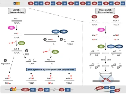
(33) One of the proposed mechanisms is that REV1 and other error-prone polymerases could be recruited by the monoubiquitylated form of PCNA, which is a heterotrimer with relevant functions during replication that is monoubiquitylated when the replication fork is stalled by DNA lesions. As an alternative, but not excluding mechanism, it has been postulated that error-prone and error-free polymerases are differentially recruited during cell cycle (Langerak et al., 2009). SHM and CSR are very particular processes in which the machinery that normally prevents and repairs DNA damage is recruited to facilitate the random introduction of mutations and the generation of SSBs or DSBs to recombine different immunoglobulin gene segments. There are similarities between SHM and CSR, even though the final outcome is different. In any case, the hallmark of both events, as mentioned above, is the expression and action of the same enzyme, AID (Figure I5).. VD J. Sμ. Cμ. Sγ3. Cδ. Cγ3. Sγ1. Cγ1. Sα1. Cα1. Sγ2. Sγ4. Cγ2. Cγ4. Somatic Hypermutation. Sε. Sα2. Cε. Cα2. Class Switch Recombination Sμ. Sγ1. AGCT. AGCT. TCGA. TCGA. AGCT. TCGA AID. MSH2. AGUT. AID. MSH6 AGUT. AGUT. TUGA. TUGA. TCGA. 3 U. AGUT. UNG. 4 AG. A. TCGA UNG. AG T TCGA. 2. U. AG. UNG. BER. 1 Replication. T. AG T. AG T. TCGA. A. *. *. *. AG. AGNT. AGCN. TCNA. TCGA. NNA. TCGA. MMR. T. AG. GA. T. Cμ. Sμ/Sγ1. Cγ1. Sγ2b. Cγ2b. T GA. T GA. *. AGNT. * *. T. MSH6. T BER. AGNT. VD J. T GA. AG. DNA synthesis by error-prone DNA polymerases. MSH2. Sα. Cδ. Cα. Figure I5. Models of Somatic Hypermutations and Class Switch Recombination. The upper gray square shows a non-mutated, non-switched, rearranged human Ig gene. The lower gray square show the Ig gene after SHM and CSR. During SHM (left part) AID deaminates cytosines to generate uracils that are recognized and processed by uracil-DNA-glycosylase. The generated abasic sites might be a template during replication and generate any mutation (1), or abasic sites can be processed by BER machinery which generates SSBs that are filled by recruitment of error prone polymerases (2). The MMR machinery can be also recruited and acts before (4) or after (3) uracil-DNA-glycosylase. MMR machinery produces gaps that are repaired by error-prone polymerases. During CSR (right part) AID targets S regions and deaminates cytosines to generate uracils. This uracils are processed by uracilDNA-glycosylase and the action of BER and MMR machineries introduce SSBs and DSBs. In this case is exemplified the recombination between Sμ and Sγ1 regions. Adapted from Longerich, Basu, Alt, & Storb, 2006.. 12.
(34) 1.3. Activation Induced Cytidine Deaminase Activation Induced Cytidine Deaminase (AID) is an enzyme that was first discovered in 1999 by Honjo and colleagues in an experiment with the murine B lymphoma clone, CH12F3-2, which undergoes robust CSR from IgM to IgA in response to IL-4, TGF-β, and CD40L. By using a cDNA substraction approach they compared the switchinduced and uninduced murine B lymphoma CH12F3-2 cells, and identified AICDA as a strongly upregulated gene during B cell activation (Muramatsu et al., 1999). AICDA, the gene that encodes human AID, is localized in the chromosome 12p13 and extents about 11kb. It is composed by five exons that give rise a protein of 198 amino acids (Muto et al., 2000). Several splicing variants of AID have been reported, but their function remains controversial (van Maldegem et al., 2009; Wu et al., 2008) AID belongs to the AID / APOBEC cytidine deaminase family of proteins. This family is composed by AID, APOBEC1, APOBEC2, the subgroup of APOBEC3 and APOBEC4 proteins.. All of them are zinc-dependent deaminases and share the. catalytic cytidine deaminase domain (Conticello, 2008). These enzymes have been related to different important roles by their catalytic activity. APOBEC1, the founder member of this family, has an important role in lipid metabolism by its RNA-editing activity. It is responsible of the apolipoprotein B (ApoB) mRNA editing, which leads to the generation of two different isoforms of ApoB with different roles in lipid transport (Chester et al., 2000). The rest of the members of the AID/APOBEC family are DNA deaminases. Interestingly, the APOBEC3 subgroup has been associated with the innate immunity against retroviruses (Harris et al., 2003). By deaminating and editing the nascent cDNAs, APOBEC3 members interfere with the reverse transcription of RNA genomes from human immunodeficiency virus (HIV) and hepatitis B virus (HBV) (Mangeat et al., 2003; Sheehy et al., 2002; Turelli et al., 2004). Meanwhile, it remains to be determined if APOBEC2 and APOBEC4 have specific and relevant functions (Bransteitter et al., 2009). 1.3.1. AID, the master regulator of antibody maturation The experiments carried out by Honjo and colleagues mentioned above, suggested for the first time a possible role of AID in activated B cells. However, the critical role of AID during B cell activation was completely elucidated through the study of two different models: knockout mice for Aid and a group of patients with a specific subset of HyperIgM Syndrome (HIGM). 13.
(35) The Aid knockout mice revealed a complete defect in class switching with a phenotype characterized by increased serum levels of IgM (Hyper-IgM) and by the presence of enlarged GCs. Subsequent experiments with splenic B cells from these AID deficient mice showed the specific impairment of CSR and SHM after different stimuli, and suggested the involvement of the catalytic activity of AID in the genetic modifications required in both processes (Muramatsu et al., 2000). At the same time, Revy and colleagues published the first evidences that linked alterations of human AID with an autosomal recessive form of HIGM. The analysis of a cohort of 18 patients with HIGM diagnosis revealed deleterious mutations scattered across the AICDA gene which generate inactive forms of AID. The phenotype observed in these patients was defective CSR, defective SHM and the presence of enlarged GCs with highly proliferating B cells, which resembled the phenotype observed in the AID knockout model (Revy et al., 2000). Thus, both models of AID deficiency, human and mice, demonstrated the absolute requirement of AID during B cell terminal differentiation and antibody affinity maturation. Interestingly, transgenic mice with constitutive and ubiquitous expression of AID develop T cell lymphomas and lung adenocarcinomas as a result of introduction of mutations in non-Ig genes by AID. Curiously, these mice do not have alterations in B cells or develop B cell malignances, which suggest the presence of additional layers of regulation to control AID activity in B cells (Okazaki et al., 2003). Cytosine deamination is the specific mechanism by which AID mediates CSR and SHM. This reaction proceeds by a direct nucleophilic attack at position 4 of the pyrimidine ring of cytosine by Zn+2 coordinated to AID (Conticello, 2008) (Figure I6). H. H. H O. AID - OH. AID. AID - OH. O. NH2. Zn. O. Zn. O. AID. 4. N. HN. HN 2. O. -. NH2. AID - OH. Zn. H. O. 6. N Cytidine. O. N. [Transition state]. O. N Uracil. Figure I6. Cytidine deamination reaction. Mechanism based on a bacterial cytidine deaminase that is homologous to APOBEC1 and AID. Zinc ions (Zn2+) coordinated with AID perform a direct nucleophilic attack at position 4 of the pyrimidine ring. Adapted from Chaudhuri & Alt, 2004.. 14. AID.
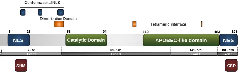
(36) The mistmatches generated by the deamination of cytosines by AID, can be detected by in an error-free manner by BER or MMR machinery. However, uracils that are not detected by DNA repair machinery can be replicated and generate transitions (from C:G to T:A). Another possibility is the processing of the mismatch through a mutagenic pathway that recruits error-prone polymerases, which can produce transitions or transversions at the deaminated site. This mutagenic pathway is the responsible of the DNA changes required during CSR and SHM, as explained above. 1.3.2. Functional Domains of AID Although AID is a small protein (24kDa), its structure contains several differentiated functional domains. At its N-terminal region it has been described a putative bipartite nuclear localization signal (NLS) (position 8 to 26) with the typical motif KR - X10–12 - K(KR)(KR), where X corresponds to any amino acid. It has been postulated that this type of sequences mediate the nuclear import through the importin α / β pathway (Freitas and Cunha, 2009; Ito et al., 2004; Kosugi et al., 2009). Near from the described classical NLS, it has been postulated a conformational nuclear localization signal. This is a non-consecutive motif of basic residues (residues19RWAK-22,34-KRR-36, 199 50-RNKN-54) that provides a positively charged surface in the folded protein (Patenaude et al., 2009). Moreover, the catalytic domain is localized between the amino acid residues 55 and 94 and possesses the conserved H(A/V)E-X24–36-PCXXC motif present in the cydine deaminase family. Meanwhile, the APOBEC-like domain (position 119 to 183) was defined by alignment with APOBEC-1, as a homologous region in the APOBEC family with uncharacterized functions. And finally, at the C-terminal region of AID, the last 15 amino acid residues correspond to a nuclear export signal (NES) that mediates the AID exclusion from the nucleus by a exportin/CRM1 dependent mechanism (Barreto and Magor, 2011; Ito et al., 2004). Additionally, other critical regions in the AID structure, based on the crystal structure of APOBEC2, have been proposed. A putative dimerization domain has been associated with the region between 46-53 amino acid position near from the catalytic domain, whereas, amino acids residues 111-113 and 168, have been associated with a putative tetrameric interface (Prochnow et al., 2007). At the functional level, two regions have been described. In the N-terminal region, a segment required for SHM has been identified (residues 13-23) , whereas the last 26 amino acids have proved to be necessary for CSR. It has been proposed that 15.
(37) these two regions are critical for the association between AID and its regulatory partners (Barreto and Magor, 2011) (Figure I7). Conformational NLS. Dimerization Domain Tetrameric interface 1. 8. 26. 94. 119. Catalytic Domain. NLS 4 - 52 Exon2. 1- 3 E1. 55. 198. 183. APOBEC-like domain 53 - 142 Exon 3. 143 - 181 Exon 4. NES 181 - 198 Exon 5. SHM. CSR. Figure I7. Functional domains of AID. In blue, AID regions linked with AID localization. In green, the domains shared by the AID/APOBEC deaminases members. In orange, putative regions for AID dimerization and tetramerization. In dark red, AID regions linked with the AID functions during antibodies affinity maturation. NLS, indicates nuclear localization signal; NES, nuclear export signal; SHM, somatic hypermutation; CSR, class switch recombination. Adapted from Barreto & Magor, 2011.. 1.3.3. AID Expression Initially, several evidences supported the notion that due to its role, AID expression was restricted to B cells from GC of the spleen and mucosal-associated lymphoid tissues (Peyer’s patches, tonsils, lymph nodes). The first analysis of AID mRNA distribution showed undetectable levels in both, T cells and different splenocyte cell populations, while a dramatic increase was observed in B cells after in vitro activation (Muramatsu et al., 1999). However, recent evidence showed that AID mRNA could be detected outside the B cell compartment under physiological and pathological conditions. As examples, AID expression has been detected in normal tissues from ovary, breast, heart, and lung, although the function of AID in these tissues remains to be elucidated. At the pathological level, AID expression has been reported in lymphomas, leukemias, and malignant epithelial cells from solid tumors. In the latter, the ectopic AID expression has been associated with the chronic inflammation triggered by different infectious agents such as Hepatitis C virus, Helicobacter pylori, Epstein Barr virus or HIV, and it has been suggested that ectopic AID expression in these contexts is mediated by aberrant NF-κB activation (Orthwein and Di Noia, 2012). Moreover, although it remains controversial, it has been proposed that AID is expressed at low levels during lymphopoiesis and it has been suggested that its expression suppresses the development of autoreactivity (Kuraoka et al., 2011; Meyers 16.
(38) et al., 2011). These findings were challenged by the analysis of a mice model in which the expression of a GFP-tagged AID allowed the identification of B cells that actively expressed AID during the immune response. An induction of AID expression in GC B cells after B cell activation was reported as well as undetectable levels of AID in bone marrow–developing B cells, which argue against the potential role of AID in selftolerance in these stages under physiological conditions (Crouch et al., 2007). More recently, AID expression in the embryonic developmental context has been reported, but these findings will be discussed later. 1.3.4. Regulation of AID expression and activity As expected, the regulation of the expression of a mutator like AID, is complex, and not only its expression requires to be restricted to B cells, but also has to display a highly specific response to particular stimuli. In addition to the regulation of AID expression, several post translational mechanisms control the activity of AID, thus maintaining the genome integrity. 1.3.5. Transcriptional Regulation of AID It has been described that there are 4 specific regulatory regions in the AICDA gene that are responsible for the regulation of AID expression, which contain binding sites for at least 19 transcription factors (both, activating and repressive factors). Region 1 is comprised by the Aicda promoter and the region immediately upstream. It contains a TATA-less promoter, and binding sites for the transcription factors Sp1, Sp3 and HoxC4. It also contains a binding site for NF-κB transcription factor which has been specifically associated with the response to the TNF-α and Toll-like receptor signaling triggered by viruses. It has also been reported an estrogen response element in region 1, but its function remains to be clarified (Nagaoka et al., 2010). Region 2 is located in the first intron and is a critical regulatory region. It confers B lineage-cell specificity on Aicda expression by containing binding sites for E-proteins and Pax5 transcription factors, both of which are necessary for B cell development, as mentioned before. This region also contains binding sites for the ubiquitous silencers cMyb and E2f, which are necessary for the transcriptional repression of AID expression (Tran et al., 2010). Region 3 is located approximately 17 kb downstream of the exon 5. Although its precise role has not been determined, it has been demonstrated that its deletion reduces AID expression. Additionally, a binding site for the transcription factor BATF, 17.
(39) which controls AID and GLT expression has been identified. Finally, Region 4 is located approximately 8 kb upstream of the TSS, and has been associated with the AID induction by environmental stimuli. It contains binding sites for NF-κB, Stat6, Smad and C/EBP proteins (Tran et al., 2010) (Figure I8). TSS Stat6 C/EBP Smad3/4 Myc NF-κB Region 4. Stat6. Sp. NF-κB HoxC4 Pax5 Region 1. 1. Myb. Pax5. E2A. Region 2. E2F. 2. BATF Region 3. Figure I8. Regulatory regions at the AICDA promoter. Black boxes indicate AID exons. Adapted from Matthews et al., 2014.. The regulation of AID expression at the transcriptional level is finally achieved by a highly strict coordination among the activating factors, which are all those elements that respond to the cytokine stimulation during B cell activation and development; and the repressing factors, constituted by ubiquitous silencers that guard the genome integrity by silencing AID expression. 1.3.6. Post transcriptional Regulation of AID After B cell activation, the increasing amount of AID mRNA is controlled by a second layer of regulation constituted by microRNAs. These small noncoding RNAs target mRNAs and inhibit their translation by altering their stability and translation efficiency. Two microRNAs have been mainly associated with AID regulation: miR-155 and miR181b. Both of them target the 3'-Untranslated Region (UTR) of the AID encoding mRNA, causing a decrease in the mRNA and protein levels of AID (Stavnezer, 2011). miR-155 is a lymphocyte-specific microRNA that has a similar expression pattern than AID during CSR. Disruption of the interaction between miR-155 and AID mRNA resulted in deregulation of AID expression that not only affects CSR and affinity maturation, but also lead to aberrant targeting of AID and the increase of chromosomal translocations between IgH and c-myc locus (Dorsett et al., 2008; Teng et al., 2008). Moreover, miR-181b was identified in a screening as a microRNA whose expression is able to impair CSR. It exhibits a different expression pattern than miR155, where the higher levels of miR-181b are observed in resting B cells and are decreased after B cell activation. This leads to the suggestion that miR-181b has a role in preventing premature AID activity (de Yébenes et al., 2008).. 18.
(40) The reduction of AID levels by microRNAs has proved to be a critical regulation point to ensure the proper AID expression and function. Altered expression of these microRNAs is associated with lymphomagenesis that mainly result from chromosomal translocations caused by aberrant AID targeting. 1.3.7. AID subcellular localization AID is a small protein that, because of its size, theoretically should diffuse passively into the nucleus. However, AID localization is mainly restricted to the cytoplasm, since the DNA mutator activity of AID demands a strict control of its nuclear entry. At present, there is evidence that supports the existence of three different mechanisms to regulate the nuclear amount of AID: active nuclear import, nuclear export, and cytoplasmic retention (Patenaude and Noia, 2010). The nuclear import is active and is mediated by the interaction with importin-α. Although AID possesses a putative bipartite NLS, experiments with deletional mutations suggest that its nuclear entry is mediated by a conformational NLS. Once in the nucleus, the amount of AID is regulated by degradation and nuclear export. The nuclear export is mediated by the recognition of the NES by the CRM1 exportin. Additionally, the cytoplasmic retention mechanism provides another layer that regulates the AID localization. This mechanism is mediated by AID interaction with Hsp-90 chaperone, which protects cytoplasmic AID from being degraded and determines the availability of functional AID (Orthwein et al., 2010; Patenaude et al., 2009) Together these mechanisms for AID compartmentalization are relevant for the regulation and the accessibility of AID to its DNA substrate, preventing off-target AID activity, and providing a cytoplasmic AID reservoir that leads to a fast nuclear translocation under appropriate stimuli (Xu et al., 2007). 1.3.8. Phosporylation of AID Several amino acid residues of AID can be phosphorylated: Ser3, Thr27, Ser38, Thr140, Ser41, Ser43 and Tyr184. Phosphorylation at Ser38 and Thr140 has been associated with AID activation, while the phosphorylation at Ser3 and Thr27 has been linked to the inhibition of AID activity. Moreover, the role of phosphorylation at residues Tyr184, Ser41 and Ser43, during CSR and/or SHM remains to be determined (Demorest et al., 2011; Pham et al., 2008; Stavnezer, 2011). Thr140, phosphorylated by Protein kinase C (PKC), has been linked to SHM. Experiments with mutants forms that are unable to become phosphorylated in this 19.
(41) residue, showed little defects on CSR but an impairment of SHM that was not related to decreased AID activity, which suggests a role of this phosphorylation in mediating key AID interactions during SHM (McBride et al., 2008) It has been reported that phosphorylation at Thr27 impairs CSR without altering AID activity, whereas phosphorylation at Ser3 impairs the association of AID with the IgH Sμ region. The role of these phosphorylation events in vivo remains to be determined (Gazumyan et al., 2011; Pasqualucci et al., 2006) The best characterized phosphorylation site for AID occurs at Ser38 (p38AID) and is carried out by protein kinase A (PKA). p38AID is detected in a small fraction of AID in vivo. Nevertheless AID Ser38 phosphorylation has proved to be critical for CSR and SHM. p38AID is enriched in the chromatin fraction of activated B cells and it is required for the interaction between AID and replication protein A (RPA), which is necessary for the targeting of AID to the IgH locus. In addition, p38AID has been related with the steps downstream DNA deamination by mediating the indirect interaction between AID and APE1, an endonuclease necessary to generate single strand breaks (SSBs) during CSR (Vuong et al., 2013). In fact, a positive feedback loop mediated by p38AID to amplify the generation of DSBs has been suggested. Thus, phosphorylation at different AID residues, more than leading to a direct alteration of AID activity, constitutes a relevant regulatory mechanism that modulates CSR and SHM through the modification of the interactions established between AID and different proteins. 1.3.9. AID interactions An additional layer of AID regulation is constituted by the proteins that target AID to the IgH locus and restrict the deamination activity mainly to the Switch and Variable regions for CSR and SHM respectively. The DNA sequence motifs, RGYW and WA, together with their reverse complementary sequences, WRCY and TW (where R=A or G, Y=C or T, W=A or T), are preferential binding sites for AID. These motifs are considered hotspots for the mutations induced by AID. However, as these motifs are degenerated and are scattered across the entire genome, they cannot account for the specificity observed during CSR and SHM. In line with this observation, it has been found that AID binds transcribed regions across the entire genome of activated B cells, as determined by chromatin immunoprecipitation (ChIP) coupled with ultrasequencing (ChIP-seq). 20.
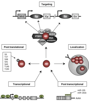
(42) experiments in mice (Yamane et al., 2011). Thus, the interaction between AID and several proteins is the main AID targeting mechanism that provides insights into how AID activity is targeted to specific Ig loci. For instance, interaction between AID and the GC associated nuclear protein (GANP) has been linked to the targeting of AID to transcribed variable regions to promote SHM, although the underlying mechanism has not been described (Maeda et al., 2010). Moreover, 14-3-3 adaptors are proteins that are upregulated during B cell activation, bind to the AID hotspots, and also interact with AID, seemingly for the targeting of AID to the S regions for CSR (Xu et al., 2010). The interaction between AID, PolII and Spt5, as mentioned above, is necessary for the recruitment of AID and explains the well characterized link between transcription and CSR and SHM. Furthermore, the splicing regulator, Polypyrimidine-tract binding protein-2 (PTBP2), has also been implicated in AID binding to S regions through its interaction with S region transcripts (Nowak et al., 2011). Interestingly, chromatin conformation also plays a role in AID recruitment. Open chromatin states defined by activation histone marks such as trimethyl histone H3 lysine 4 (H3K4me3) and hyperacetylated H3K9 (H3K9Ac), are linked with CSR and its importance is highlighted by experiments in which the activity of the enzymes responsible of these histone marks are altered, leading to an impairment of CSR (Daniel et al., 2010; Wang et al., 2009). Together, the coordination of different regulatory mechanisms ensure the proper expression and targeting of AID, which is important not only for proper antibody maturation and immune response, but also to guarantee the genome integrity during AID function. The main regulatory mechanisms of AID are summarized in the Figure I9. 1.3.10. AID roles beyond CSR and SHM in B cells In recent years, new potential AID roles have been proposed since the detection of AID expression in non-B cell contexts and its association with the regulation of DNA methylation, an important epigenetic mechanism.. 21.
(43) Targeting. VDJ. Sμ. Cμ. Sx. Cx. PolII Spt5 14-3-3 PTBP2 AID GANP. RNA exosome. Post translational S3 T27 S38 S41 S43 T140 Y184. Localization. AID AID. P. AID AID. AID. AID. AID AID. Transcriptional. Post transcriptional miR-155 miR-181b. 3. 1. 2. 4. AAA. Figure I9. Regulation of AID expression and activity. Different transcription factors regulate AID expression. At the post-transcriptional level, specific micro-RNAs regulate the availability of AID mRNA. The generated AID protein is then controlled by phosphorylation and subcellular localization mediated by the localization signals contained in its structure and by specific protein interactions. Furthermore, protein interactions also control the AID targeting to V and S regions. Adapted from Matthews et al., 2014.. Development of primordial germ cells (PGCs) during embryogenesis, which are the precursors of spermatocytes and oocytes, requires rapid DNA demethylation to the erasure of paternal imprints (Sasaki and Matsui, 2008). AID is expressed in PGCs, and the observation that AID deficient-PGCs exhibit higher levels of global methylation than their WT counterparts, led to the suggestion that AID may participate in active DNA demethylation mechanisms (Popp et al., 2010). Moreover, in vitro reprogramming of somatic cells to produce induced pluripotent stem cells (iPS), which is another model that requires rapid DNA demethylation, revealed that AID is necessary for promoter demethylation and expression of the critical pluripotency genes, OCT4 and NANOG (Bhutani et al., 2010). Furthermore, the reprogramming of Aid-null differentiated cells to induced pluripotent stem cells showed failed stabilization of the pluripotent state due to hypermethylation of pluripotency associated genes (Kumar et al., 2013), which reinforces the role of AID in 22.
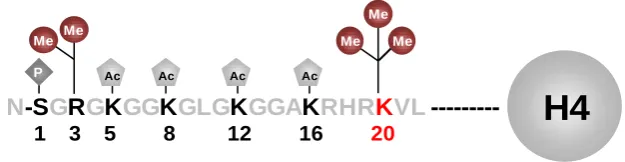
(44) the DNA demethylation required for cell reprogramming. In these systems, the proposed demethylation mechanism is the direct deamination of 5-methylcytosines (5mC) by AID to produce thymines which are later replaced by unmethylated cytosines after the action of BER machinery. Another supporting evidence of the role of AID in DNA demethylation is the observation that cooperative action of AID and ten-eleven translocation 1 (TET1) protein. can. mediate. DNA. demethylation.. It. has. been. reported. that. 5-. hydroxymethylcytosines (5hmC), the product of the catalytic activity of TET1, can be deaminated by AID producing 5-hydroxymethyluracil (5hmU), which in turn can be detected and processed by thymine DNA glycosylase (TDG) and finally replaced by unmethylated cytosines. The ability of AID to deaminate 5hmC was also demonstrated in vivo in mice brain that overexpress AID, nevertheless whether this mechanism take place under physiological conditions remains to be determined. In either case, no AID expression has been detected in mice brain, while the expression of apobec1 is detected (Guo et al., 2011). Despite the strong evidence that links AID with epigenetic changes, such as DNA demethylation, it seems that this role is restricted to specific scenarios. In parallel, biochemical analysis has revealed substantially reduced AID activity on 5mC relative to cytosines, and no detectable deamination of 5hmC, which argue against the proposed function of AID in DNA demethylation (Nabel et al., 2012). Thus, it is clear that there are several unresolved issues about AID function and regulation. 1.4. Epigenetic regulation The concept of Epigenetics was formally introduced in 1940s by Conrad Waddington as the "study of the causal interactions between genes and their products which bring the phenotype into being". Since then, the epigenetic field has experimented important advances and later on epigenetics was redefined as “the study of mitotically and/or meiotically heritable changes in gene function that cannot be explained by changes in DNA sequence" (Russo et al., 1996). More recently, Adrian Bird has proposed that epigenetic events can be defined as "the structural adaptation of chromosomal regions so as to register, signal or perpetuate altered activity states" (Bird, 2007). The epigenome is the set of epigenetic modifications that occur along the genomic sequence of an individual. Given that the set of epigenetic marks of a cell type is determined by the set of transcription factors, upstream signaling pathways and extracellular signals, an organism will be characterized for different epigenomes for. 23.
Figure


+7


Documento similar
In the other point of view, we have done a systematic comparison between the two cell types that express FMRFa in the VNC, Ap4/FMRFa and SE2/FMRFa neurons, and we have
The analysis of the biological effects of gold nanoparticles generated by biological reduction of HAuCl 4 gold precursor in HeLa cells and the comparison with
SECTION 3 - The role of RINGO proteins in the brain 123 RingoA mRNA is expressed in neural stem cells and proliferating progenitor cells 123 Analysis of neural stem cells
scRNAseq analysis of the organoids has revealed clusters with distinct gene expression profiles and, together with the use of γ- secretase inhibitors, has provided
Scarcity: The resources available Albani, are not very difficult to achieve, since with regard to human resources, with a good selection and training of its employees
We have extended this analysis to the suboesophageal Capa cells (SECA cells), and show that expression of the same terminal identity gene Capa can be achieved from different
Splicing in SDG24 resulted in different spatial expression patterns, being the SDG24.1 specifically expressed in root cells, and to the nucleus of the central cell in the mature
During the present thesis we have established a robust protocol of gene targeting for different types of hematopoietic cells, mainly lymphoblastic cell lines (LCLs) and
