Policy Statement
Policy statements and clinical policies are the official policies of the American College of Emergency Physicians and, as such, are not subject to the same peer review process as articles appearing in the journal. Policy statements and clinical policies of ACEP do not necessarily reflect the policies and beliefs of Annals of Emergency Medicineand its editors.
Ultrasound Guidelines: Emergency, Point-of-Care and Clinical Ultrasound Guidelines in Medicine
Revised and approved by the ACEP Board of Directors with current title June 2016 Revised and approved by the ACEP Board of Directors October 2008
Originally approved by the ACEP Board of Directors with title“Emergency Ultrasound Guidelines”June 2001
0196-0644/$-see front matter
Copyright © 2016 by the American College of Emergency Physicians.
Ultrasound Guidelines: Emergency, Point-of-Care and Clinical Ultrasound Guidelines in Medicine
[Ann Emerg Med. 2017;69:e27-e54.]
SECTION 1 - INTRODUCTION
Ultrasound (US) has become an integral modality in emergency care in the United States during the last two decades. Since the last update of these guidelines in 2008, US use has expanded throughout clinical medicine and established itself as a standard in the clinical evaluation of the emergency patient. There is a wide breadth of recognized emergency US applications offering advanced diagnostic and therapeutic capability benefit to patients across the globe. With its low capital, space, energy, and cost of training requirements, US can be brought to the bedside anywhere a clinician can go, directly or remotely. The use of US in emergency care has contributed to improvement in quality and value, specifically in regards to procedural safety, timeliness of care, diagnostic accuracy, and cost reduction. In a medical world full of technological options, US fulfills the concept of“staged imaging,”where the use of USfirst can answer important clinical questions accurately without the expense, time, or side effects of advanced imaging or invasive procedures.
Emergency physicians have taken the leadership role for the establishment and education of bedside, clinical, point-of-care US use by clinicians in the United States and around the world. Ultrasonography has spread throughout all levels of medical education, integrated into medical school curricula, through residency, to postgraduate education of physicians, and extended to other providers
such as nursing, advanced practice professionals, and prehospital providers. US curricula in undergraduate medical education is growing exponentially due to the leadership and advocacy of emergency physicians. US in emergency medicine (EM) residency training has now been codified in the Accreditation Council for Graduate Medical Education (ACGME) Next Accreditation System (NAS). Emergency US specialists have created the foundation of a subspecialty of ultrasonography that provides the expertise for establishing clinical practice, educating across the educational spectrum, and researching the wide range of applications of ultrasonography. Within healthcare institutions and healthcare systems, emergency physicians are now leading institutional clinical US programs that have used this guideline as a format for multidisciplinary programs.
These guidelines reflect the evolution and changes in the evolving world of emergency medicine and the growth of US practice. Themes of universality of practice, educational innovation, core credentialing, quality improvement, and value highlight this new edition of the guidelines. The ultimate mission of providing excellent patient care will be enhanced by emergency physicians and other clinicians being empowered with the use of US.
SECTION 2- SCOPE OF PRACTICE
Emergency Ultrasound (EUS) is the medical use of US technology for the bedside evaluation of acute or critical medical conditions.1It is utilized for diagnosis of any emergency condition, resuscitation of the acutely ill, critically ill or injured, guidance of procedures,
monitoring of certain pathologic states and as an adjunct to therapy. EUS examinations are typically performed, interpreted, and integrated into care by emergency physicians or those under the supervision of emergency physicians in the setting of the emergency department (ED) or a non-ED emergency setting such as hospital unit, out-of-hospital, battlefield, space, urgent care, clinic, or remote or other settings. It may be performed as a single examination, repeated due to clinical need or deterioration, or used for monitoring of physiologic or pathologic changes.
Emergency US is synonymous with the terms clinical, bedside, point-of-care, focused, and physician performed, but is part of a larger field of clinical ultrasonography. In this document, EUS refers to US performed by emergency physicians or clinicians in the emergency setting, while clinical ultrasonography refers to a multidisciplinaryfield of US use by clinicians at the point-of-care.2 Table 1 summarizes relevant US definitions in EUS.
Other medical specialties may wish to use this document if they perform EUS in the manner described above. However, guidelines which apply to US examinations or procedures performed by consultants, especially
consultative imaging in US laboratories or departments, or in a different setting may not be applicable to emergency physicians.
Emergency US is an emergency medicine procedure, and should not be considered in conflict with exclusive
“imaging” contracts that may be in place with consultative US practices. In addition, emergency US should be reimbursed as a separate billable procedure.3 (SeeSection 6- Value and Reimbursement)
EUS is a separate entity distinct from the physical examination that adds anatomic, functional, and physiologic information to the care of the acutely-ill patient.4 It provides clinically significant data not
obtainable by inspection, palpation, auscultation, or other components of the physical examination.5 US used in this clinical context is also not equivalent to use in the training of medical students and other clinicians in training looking to improve their understanding of anatomic and
physiologic relationships of organ systems.
EUS can be classified into the following functional clinical categories:
1. Resuscitative: US use as directly related to an acute resuscitation
2. Diagnostic: US utilized in an emergent diagnostic imaging capacity
3. Symptom or sign-based: US used in a clinical pathway based upon the patient’s symptom or sign (eg, shortness of breath)
4. Procedure guidance: US used as an aid to guide a procedure
5. Therapeutic and Monitoring: US use in therapeutics or in physiological monitoring
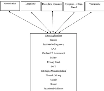
Within these broad functional categories of use, 12 core emergency US applications have been identified as Trauma, Pregnancy, Cardiac /Hemodynamic assessment, Abdominal aorta, Airway/Thoracic, Biliary, Urinary Tract, Deep Vein Thrombosis (DVT), Soft-tissue/Musculoskeletal (MSK), Ocular, Bowel, and Procedural Guidance. Evidence for these
Table 1. Relevant Ultrasound Definitions.
Resuscitative US use directly related to a resuscitation Diagnostic US utilized a diagnostic imaging capacity
Symptom or sign-based US used in a clinical pathway based upon the patient’s symptoms or sign (eg, shortness of breath) Therapeutic and Monitoring US use in therapeutics or physiological monitoring
Procedural guidance US used as an aid to guide a procedure
Consultative Ultrasound A written or electronic request for an US examination & interpretation for which the patient is transported to a laboratory or imaging department outside of the clinical setting.
Emergency Ultrasound Performed and interpreted by the provider as an emergency procedure and directly integrated into the care of the patient
Clinical Ultrasound US used in the clinical setting, distinct from the physical examination, that adds anatomic, functional and physiologic information to the care of the acutely ill patient.
core applications may be found inAppendix 1. The criteria for inclusion for core are widespread use, significant evidence base, uniqueness in diagnosis or decisionmaking, importance in primary emergency diagnosis and patient care, or technological advance.
Alternatively, symptom and sign based US pathways, such as Shock or Dyspnea, may be considered an integrated application based on the skills required in the pathway. In such pathways, applications may be mixed and utilized in a format and order that maximizes medical decisionmaking, outcomes, efficiency and patient safety tailored to the setting, resources, and patient characteristics. See Figure 1.
Emergency physicians should have basic education in US physics, instrumentation procedural guidance, and Focused Assessment with Sonography in Trauma (FAST) as part of EM practice. It is not mandatory that every clinician performing emergency US examinations utilize or be expert in each core application, but it is understood that each core application is incorporated into common emergency US practice nationwide. The descriptions of these examinations may be found in the ACEP policy, Emergency Ultrasound Imaging Criteria Compendium.6 Many other US applications or advanced uses of these applications may be used by emergency physicians. Their non-inclusion as a core application does not diminish their importance in practice nor imply that emergency
physicians are unable to use them in daily patient care. Each EUS application represents a clinical bedside skill that can be of great advantage in a variety of emergency patient care settings. In classifying an emergency US a single application may appear in more than one category
and clinical setting. For example a focused cardiac US may be utilized to identify a pericardial effusion in the diagnosis of an enlarged heart on chest x-ray. The focused cardiac US may be utilized in a cardiac resuscitation setting to differentiate true pulseless electrical activity from profound hypovolemia. The focused cardiac US can be used to monitor the heart during resuscitation in response tofluids or medications. If the patient is in cardiac tamponade, the cardiac US can also be used to guide the procedure of pericardiocentesis. In addition, the same focused cardiac study can be combined with one or more additional emergency US types, such as the focused abdominal, the focused aortic or the focused chest US, into a clinical algorithm and used to evaluate a presenting symptom complex. Examples of this would be the evaluation of patients with undifferentiated non-traumatic shock or the focused assessment with sonography in trauma (FAST), or extended FAST examination in the patient presenting with traumatic injury. See Figure 1.
Ultrasound guided procedures provide safety to a wide variety of procedures from vascular access (eg, central venous access) to drainage procedures (eg, thoracentesis
pericardiocentesis, paracentesis, arthrocentesis) to localization procedures like US guided nerve blocks. These procedures may provide additional benefits by increasing patient safety and treating pain without the side effects of systemic opiates.
Other US applications are performed by emergency physicians, and may be integrated depending on the setting, training, and needs of that particular ED or EM group. Figure 2lists other emergency US applications.
Other Settings or Populations
Pediatrics. US is a particularly advantageous diagnostic tool in the management of pediatric patients, in whom radiation exposure is a significant concern. EUS
applications such as musculoskeletal evaluation for certain
Figure 1. ACEP 2016 Emergency US Guidelines Scope of Practice.
fractures (rib, forearm, skull), and lung for pneumonia may be more advantageous in children than in adults due to patient size and density.7US can be associated with increased procedural success and patient safety, and decreased length of stay.8,9 While most US modalities in the pediatric arena are the same as in adult patients (the EFAST exam for trauma, procedural guidance), other modalities are unique to the pediatric population such as in suspected pyloric stenosis and intussusception, or in the child with hip pain or a limp).10-12Mostly recently, EUS has been formally incorporated into Pediatric EM
fellowship training.13-14
Critical Care.EUS core applications are being integrated into cardiopulmonary and non-invasive hemodynamic monitoring into critical care scenarios.15-16 Dual-trained physicians in emergency medicine and critical care are leading the application, education, and research of US for critically ill patients, and have significant leadership in advancing US concepts in multidisciplinary critical care practice. Advanced cardiopulmonary US applications are being integrated into critical care practice.
Prehospital.There is increasing evidence that US has an increasing role in out-of-hospital emergency care.17-18 Challenges to the widespread implementation of out-of-hospital US include significant training and equipment requirements, and the need for careful physician oversight and quality assurance. Studies focusing on patient
outcomes need to be conducted to further define the role of out-of-hospital US and to identify settings where the benefit to the patient justifies the investment of resources necessary to implement such a program.19
International arena including field, remote, rural, global public health and disaster situations. US has become the primary initial imaging modality in disaster care.20-24 US can direct and optimize patient care in domestic and international natural disasters such as tsunami, hurricane, famine or man-made disasters such as battlefield or refugee camps. US provides effective advanced diagnostic technology in remote geographies such as rural areas, developing countries, or small villages which share the common characteristics of limited technology (ie, x-ray, CT, MRI), unreliable electrical supplies, and minimally trained health care providers. US use in outer space is unique as the main imaging modality for space exploration and missions.25-26 Ultrasound has also been used in remote settings such as international exploration, mountain base camps, and cruise ships.27 The increasing portability of US machines with increasing image resolution has expanded the use of emergent imaging in such settings. See ACEP linked resources at www.globalsono.org.
Military and Tactical.The military has embraced the utilization of US technology in austere battlefield
environments.28-29It is now routine for combat support hospitals as well as forward surgical teams to deploy with next generation portable ultrasonography equipment. Clinical ultrasonography is often used to inform decisions on mobilization of causalities to higher echelons of care and justify use of limited resources.
Within the last decade, emergency physicians at academic military medical centers have expanded
ultrasonography training to clinical personnel who practice in close proximity to the point of injury, such as combat medics, special operations forces, and advanced practice professionals.30The overarching goal of these training programs is to create a generation of competent clinical sonologists capable of practicing “good medicine in bad places.” The military is pursuing telemedicine-enabled US applications, automated US interpretation capabilities, and extension of clinical ultrasonography in additional areas of operation, such as critical care air evacuation platforms.31
SECTION 3 – TRAINING AND PROFICIENCY There is an evolving spectrum of training in clinical US from undergraduate medical education through post-graduate training, where skills are introduced, applications are learned, core concepts are reinforced and new
applications and ideas evolve in the life-long practice of US in emergency medicine.32-33
Competency and Curriculum Recommendations Competency in EUS requires the progressive
development and application of increasingly sophisticated knowledge and psychomotor skills for an expanding number of EUS applications. This development parallels the performance of any EUS exam.
includes knowledge of each particular exam accuracy, as well as proper documentation, quality assurance, and EUS reimbursement. See ACEP linked resources athttp://www. acep.org/sonoguide.
An EUS curriculum requires considerable faculty expertise, dedicated faculty time and resources, and departmental support. These updated guidelines continue to provide the learning objectives (SeeAppendix 2), educational methods, and assessment measures for any EUS residency or practice-based curriculum. As part of today’s effort to reinvent medical education, all educators are now faced with the challenge of creating curricula that provide for individualized learning yet result in the standardized outcomes such as those outlined in current residency milestones.34
Innovative Educational Methods and Assessment Measures
As a supplement to traditional EUS education already described in previous guidelines, recent online and technological innovation is providing additional individualized educational methods and standardized assessment measures to meet this challenge.32,35-36Free open access medical (FOAM) education podcasts and narrated lectures provide the opportunity to create theflipped EUS classroom.37-40For the trainee, asynchronous learning provides the opportunity to repeatedly review required knowledge on demand and at their own pace. For educators, less time may be spent providing recurring EUS didactics, and more time dedicated to higher level tasks such as teaching psychomotor skills and integration of examfindings into patient and ED management. Both EUS faculty and trainees together may identify potential FOAM resources. However, EUS faculty must now take the new role of FOAM curator. New online resources must be carefully reviewed to ensure that each effectively teaches the objectives in these guidelines before being introduced into an EUS curriculum.
Similar to knowledge learning, there are new educational methods to teach the required psychomotor skills of EUS. The primary educational method continues to be small group hands-on training in the ED with EUS faculty, followed by supervised examination performance with timely quality assurance review. Simulation is currently playing an increasingly important role as both an EUS educational method and assessment measure.36Numerous investigators have demonstrated that simulation results in equivalent image acquisition, interpretation, and operator confidence in comparison to traditional hands-on training.41-42US simulators provide the opportunity for deliberate practice of a new skill in a safe environment prior to actual clinical performance. The use of simulation for deliberate practice
improves the success rate of invasive procedures and reduces patient complications.43-44Additionally, simulation has the potential to expose trainees to a wider spectrum of pathology and common variants than typically encountered during an EUS rotation. Blended learning created by the
flipped classroom, live instructor training, and simulation provide the opportunity for self-directed learning, deliberate practice and mastery learning.45-47
Simulation also provides a valid assessment measure of each component of EUS competency. Appropriately designed cases assess a trainee’s ability to recognize indications, demonstrate image acquisition and interpretation, as well as apply EUSfindings to patient and ED management.42These proven benefits and the reduction in direct faculty time justify the cost of a highfidelity US simulator. Furthermore, costs may be shared across departments.
Documenting Experience and Demonstrating Proficiency
Traditional number benchmarks for procedural training in medical education provide a convenient method for documenting the performance of a reasonable number of exams needed for a trainee to develop competency.48-49 However, learning curves vary by trainee and application.49 Individuals learn required knowledge and psychomotor skills at their own pace. Supervision, opportunities to practice different applications and encounter pathology also differ across departments.
Therefore, in addition to set number benchmarks individualized assessment methods need to be utilized. Recommended methods include the following: real time supervision during clinical EUS, weekly QA teaching sessions and image review, ongoing QA exam feedback, standardized knowledge assessments, small group Observed Structured Clinical Examinations (OSCEs), one-on-one standardized direct observation tools (SDOTs), simulation assessments and other focused educational tools.36 Ideally these assessment measures are completed both at the beginning and the end of a training period. Initial assessment measures identify each trainee’s unique needs, providing the opportunity to modify a local curriculum as needed to create more individualized learning plans. Final assessment measures demonstrate current trainee
competency and future learning needs, as well as identify opportunities for improvement in local EUS education.
applicable to new applications. For example, experience with FAST provides a springboard to learning resuscitation, genitourinary, and transabdominal pelvic EUS.
Overall EUS trainees should complete a benchmark of 150-300 total EUS exams depending on the number of applications being utilized. For example, an academic department regularly performing greater than six
applications may require residents to complete more than 150 exams, while a community ED with practicing physicians just beginning to incorporate EUS with FAST and vascular access should require less.
If different modalities such as endovaginal technique are being used for an application, the minimum may need to include a substantial experience in that technique. We would recommend a minimum of 10 examinations in the other technique (eg, endocavitary for early pregnancy) with the assumption that educational goals of anatomic, pathophysiology, and abnormal states are identified with all techniques taught.
Procedural US applications require fewer exams given prior knowledge, psychomotor skills, and clinical experience with traditional blind technique. Trainees should completefive quality reviewed US-guided procedure examinations or a learning module on an US-guided procedures task trainer.
Training exams need to include patients with different conditions and body types. Exams may be completed in different settings including clinical and educational patients in the ED, live models at EUS courses, utilizing US simulators, and in other clinical environments. Abnormal or positive scans should be included in a significant number of training exams used to meet credentialing requirements. Image review or simulation may be utilized for training examinations in addition to patient encounters when adequate pathology is not available for the specific application. In-person supervision is optimal during introductory education but is not required for residency or credentialing examinations after initial didactic training.
During benchmark completion, all EUS exams should be quality reviewed for technique and accuracy by EUS faculty. Alternatively, an EUS training portfolio of exam images and results may be compared to other diagnostic studies and clinical outcomes in departments where EUS faculty are not yet available. After initial training, continued quality assurance of EUS exams is recommended for a proportion (5-10%) of ongoing exams to document continued competency.
Recently, several secure online quality assurance workflow systems have become commercially available (SeeSection 5-Quality and US Management). Current systems greatly enhance trainee feedback by providing for more timely review of still images and video loops, customized application
and feedback forms, typed and voice feedback, as well as storage and export of data within a relational database.
Training Pathways
There are two recommended pathways for clinicians to become proficient in EUS. SeeFigure 3. The majority of emergency physicians today receive EUS training as part of an ACGME-approved EM residency. A second practice-based pathway is provided for practicing EM physicians and other EM clinicians who did not receive EUS training through completion of an EM residency program.
These updated EUS guidelines continue to provide the learning objectives, educational methods and assessment measures for either pathway. Learning objectives for each application are described in Appendix 2.
Residency-Based Pathway
EUS has been considered a fundamental component of emergency medicine training for over two decades.50-52The ACGME mandates procedural competency in EUS for all EM residents as it is a“skill integral to the practice of Emergency Medicine”as defined by the 2013 Model of the Clinical Practice of EM.53The ACGME and the American Board of Emergency Medicine (ABEM) recently defined twenty-three sub competency milestones for emergency medicine residency training.34Patient Care Milestone twelve (12) describes the sequential learning process for EUS and should be considered a guideline in addition to other assessment methods mentioned in this guideline.Appendix 3 provides recommendations for EM residency EUS education.
Upon completion of residency training, emergency medicine residents should be provided with a standardized EM Resident EUS credentialing letter. For the EUS faculty, ED Director or Chairperson at the graduate’s new institution, this letter provides a detailed description of the EUS training curriculum completed, including the number of quality reviewed training exams completed by application and overall, and performance on SDOTs and simulation assessments.
Practice Based Pathway
physics and knobology is required prior to training in individual applications. Pre-course and post-course online learning may be utilized to reduce the course time spent on traditional didactics and facilitate later review. Small group hands-on instruction with EUS faculty on models,
simulators, and task trainers provides experience in image acquisition, interpretation, and integration of EUS exam
findings into patient care. SeeAppendix 4.
Preceptorships typically lasting 1-2 weeks at an institution with an active EUS education program have also been
utilized successfully to train practicing physicians. Each preceptorship needs to begin with a discussion of the trainees’ unique educational needs, hospital credentialing goals as well asfinancial support for faculty teaching time. Then the practicing physician participates in an appropriately tailored curriculum typically in parallel with ongoing student, resident, fellow and other educational programming.
Similar to an EM Resident EUS credentialing letter, course and preceptorship certificates should include a description of the specific topics and applications reviewed,
Residency Training Practicing Physician Didactics
Experiential
Proficiency
Credentialing
Ongoing Review and Education
New Applications
Ultrasounds are obtained with documentation and reviewed to meet ACEP emergency ultrasound proficiency guidelines. Ultrasound available for departmental and hospital examination.
Residency Director and/or Ultrasound Coordinator certifies ultrasound training categorized by the ACEP emergency ultrasound proficiency guidelines and ABEM “The Model of the Clinical Practice of Emergency Medicine”
Attends introductory emergency ultrasound course or courses that cover core emergency US applications
Attends residency curriculum covering emergency ultrasound curriculum
New applications adopted after CME, research, or other training.
Performs ultrasounds under
supervision over-reads, gold standards confirmatory testing or patient outcome review within departmental ultrasound plan
Training in residency per Emergency Medicine Residency Ultrasound Guidelines and ACGME Milestones
Acquired at local hospital setting within departmental privileges.
Quality review of ultrasound performed continuously.
CME attended in accordance with specialty guidelines.
total number of training exams completed with expert supervision, performance on other course assessment measures such as SDOTs or simulation cases, as well as the number of CME hours earned. These certificates are then given to local EUS faculty or ED Director/Chairperson to document training.
Advanced Practice Providers, Nursing, Paramedics, and other EM clinicians
In many practice environments, EUS faculty often provide clinical US training to other to non-physician staff including Advanced Practice Professionals, Nurses, Paramedics, Military Medics and Disaster Response Team members. The recommendations in these guidelines should be utilized by EUS faculty when providing such training programs. Pre-course preparation needs to include discussions with staff leadership to define role-specific learning needs and applications to be utilized. Introductory US physics, knobology, and relevant anatomy and pathophysiology are required prior to training in targeted applications.
For Advanced Practice Providers and other clinicians practicing in rural and austere environments where direct EUS trained EM physician oversight is not available, EUS training needs to adhere to the recommendations in these guidelines. Specifically, comprehensive didactics and skills training, as well as minimum benchmarks need to be completed prior to independent EUS utilization. Beyond this initial training, EUS faculty are needed to provide ongoing quality assurance review. Telemedicine may provide the opportunity for real time patient assessment, assistance with image acquisition, and immediate review of patient images.
Ongoing Education
As with all aspects of emergency medicine, ongoing education is required regardless of training pathway. The amount of education needed depends on the number of applications being performed, frequency of utilization, the local practice of the individual clinician and other
developments in EUS and EM. Individual EUS credentialed physicians should continue their education with a focus on US activities as part of their overall educational activities. Educational sessions that integrate US into the practice of EM are encouraged, and do not have to be didactic in nature, but may be participatory or online. Recommended EUS educational activities include conference attendance, online educational activities, preceptorships, teaching, research, hands-on training, program administration, quality assurance, image review, in-service examinations, textbook and journal readings, as well as morbidity and mortality conferences inclusive of US cases. US quality improvement is an example of an activity
that may be used for completion of the required ABEM Assessment of Practice Performance activities.
Fellowship Training
Fellowships provide the advanced training needed to create future leaders in evolving areas of medicine such as clinical US. This advanced training produces experts in clinical US and is not required for the routine utilization of EUS.
An EUS fellowship provides a unique, focused, and mentored opportunity to develop and apply a deeper comprehension of advanced principles, techniques, applications, and interpretativefindings. Knowledge and skills are continually reinforced as the fellow learns to effectively educate new trainees in EUS, as well as clinicians in other specialties, and practice environments. A methodical review of landmark and current literature, as well as participation in ongoing research, creates the ability to critically appraise and ultimately generate the evidence needed for continued improvements in patient care through clinical US. Furthermore, fellowship provides practical experience in EUS program management including quality assurance review, medical legal documentation, image archiving, reimbursement, equipment maintenance, and other administrative duties of an EUS program director.
Recommendations for fellowship content, site
qualifications, criteria for fellowship directors, and minimum graduation criteria for fellows have been published by national EUS leadership and ACEP Emergency Ultrasound Fellowship Guidelines.54-55Each fellowship program’s structure and curriculum will vary slightly based on local institution and department resources. At all fellowship programs, mentorship and networking are fundamental to a fellow’s and program’s ultimate success. Both require significant EUS faculty time for regular individual instruction as well as participation in the clinical US community locally and nationally. Hence, institution and department leadership support is essential to ensuring an appropriate number of EUS faculty, each provided with adequate non-clinical time.
For the department, a fellowship speeds the development of an EUS program. Fellowships improve EM resident training resulting in increased performance of EUS examinations.56 Furthermore, a fellowship training program may have a significant positive impact on overall EUS utilization, timely quality assurance review, faculty credentialing, billing revenue, and compliance with documentation.57For an institution, an EUS fellowship provides a valuable resource for other specialties just beginning clinical US programs. Collaborating with EUS faculty and fellows, clinicians from other
US in Undergraduate Medical Education
Emergency Medicine has again taken a lead role in efforts to improve Undergraduate Medical Education (UME) through the early integration of clinical US.58-62 During the preclinical years, US has been demonstrated to be an effective educational method to reinforce student understanding of anatomy, physical examination skills, pathology and bedside diagnostic skills.63-68During the clinical years, students are then better able to utilize US for clinical diagnosis and on specific rotations. US exposure in UME can provide a solid knowledge base for individuals to build upon and later utilize as US is integrated into their clinical training.
Integrating US into UME
Integration of US into pre-clinical UME often begins with medical student and faculty interest. By working closely with a medical school’s curriculum committee, US may then be incorporated as a novel educational method to enhance learning within existing preclinical courses. Although dedicated US specific curriculum time is not often available in UME, considerable clinical US faculty time and expertise is still required for effective integration of US into existing medical school courses. Widespread clinical US utilization by different specialties within a medical school’s teaching hospitals, and education within Graduate Medical Education programs, provides initial faculty expertise, teaching space, and US equipment. Ongoing education then requires local departmental and medical school leadership support, as well as continued organized collaboration between faculty from participating specialties.
Innovative educational methods again provide the opportunity for clinical US faculty to focus on small group hands-on instruction as described in the innovative education section.60,64,69-70
Many academic departments that currently offer clinical rotations within Emergency Medicine already include an introduction to EUS as a workshop, or a set number of EUS shifts. Dedicated EUS elective rotations provide an additional opportunity for medical students interested in Emergency Medicine and other specialties utilizing clinical US to participate in an EUS rotation adapted to their level of training and unique career interests. See Appendix 5for recommendations for EUS and Clinical US medical school rotations.
US in UME continuing into Clinical US in GME UME US experience should prepare new physicians to more rapidly utilize clinical US to improve patient care
during graduate medical education (GME) training. Medical students today therefore should graduate with a basic understanding of US physics, machine operation, and common exam protocols such as US guided vascular access. Medical students matriculating from a school with an integrated US curriculum, as well as those completing an elective clinical US rotation, should be provided with a supporting letter similar in regards to didactics, hands-on training, and performed examinations. Although all trainees need to complete the EUS residency milestones, trainees with basic proficiency in clinical US from UME training may progress more rapidly and ultimately achieve higher levels of EUS expertise during GME. Additionally, these residents may provide considerable EUS program support as peer-to-peer instructors, residency college leaders, investigators and potentially future fellows.
SECTION 4 – CREDENTIALING AND PRIVILEGING
Implementing a transparent, high quality, verifiable and efficient credentialing system is an integral component of an emergency US program. An emergency US director, along with the department leadership, should oversee policies and guidelines pertaining to emergency US. The department should follow the specialty- specific guidelines set forth within this document for their credentialing and privileging process.
Pertaining to clinician performed US, the American Medical Association (AMA) House of Delegates in 1999 passed a resolution (AMA HR. 802) recommending hospitals’credentialing committees follow specialty-specific guidelines for hospital credentialing decisions related to US use by clinicians.71This resolution affirms that US imaging is within the scope of practice of appropriately trained physician specialists and provides clear support for hospital credentialing committees to grant emergency US (EUS) privileging based on the specialty-specific guidelines contained within this document without the need to seek approval from other departments. Furthermore, HR 802 states that opposition that is clearly based on financial motivation meets criteria tofile an ethical complaint to the AMA.
include assessment of current competence. The EM leadership will, with the input of department members, determine the means by which each emergency physician will maintain competence and skills and the mechanism by which each physician is monitored.
EM departments should list emergency US within their core emergency medicine privileges as a single separate privilege for “Emergency US” or US applications can be bundled into an “US core” and added directly to the core privileges. EM should take responsibility to designate which core applications it will use, and then track its emergency physicians in each of those core applications. To help integrate physicians of different levels of sonographic competency (graduating residents, practicing physicians, fellows and others), it is recommended that the department of emergency medicine create a credentialing system that gathers data on individual physicians, which is then communicated in an organized fashion at predetermined thresholds with the institution-wide credentialing
committee. This system focuses supervision and approval at the department level where education, training, and practice performance is centered prior to institutional final review. As new core applications are adopted, they should be granted by an internal credentialing system within the department of emergency medicine.
Eligible providers to be considered for privileging in emergency ultrasonography include emergency physicians or other providers who complete the necessary training as specified in this document via residency training or practice based training (see Section 3 - Training and Proficiency). After completing either pathway, these skills should be considered a core privilege with no requirement except consistent utilization. At institutions that have not made EUS a core privilege, submission of 5-10% of the initial requirement for any EUS application is sufficient to demonstrate continued proficiency.
Sonographer certification or emergency US certification by external entities is not an expected, obligatory or encouraged requirement for emergency US credentialing.72 Physicians with advanced US training or responsibilities may be acknowledged with a separate hospital credential if desired.
Regarding recredentialing or credentialing at a new health institution or system, ACEP recommends that once initial training in residency or by practice pathway is completed, credentialing committees recognize that training as a core privilege, and ask for proof of recent updates or a short period of supervision prior to granting full privileges.
In addition to meeting the requirements for ongoing clinical practice set forth in this document, physicians
should also be assessed for competence through the CQI program at their institution. (SeeSection 5-Quality and US Management) The Joint Commission (TJC) in 2008 implemented a new standard mandating detailed evaluation of practitioners’professional performance as part of the process of granting and maintaining practice privileges within a healthcare organization.73 This standard includes processes including the Ongoing Professional Practice Evaluation (OPPE) and the Focused Professional Practice Evaluation (FPPE). Specific to FPPE and US credentialing, for infrequently performed US examinations, FPPE monitoring can be performed on a pre-determined number of examinations (ie, review of the diagnoses made on the
first 10 or 20 of a particular US examination). The FPPE process should: 1. Be clearly defined and documented with specific criteria and a monitoring plan; 2. Be offixed duration; and 3. Have predetermined measures or conditions for acceptable performance. OPPE can incorporate EUS quality improvement processes. US directors should follow these guidelines when setting up their credentialing and privileging processes.
SECTION 5 QUALITY AND US MANAGEMENT In order to ensure quality, facilitate education, and satisfy credentialing pathways, a plan for an emergency US quality assurance (QA) and improvement program should be in place. This plan should be integrated into the overall ED operations.
The facets of such a program are listed below. Programs should strive for meeting these criteria, and may seek accreditation through the Clinical Ultrasound Accreditation Program (CUAP).74
Emergency US Director
The emergency US director is a board-eligible or certified emergency physician who has been given administrative oversight over the emergency US program from the EM Chairperson, director or group. This may be a single or group of physicians, depending on size, locations, and coverage of the group.
Specific responsibilities of an US director and associates may include:
- Developing and ensuring compliance to overall program goals: educational, clinical, financial, and academic. - Selecting appropriate US machines for clinical care
setting and developing and monitoring maintenance care plan to ensure quality and cleanliness
- Designing and implementing in-house and/or out-sourced educational programs for all providers involved in the credentialing program.
- Monitoring and documenting individual physician privileges, educational experiences, and US scans, - Developing, maintaining, and improving an adequate
QA process in which physician scans are reviewed for quality in a timely manner and from which feedback is generated.
The emergency US director must be credentialed as an emergency physician and maintain privileges for emergency US applications. If less than two years in the position of US director, it is recommended that the director have either: 1) graduated from an emergency US fellowship, 2)
participated in an emergency US management course, or 3) completed an emergency US preceptorship or mini-fellowship. If part of a multihospital group, there must be consideration of local US directors with support from overall system US director. Institutional and departmental support should be provided for the administrative
components listed above.
Supervision of US Training and Examinations Ultrasound programs in clinical specialties have a continuing and exponential educational component encompassing traditionally graduate and post-graduate medical training, but now undergraduate, APP, prehospital, remote, and other trainees are seeking training. Policies regarding the supervision and responsibility of these US examinations should be clear. (SeeSections 2, 3, and4)
US Documentation
Emergency US is different from consultative US in other specialties as the emergency physician not only performs but also interprets the US examination. In a typical hospital ED practice, USfindings are immediately interpreted, and should be communicated to other physicians and services by written reports in the ED medical record. Emergency US documentation reflects the nature of the exam, which is focused, goal-directed, and performed at the bedside contemporaneously with clinical care. This documentation may be preliminary and brief in a manner reflecting the presence or absence of the relevant findings.
Documentation as dictated by regulatory and payor entities may require more extensive reporting including indication, technique, findings, and impression. Although EMRs are quickly becoming the norm, documentation may be handwritten, transcribed, templated, or computerized. Regardless of the documentation system, US reports should be available to providers to ensure timely availability of
interpretations for consultant and health care team review.75 Ideally, EMR systems should utilize effective documentation tools to make reporting efficient and accurate.
During out-of-hospital, remote, disaster, and other scenarios, US findings may be communicated by other methods within the setting constraints. Incidental findings should be communicated to the patient or follow-up provider. Discharge instructions should reflect any specific issues regarding USfindings in the context of the ED diagnosis. Hard copy (paper, film, video) or digital US images are typically saved within the ED or hospital archival systems. Digital archival with corresponding documentation is optimal and recommended.76Finally, documentation of emergency US procedures should result in appropriate reimbursement for services provided.77-78 (See Section 6– Value and Reimbursement)
Quality Improvement Process
Quality improvement (QI) systems are an essential part of any US program. The objective of the QI process is to evaluate the images for technical competence, the
interpretations for clinical accuracy, and to provide feedback to improve physician performance.
Parameters to be evaluated might include image resolution, anatomic definition, and other image quality acquisition aspects such as gain, depth, orientation, and focus. In addition, the QI system should compare the impression from the emergency US interpretation to patient outcome measures such as consultative US, other imaging modalities, surgical procedures, or patient clinical outcome.
The QI system design should strive to provide timely feedback to physicians. Balancing quality of review with provision of timely feedback is a key part of QA process design. Any system design should have a data storage component that enables data and image recall.
A process for patient callback should be in place and may be incorporated into the ED’s process for calling patients back. Callbacks should occur when the initial image interpretation, upon QA review, may have been questionable, inappropriate and of clinical significance. In all cases, the imaging physician is informed of the callback and appropriate counseling/training is provided.
Due to the necessities of credentialing, it is prudent to expect that all images obtained prior to a provider attaining levels sufficient for credentialing should be reviewed.
assurance should be determined by the US director and should strive to protect patient safety and maintain
competency. While this number can vary, a goal of 10% may be reasonable, adjusted for the experience of the providers and newness of the US application in that department.
The general data flow in the QA system is as follows: 1. Images obtained by the imaging provider should be
archived, ideally on a digital system. These images may be still images or video clips, and should be representative of the USfindings.
2. Clinical indications and US interpretations are documented on an electronic or paper record by the imaging provider.
3. These images and data are then reviewed by the US director or his/her designee.
4. Reviewers evaluate images for accuracy and technical quality and submit the reviews back to the imaging provider.
5. Emergency US studies are archived and available for future review should they be needed.
QA systems currently in place range from thermal images and log books to complete digital solutions. Finding the system that works best for each institution will depend on multiple factors, such as machine type, administrative and financial support, and physician compliance. Current digital management systems offer significant advantages to QA workflow and are recommended.
US QA may also contribute to the ED’s local and national QI processes. US QA activities may be included in professional practice evaluation, practice performance, and other quality improvement activities. Measures such as performance of a FAST exam in high acuity trauma, detection of pregnancy location, use of US for internal jugular vein central line cannulation may be the initial logical elements to an overall quality plan. In addition, US QA databases may contribute to a registry regarding patient care and clinical outcomes.
US programs that include multiple educational levels and various types of providers should implement processes to integrate QA into the education process as well as the departmental or institutional quality framework.
Technology allowing remote guidance and review may be integrated into the US QA system.
US Machines, Safety, and Maintenance
Dedicated US machines located in the ED for use at all times by emergency physicians are essential. Machines should be chosen to handle the rigors of the multi-user, multi-location practice environment of the ED.79 Other issues that should be addressed regarding emergency US equipment include: regular in-service of personnel using
the equipment and appropriate transducer care, stocking and storage of supplies, adequate cleaning of external and internal transducers with respect to infection control, maintenance of US machines by clinical engineering or a designated maintenance team, and efficient
communication of equipment issues. Ultrasound providers should follow common.
ED US safety practices including ALARA, probe decontamination, and machine maintenance.
Risk Management
US can be an excellent risk reduction tool through 1) increasing diagnostic certainty, 2) shortening time to definitive therapy, and 3) decreasing complications from procedures that carry an inherent risk for complications.80 An important step to managing risk is ensuring that physicians are properly trained and credentialed according to national guidelines such as those set by ACEP. Proper quality assurance and improvement programs should be in place to identify and correct substandard practice. The greatest risk in regards to emergency US is lack of its use in appropriate cases.
The standard of care for emergency US is the performance and interpretation of US by a credentialed emergency physician within the limits of the clinical scenario. Physicians performing US imaging in other specialties or in different settings have different goals, and documentation requirements, and consequently should not be compared to emergency US. As emergency US is a standard emergency medicine procedure, it is included in any definition of the practice of emergency medicine with regards to insurance and risk management.
SECTION 6 – VALUE AND REIMBURSEMENT
Value in health care has been defined as outcomes that matter to patients relative to cost.81The value of clinical US is maximized when time spent by the clinician prevents costly imaging, invasive therapeutics, unnecessary
consultations and produces accessible real-time results for the patient and the health care system.
Value is added to the medical system when US imaging increases patient health or decreases the cost to achieve that same level of patient heath. Clinical US contributes to patient health in several ways:
1. Improving patient safety by reducing medical errors during procedures
2. Increasing patient satisfaction
Reimbursement for US derives from Current Procedural Terminology (CPT) codes and their respective relative value units (RVUs). The reimbursements for US are calculated on work performed by entities within the healthcare system, with some going to physicians and some going to hospital entities.3The current system assumes a similar workflow for all US. The evolution of clinician-performed or clinical US has changed the workflow for many clinicians.
The current workflow for clinical US differs widely from the historical workflow. While consultative US centers on providing a work product for the interpreting physician, clinical US centers on the patient. The clinician evaluating the patient utilizes US at the patient’s bedside to answer a focused question or guide an invasive procedure. The bedside physician takes over tasks that are attributed to the hospital’s practice expense such as bringing the unit to the bedside, obtaining US images, and archiving images for the medical record. Figure 4 shows the workflow in the model of clinical US.
In addition to workflow differences, clinical bedside US has low expenses related to capital equipment, physical plant and supplies. The US machine is a less expensive mobile unit located in the ED and moved to the patient’s bedside. Hospitals are turning to lower cost archiving alternatives to PACS, US management systems (also known as middleware or workflow solutions) or cloud based software solutions which allow readily accessible digitally archived images.
CPT values physician work (wRVU) required for common emergency US at approximately 40% of the global RVU (total professional plus total technical). Active emergency US programs allow the hospital to bill technical fees which support the cost of the machine, supplies, and arriving/quality assurance software.
Efficiencies gained by incorporating bedside US imaging in the care of emergency medicine patients can produce an overall cost savings to the health care system. Clinical point-of-care ultrasound may provide significant benefits by reducing the needs for hospitalization, improved diagnosis and improved outcomes. With these benefits, shared savings should be attributed appropriately to the entity which affected the change.
A more detailed calculation of work depends on the specific clinical system organization and division of labor/ resources. Future alternative payment structures such as value based purchasing, bundled payments, or accountable care organizations (ACOs) should appropriately factor the resources, efficiency and value of clinical based US into the value and reimbursement of emergency medical care.
SECTION 7- CLINICAL US LEADERSHIP IN HEALTHCARE SYSTEMS
Increasingly, many specialties have an interest in utilizing US in their clinical practice across diverse patient care settings. Consequently, there is a need for direction, leadership, and administrative oversight for hospital systems to efficiently deliver this technology in an organized and
coordinated manner. Emergency physicians by nature have a broad scope of practice and interact with essentially all specialties and are thus uniquely positioned to take this role. Specifically, healthcare and hospital systems should:
1. Consider clinical, point-of-care ultrasonography separate from consultative imaging
2. Use these guidelines for design of institutional clinical US programs, and
3. Strongly consider experienced emergency physician US leaders for system leadership in clinical, point-of-care ultrasonography.
There are many approaches to institutional oversight of multidisciplinary US programs including consensus from major utilizers, the formation of a governing body such as a clinical US steering committee or the creation of the position of an institutional clinical US director, who has a broad understanding of all the uses of clinical US. Specific items to consider which require leadership and coordination include policy development, equipment purchase, training and education, competency assessment and credentialing, quality assurance, and value/reimbursement.
Inherently, there will be a large number of requests for point-of-care US equipment. There may be significant advantages to standardizing or coordinating hardware and software when possible so that providers may share equipment across departments. This standardization may allow purchasing and cost saving advantages due to bulk deals and offers advantages in training and machine familiarity (eg, resuscitation areas). Standardization may have some negative effects with vendor exclusivity in regards to advancement in technologies and feature availability which may benefit individual settings.
In academic and community centers there will be a need for educating all levels of trainees. Ideally, education for each individual specialty should come from within that specialty. In the situation where education is needed, but there are no leaders within a specific specialty, then the training may fall to the director or committee as described above. In these cases, the director should work with the leadership within each specific specialty to make sure the training meets the specific need of that department.“Train the trainer”programs should be encouraged. It is crucial to develop multiple leaders within the hospital to meet the ever-increasing educational needs. Once these leaders are established it will be useful to have the committee or director to oversee and coordinate to make sure the education is consistent across specialties, and that resources and work effort are shared and not duplicated.
Credentialing for each specialty should follow national guidelines and be specialty specific.71 However if national training guidelines for specialties do not exist, the director
or committee should work to create general credentialing guidelines based on the ACEP structure, that are flexible enough to work with each specialty to meet their needs for specific applications.
Quality assurance should be organized and runs within a department; however, frequently, there are not leaders with the time, qualifications, and/or interest in providing this service and need. In these cases, the director or committee should develop a plan to meet this need. Institutions must provide appropriate resources to system-wide Clinical US programs to allow efficient operations including hardware (US machines) and software such as US management programs. (See Section 5–Quality and US Management)
Clinical US in hospital and health care systems can be coordinated with successful initiation, maturation, and continual operation of a well-developed plan led by knowledgeable physicians with point-of-care experience. Coordination of specialties, equipment, software,
education, quality review, and reimbursement are essential elements of such programs.
SECTION 8- FUTURE ISSUES
Recent technological advances have improved access and overall US imaging. Wireless transducers, handheld systems and app based imaging connected via smart device are all reality.82-85These enhancements represent novel and exciting forms of US technology that expand the
availability of US to new clinical settings due to increased portability and relative affordability. These new devices are currently being evaluated in a variety of clinical settings and more diverse situations that had not previously been possible.
Telesonography is a rapidly developing model which allows transfer of US images and video from remote locations to obtain consultation and treatment recommendations.80,86Recent advances in US and informatics allow remote experts to direct on-site less experienced ultrasonographers to obtain and interpret images that can impact patient care in real-time. An expert US mentor could potentially guide distant untrained health care providers geographically dispersed over multiple locations around the world. This paradigm may be utilized across all applications including procedural assistance. The practice of remote telesonography has the potential to improve quality of care in underserved communities in both domestic and international settings.
These automated protocols may become the great equalizers by allowing a relative novice access to the same diagnostic information others have spent years training to attain. Finally, transducer technology will continue to change, including high-resolution transducers that optimize sonographic windows, integrated probe/machine devices, and devices that use existing and new computer
connections. Continuous advancements will allow
clinicians to utilize US technology more and more and to limit inherent limitations and obstacles to use.
Other health care providers are also now realizing the utility of clinical US in their daily practice. Advanced practice professionals, nurses, emergency medical service personnel and others recognize the potential in their practice settings and desire to learn appropriate
applications. Emergency physicians will continue to work with our colleagues at local, regional and national levels to help educate and establish appropriate training and practice standards for the safety of our patients. Leadership, supervision, and collaboration with other point-of-care specialists will continue to be critical to assure the safe, effective use of clinical US.
Advanced users of US in emergency, clinical, and point-of-care US have been creating a subspecialty of expert ultrasonographers who provide education, research, and advanced clinical practice with US. In addition, quality programs such as the Clinical Ultrasound Accreditation Program will provide leadership to EDs who can meet the criteria in this document.
As emergency US moves forward, continued high quality research in the field needs to occur. Future methodological improvements focused on patient
outcomes are crucial for the advancement of point-of-care US within medicine. Multi-center studies producing higher level of evidence will allow the continued growth and appropriate use of US in emergency care. The future, while undeniably bright still requires much effort on the part of us all.
SECTION 9 – CONCLUSION
ACEP endorses the following statements on the use of emergency clinical, point-of-care US:
1. Emergency, clinical point-of-care ultrasound performed, interpreted, and integrated into clinical care by
emergency physicians is a fundamental skill in the practice of emergency medicine.
2. The scope of practice of emergency US can be classified into categories of resuscitation, diagnostic, symptom or sign-based, procedural guidance, and monitoring/therapeutics in which a variety of emergency US applications exists, including the core
applications of trauma, pregnancy, abdominal aorta, cardiac/HD assessment, biliary, urinary tract, deep venous thrombosis, thoracic-airway, soft-tissue/ musculoskeletal, ocular, bowel and procedural guidance.
3. Training and proficiency requirements should include didactic, experiential and integrative components as described within this document.
4. Emergency US training in emergency medicine residency programs should be fully integrated into the curriculum and patient care experience.
5. Emergency US should be considered a core credential for emergency physicians undergoing privileging in modern healthcare systems without need for external certification.
6. US QA and management require appropriate resources including physician direction, dedicated US machines, digital US management systems, and resources for QA.
7. Healthcare clinical point-of-care ultrasound programs optimally led by emergency physicians should be supported with resources for leadership, quality improvement, training, hardware and software acquisition and maintenance.
8. Emergency US is an independent procedure that should be reimbursed and valued, independent of the ED history, physical examination, and medical decisionmaking.
9. Emergency physicians with advanced US expertise should contribute leadership in clinical
ultrasonography at the departmental, institutional, system, national, and international level.
10. Evolving technological, educational, and practice advancements may provide new approaches,
efficiencies, and modalities in the care of the emergent patient.
APPENDIX 1. EVIDENCE FOR CORE EMERGENCY ULTRASOUND APPLICATIONS
Trauma
The use of US in a trauma patient is typically for the detection of abnormal fluid or air collection in the torso. This application applies to both blunt and penetrating trauma in all ages. Perhaps the first bedside US technique studied in the hands of non-radiologists was the focused assessment with sonography in trauma (FAST)
bleeding in blunt trauma, and 91% sensitive and 100% specific in penetrating trauma.88 A retrospective review of patients with penetrating thoracic trauma demonstrated 100% sensitivity for the detection of pericardial effusion and more rapid diagnosis and management when US was employed in their assessment.89 Recently, a prospective randomized controlled study assessed 262 blunt trauma patients managed using the FAST exam as a diagnostic adjunct vs. no FAST exam. Patients randomized to the FAST exam group had more rapid disposition to the operating room, required fewer CT scans, and incurred shorter hospitalizations, fewer complications, and lower charges than those in whom the FAST was not
performed.90 During the last decade, pneumothorax has been added to the FAST exam as the EFAST
examination.91 FAST examination also may have an effect on the utilization of ionizing radiation tests.92
Pregnancy
Use of emergency US in pelvic disorders centers on the detection of intrauterine pregnancy (IUP), detection of ectopic pregnancy, detection of fetal heart rate in all stages of pregnancy, dating of the pregnancy, and detection of significant free fluid. Bedside pelvic US during the first trimester of pregnancy can be used to exclude ectopic pregnancy by demonstrating an intrauterine pregnancy. Studies of EP-performed US in this setting have
demonstrated sensitivity of 76-90% and specificity of 88-92% for the detection of ectopic pregnancy.77-78,93-94In one study, EPs were able to detect an intrauterine pregnancy in 70% of patients with suspected ectopic pregnancy (first trimester pregnancy with abdominal pain or vaginal bleeding).93When intrauterine fetal anatomy was visualized at the bedside, ectopic pregnancy was ruled out with a negative predictive value of essentially 100%. When bedside US evaluation was incorporated into a clinical algorithm for the evaluation of patients with suspected ectopic pregnancy, the incidence of discharged patients returning with ruptured ectopic pregnancy was significantly reduced.95Pelvic US by emergency physicians also save resources including length of stay and consultative imaging.96
Abdominal Aortic Aneurysm (AAA)
The use of emergency US of the aorta is mainly for the detection of AAA, though aortic dissection may
occasionally be detected. Although CT scan and MRI often serve as the criterion standard for AAA assessment, US is frequently used by radiology departments as a screening modality as well. In the ED, bedside US demonstrates
excellent test characteristics when used by emergency physicians to evaluate patients with suspected AAA. One study of 68 ED patients with suspected AAA demonstrated sensitivity, specificity, positive and negative predictive values of 100%.97In another, 125 patients were assessed by EPs. Sensitivity was 100%, specificity 98%, positive predictive value 93% and negative predictive value 100% in this study.98In both studies, CT scan, radiology US, MRI, and operativefindings served as a combined criterion standard.
Emergent Echocardiography and Hemodynamic Assessment
Emergent cardiac US can be used to assess for pericardial effusion and tamponade, cardiac activity, a global
assessment of contractility, and the detection of central venous volume status. One early study of bedside echocardiography by EPs demonstrated 100% sensitivity for the detection of pericardial effusion in the setting of penetrating chest trauma. In this series, patients evaluated with US were diagnosed and treated more rapidly when US was employed in their assessment.89,99Test characteristics of EP-performed echocardiography (when compared to expert over-read of images) for effusion include sensitivity of 96-100%, specificity 98-100%, positive predictive value 93-100% and negative predictive value 99-100%. The prognostic value of EP-performed bedside
echocardiography has been well-established.100 In one study of 173 patients in cardiac arrest, cardiac standstill on US was 100% predictive of mortality, regardless of electrical rhythm (positive predictive value of 100%).101 US has been incorporated into the resuscitation of the critically ill and arrest patient. In the assessment of patients with undifferentiated hypotension, EP assessment of cardiac contractility correlated well and has improved diagnostic accuracy (R¼0.84).102-104Emergent cardiac US has expanded to the use of heart failure and dyspnea.105-106 In addition hemodynamic assessment with US for preload, cardiac function, and afterload has become an accepted diagnostic and monitoring tool.107-116
Hepatobiliary System
into the context of an individual patient’s clinical picture (presence of fever, tenderness, laboratory evaluation, etc.). The test characteristics for gallstone detection by bedside US are: sensitivity 90-96%, specificity 88-96%, positive predictive value 88-99% and negative predictive value 73-96%.118-121A retrospective review of 1252 cases of suspected cholecystitis demonstrated that bedside emergency physician US vs radiology US evaluation decreased length of stay by 7% (22 minutes) overall, and up to 15% (52 minutes) when patients were evaluated during evening or nighttime hours.122
Urinary Tract
The use of emergency US in the urinary tract is for detection of hydronephrosis and bladder status. The detection of hydronephrosis on bedside US, when combined with urinalysis and clinical assessment, may be helpful in differentiating patients with acute renal colic.123-124Bedside renal US by experienced EPs has demonstrated sensitivity of 75-87% and specificity of 82-89% when compared with CT scan.125-126Urinary tract US has also been shown similar to radiology US and CT imaging for imaging for patients with suspected renal colic.127
Deep Vein Thrombosis
The use of emergency US for detection of DVT has centered on the use of multilevel compression US on proximal veins, especially in the lower extremity.128,129 A number of ED studies have examined the test
characteristics of EP-performed limited venous
compression sonography for the evaluation of DVT. A recent systematic review of six studies, (with a total of 132 DVTs in 936 patients) found a pooled sensitivity and specificity of 95% and 96%, respectively.41,130One study demonstrated more rapid disposition for patients
undergoing bedside US for DVT assessment compared with radiology department DVT assessment (95 vs. 225 minutes).131
Soft Tissue/Musculoskeletal
The use of emergency US in soft-tissue has focused on soft-tissue infection, foreign bodies, and cutaneous masses. Although a host of musculoskeletal applications of bedside US have been studied by EPs, among the most common and best described is the assessment of cellulitis and abscess at the bedside. Ultrasound has been shown to improve the clinical assessment of patients with cellulitis and possible abscess in several studies.132 In one study of 105 patients with suspected abscess, US demonstrated sensitivity of 98%, specificity 88%, positive predictive value 93% and
negative predictive value 97% compared with needle aspiration.132,133Another study demonstrated that bedside US altered the management of patients with cellulitis (and no clinically obvious abscess) in 56% of cases.134 These patients were found to have abscesses or require surgical evaluations which were not evident on clinical examination alone. Fractures have been identified in series and
prospective studies with good accuracy.135-136Tendons injuries and joint effusions have been studied with excellent clarity.137-139
Thoracic-Airway
The use of emergency US in the thorax has been for the detection of pleural effusion and pneumothorax, interstitial and inflammatory disorders.140-144Bedside US for the evaluation of thoracic disorders was described in the 1990s in European critical care settings. Since then, emergency physicians have utilized the technology for the detection of pneumothorax and other acute pathology. In the setting of blunt thoracic trauma, EP-performed US demonstrated sensitivity of 92-98%, specificity 99%, positive predictive value 96-98% and negative predictive value 99% compared with CT scan or air release during chest tube placement.145 In the last decade, tracheal and airway assessment and endotracheal guidance has been studied with US. Recent cardiac resuscitation guidelines have included tracheal US as an alternative confirmatory test in cardiac arrest.146-152
Ocular
The use of emergency US in the eye has described for the detection of posterior chamber and orbital pathology. Specifically US has been described to detect retinal detachment, vitreous hemorrhage, and dislocations or disruptions of structures.153-156In addition the structures posterior to the globe such as the optic nerve sheath diameter may be a reflection of other disease in the central nervous system.
Bowel
Abdominal US can aid in the diagnosis a wide array of bowel pathology. Appendicitis is the most common surgical emergency of the abdomen and has traditionally been diagnosed by CT; however trained emergency physicians have been capable of diagnosing appendicitis with point-of-care US with 60-96% sensitivity and 68-98%