Embryonic Cerebrospinal Fluid
Regulates Neuroepithelial Survival,
Proliferation, and Neurogenesis in
Chick Embryos
A´ NGEL GATO,1,2*J.A. MORO,1,2M.I. ALONSO,1,2D. BUENO,3 A. DE LA MANO,1ANDC. MARTI´N1
1Departamento de Anatomı´a y Radiologı´a, Facultad de Medicina, Universidad de
Valladolid, Valladolid, Spain
2Laboratorio de Desarrollo y Teratologı´a del Sistema Nervioso, Instituto de
Neurociencias de Castilla y Leo´n, Universidad de Valladolid, Valladolid, Spain
3Departament de Gene`tica, Facultat de Biologia, Universitat de Barcelona,
Barcelona, Catalonia, Spain
ABSTRACT
Early in development, the behavior of neuroepithelial cells is controlled by several factors, which act in a developmentally regulated manner. Dif-fusible factors are secreted locally by the neuroepithelium itself, although other nearby structures may also be involved. Evidence suggests a physio-logical role for the cerebrospinal fluid in the development of the brain. Here, using organotypic cultures of chick embryo neuroepithelial explants from the mesencephalon, we show that the neuroepithelium in vitro is not able to self-induce cell survival, replication, and neurogenesis. We also show that the embryonic cerebrospinal fluid (E-CSF) promotes neuroepithelial stem cell survival and induces proliferation and neurogenesis in mesencephalic explants. These data strongly suggest that E-CSF is involved in the regu-lation of neuroepithelial cells behavior, supporting the hypothesis that this fluid plays a key role during the early development of the central nervous system. ©2005 Wiley-Liss, Inc.
Key words: brain development; neural tube; neural differentia-tion; neuroepithelium tissue culture; mesencepha-lon; neural stem cell
In vertebrates, early brain development takes place at the expanded anterior end of the neural tube. The devel-opment of a complex structure such as the central nervous system (CNS) from a relatively simple primordium in-volves the simultaneous and interdependent action of var-ious developmental mechanisms, including morphoge-netic mechanisms, the establishment of positional identities, and the complex process of histogenesis. The individual regulation and coordination among these pro-cesses are only partially known.
During early stages of development, the brain wall is formed by a pseudostratified neuroepithelium composed of rapidly proliferating precursors that maintain con-nections to both the ventricular and the pial surface (Panchision and McKay, 2002). This neuroepithelial
wall also encloses the brain cavity, which is filled with the E-CSF.
It has been demonstrated that some of the molecular mechanisms involved in the control of the cellular behav-ior and patterning of the CNS come from the
neuroepithe-*Correspondence to: A´ ngel Gato, Departamento de Anatomı´a, Facultad de Medicina, Universidad de Valladolid, C/Ramo´n y Cajal 7, E-47005-Valladolid, Spain. Fax: 00983423022.
E-mail: [email protected]
Received 22 July 2004; Accepted 10 December 2004 DOI 10.1002/ar.a.20185
Published online 31 March 2005 in Wiley InterScience (www.interscience.wiley.com).
lium itself. Thus, many studies on the development of the CNS have focused exclusively on neuroepithelial tissue. The pattern of gene expression in brain vesicles, whose spatial and temporal dynamics are developmentally reg-ulated, and the action of many of these genes on the patterning of CNS structures have been established. For example, the differential expression of several Hox genes in the hindbrain is responsible for the particular rhom-bomere identity (Duboule, 1994; Gaufo et al., 2003) and the expression of other transcription factors, e.g.,emx,otx, anden, among others, accounts for the patterning of the cephalic mid- and forebrain (Bally-Cuif and Bonicelli, 1997).
On the other hand, several groups of cells that act as organizing centers have also been identified within the neuroepithelium, namely, the floor plate, the mesence-phalic-rhombencephalic isthmus, and the most anterior part of the telencephalic vesicles, and in adjacent struc-tures such as the notochord (Placzek et al., 1991, 1993; Yamada et al., 1991; Roelink et al., 1994; Crossley and Martin, 1995; Bueno et al., 1996; Crossley et al., 1996). These organizing centers produce diffusible molecules, e.g., growth factors and morphogens, that are recognized by the receptors of cells within the organizing center or at a certain distance from it. These secreted molecules are involved in the establishment of positional identities and in the patterning of structures during CNS growth and development. Many of the molecules that exert their ac-tions from these organizing centers have been identified, and they include several members of thefgf family and
shh, among others (Bueno et al., 1996; Shamim et al., 1999; Vaccarino et al., 1999a, 1999b; Toresson et al., 2000; Garda et al., 2001; Panchision and McKay, 2002).
During histogenesis, neuroepithelial cells show a highly dynamic cellular behavior. Initially, the neuroepithelial wall is mainly built up of neural precursors, which readily fulfill two criteria for stem cells: capacity for self-renewal and multipotency. The brain neuroepithelium quickly evolves from an initial and intense proliferation phase, during which neuroepithelial stem cells are driven to in-crease the number of progenitor cells, to a differentiation phase, which drives neuroepithelial stem cells first to neu-rogenesis and later to gliogenesis (Panchision and McKay, 2002).
Except for the known effect of the notochord on the floor plate of the neural tube through the secretion of shh
(Echelard et al., 1993; Martı´ et al., 1995), several reports indicate that the neuroepithelium shows autonomous be-havior, which is exerted by the autocrine and/or paracrine secretion of diffusible molecules (Vaccarino et al., 1999b). Certain growth factors have been implicated in the control of neuroepithelial stem cell proliferation, neurogenesis, and gliogenesis, e.g., FGF2 and EGF, which may be in-volved in the control of replication and neurogenesis at early developmental stages (Gensburger et al., 1987; Mur-phy et al., 1990; Kilpatrick and Bartlet, 1993; Tropepe et al., 1999; Vaccarino et al., 1999a, 1999b; Panchision and McKay, 2002; Rajan et al., 2003).
However, the architecture of the brain primordium re-veals another component, the cavity of brain vesicles, which is filled by E-CSF, a protein-rich fluid. The role of this fluid has not been as deeply analyzed as that of the rest of the components involved in brain development at early developmental stages (i.e., the neuroectoderm itself
and the organizing centers), although it may influence neuroepithelial stem cells. In this way, several studies have demonstrated that, during development, the most prevalent components of the E-CSF are proteins. More-over, the fact that the protein fraction of the CSF is more complex in embryos than in adults, and that it is detected at higher concentrations, has led some authors to suggest that the E-CSF is involved in the regulation of neuroepi-thelial cell behavior (Dziegielewska et al., 1980b, 2000; Ojeda and Piedra, 2000; Miyan et al., 2003; Gato et al., 2004). In addition, it has been shown that E-CSF plays a key role in the expansion of the embryonic brain primor-dium at the earliest stages of development (Desmond and Jacobson, 1977; Desmond, 1985; Gato et al., 1993; Alonso et al., 1998, 1999; Desmond and Levitan, 2002). CSF in-fluences brain development and cortical histogenesis at fetal and early posthatching stages (Miyan et al., 2003). Moreover, an altered CSF in some congenital malforma-tions has been associated with variamalforma-tions in cortical his-togenesis (Mashayekhi et al., 2002; Owen-Lynch et al., 2003).
The aim of this study is to demonstrate that the diffus-ible molecules of the E-CSF have a direct role in the survival, proliferation, and differentiation of the neuroep-ithelial cells. To test this hypothesis, we developed an organotypic tissue culture technique for explants of the mesencephalic neuroepithelium that allowed us to ana-lyze the cellular behavior of these explants in a simplified system. This culture system maintains the neuroepithe-lium architecture with its intrinsic cell-cell interactions and eliminates the influences exerted by other embryonic structures. First, to assess the validity of this technique, we compared the cellular behavior of these explants cul-tured with FCS-supplemented medium and that of em-bryos maintained in vivo. Second, to test whether the cellular behavior of neuroepithelial cells is autonomous or depends, at least partially, on stimuli other than those of the neuroepithelium itself, we compared the cellular be-havior of these explants cultured in vitro with a chemi-cally defined serum-free medium and that of explants cultured with FCS-supplemented medium and the em-bryos maintained in vivo. Third, to test whether the E-CSF conditions the cellular behavior of neuroepithelial cells, we compared the data obtained in the above exper-iments with data obtained after culturing mesencephalic neuroepithelial explants with E-CSF-supplemented me-dium.
Here, we demonstrate that the early mesencephalic neuroepithelium cannot self-stimulate its basic cellular behavior in vitro, and that normal behavior is, to a great extent, promoted by extraneural signals that seem to be present in the E-CSF as diffusible factors.
MATERIALS AND METHODS Obtaining Embryonic Cerebrospinal Fluid
Fertile chicken eggs were incubated at 38°C in a humid-ified atmosphere to obtain chick embryos at developmen-tal stage HH25 (Hamburger and Hamilton, 1951). After dissecting the embryos out of extraembryonic membranes, the E-CSF was aspirated as previously described (Gato et al., 2004). To minimize protein degradation, E-CSF sam-ples were kept at 4°C, aliquoted, lyophilized, and frozen at
Organotypic Cultures of Mesencephalic Neuroectoderm
Chick embryos at HH20 stage were removed from the egg and placed in a Petri dish with sterile saline. After dissecting the embryos out of the extraembryonic mem-branes, the ectoderm covering the dorsal part of the mes-encephalic vesicle was removed under dissecting micro-scope control using a thin tungsten needle, and the mesenchyme adhering to the explant were eliminated as far as possible by microdissection. We avoided doing this by enzymatic digestion so as to preserve the integrity of the extracellular matrix of the neuroepithelial tissue itself and its basal membrane, which might be important in affecting behavior. Nevertheless, the behavior of the ex-plants cultured in a defined medium with no contribution from exogenous factors showed that the tiny fragments of mesenchyme adhering to the explants were unable to modify substantially the way neuroepithelial cells behave. The dorsal region of mesencephalon was then cut off with microscissors following a craneocaudal sectioning line that was dorsal to the diencephalon-mesencephalon fold and the mesencephalon-rhombencephalon isthmus (im-portant organizing center); consequently, the explants comprised the roof plate of the midbrain and the neuro-epithelium lateral to it, with a size ranging from 7 to 10 mm2. During the sectioning of the explant, its edges stuck
together, maintaining their basal-apical orientation; in addition, the roof plate differentiated from the rest of the neuroepithelium due to its greater transparency, permit-ting the explant’s anterior-posterior orientation. Follow-ing our adaptation of Trowell’s technique for organotypic tissue cultures (Brunet et al., 1993), the explants were transferred to a culture well containing a chemically de-fined serum-free medium (DMEM: F12, Sigma) supple-mented with 1% ascorbic acid and were carefully washed three times in serum free-medium. Small rectangles of Millipore filters (0.8 m pore size) previously boiled in distilled and deionized water were equilibrated in serum-free medium for 15 min, and the explants were placed on top of the filter with the apical surface of the neuroepithe-lium in close contact with the dark surface of the filter. To avoid the detachment of the explants, they were periph-erally fixed to the filter with the tungsten needle, orient-ing the axis of the roof plate parallel to the large axis of the filter paper. Finally, they were cultured at 37°C with 5% CO2for 24 hr (which corresponded chronologically to stage
HH23).
Cultured explants and control embryos were processed to monitor several parameters of neuroepithelial stem cell behavior, i.e., BrdU incorporation, apoptosis, and neuro-nal differentiation in various sets of experiments. In all cases, the study was made with histological samples taken from the central area of the explants to standardize re-sults and to avoid damage to peripheral tissue during handling of the explants. The experiments were as follows. First, to establish the normal pattern of BrdU incorpo-ration, apoptosis and neuronal differentiation at the be-ginning and at the end of the period analyzed, control embryos were maintained in vivo until developmental stages HH20 or HH23. For BrdU incorporation, mesence-phalic neuroepithelial explants obtained from embryos of developmental stages HH20 and HH23 were cultured in vitro in the presence of BrdU for 1 hr.
Second, to test the validity of the organotypic culture technique in the absence of other structures that surround the explant in vivo, mesencephalic neuroepithelial ex-plants were cultured for 24 hr with a defined culture medium supplemented with 7% fetal calf serum (FCS).
Third, to test the developmental autonomy of the mes-encephalic neuroepithelial stem cells compared with ex-traneural surrounding signals during the developmental stages analyzed, neuroepithelial explants were cultured for 24 hr with only a defined culture medium.
Fourth, to test the trophic role of the E-CSF in neuro-epithelial stem cell behavior, neuroneuro-epithelial explants were cultured for 24 hr with a defined culture medium supplemented with E-CSF at 1/7 v/v. Although these or-ganotypic cultures were prepared with mesencephalic neuroectoderm from embryos cultured in vivo until HH20 and left to develop for 24 hr (corresponding chronologically to stage HH23), the E-CSF was obtained from embryos at stage HH25 to obtain a reasonable amount of this fluid (approximately 5 l/embryo). This temporal mismatch may not significantly affect the in vitro behavior of the mesencephalic neuroepithelial explants, as the protein composition of the E-CSF is not qualitatively modified between HH20 and HH25 (Gato et al., 2004).
Finally, to test the presence of diffusible molecules in the E-CSF affecting neuroepithelial stem cell behavior, heparin acrylic microbeads (100 –200 m diameter; Sigma) were soaked in E-CSF for 8 hr at 4°C. They were briefly rinsed in serum-free medium to remove excess E-CSF and placed between the filter paper and the ex-plants. These neuroepithelial explants were cultured for 24 hr in a serum-free medium.
BrdU and3-Tubulin Determination
Determination of BrdU incorporation into cell nuclei was performed by adding BrdU to the culture medium at a final concentration of 5M for 1 hr at the end of the organotypic culture. Immediately after this, the explants were fixed in Carnoy for 20 min, dehydrated in an alcohol series, passed through xylene, and embedded in paraffin. After cutting the tissues transversally to the roof plate, they were deparaffinized and BrdU was detected following standard procedures. The sections were incubated in a solution containing a monoclonal antibody to BrdU (Dako) at 1/100 for 30 min at RT. To detect the primary antibody, the avidin-extravidin system conjugated to peroxidase (mouse antirabbit 1/20 for 30 min and extravidin 1/20 for 10 min; Sigma) was used and staining was developed with DAB. For visualizing and photographing the prepara-tions, we used a Nikon microphot-FXA photomicroscope. A quantitative analysis of nuclear BrdU incorporation was performed by counting the number of BrdU-positive nuclei in 34 microscopic fields of 1,400 m2, taken from the
central region of each explant, and from 5 different ex-plants. The average of each condition and the standard error were plotted, and their significance was tested by an unpaired two-tailed Student’st-test.
To detect early neuronal differentiation, we monitored
developmental stage HH23. For visualization and photo-graphing of the preparations, we used a confocal micro-scope (Zeiss LSM-310). A quantitative analysis of 3-tu-bulin localization was performed by counting the number of neuroepithelial cells with immunostained cytoplasm in 20 microscopic fields of 1,900m2, taken from the central
area of each explant, and from 4 different explants. The average of each condition and the standard error were plotted, and their significance was tested by an unpaired two-tailed Student’st-test.
TUNEL Assay
Apoptotic cells were detected by the TUNEL assay on sagittal paraffin sections from formalin-fixed sections. Ap-optotic cells were detected using the Apoptosis Detection System Fluorescein Kit (Promega) following the manufac-turer’s instructions. Visualization was made with a confo-cal microscope (Zeiss LSM-310). We did not perform any quantitative analysis of apoptotic cells, as only the ex-plants cultured in serum-free medium showed a great number of positive cells.
RESULTS
Organotypic In Vitro Tissue Culture of Neuroepithelial Explants
The technique of organotypic in vitro tissue culture of chick neuroepithelium explants allowed us to analyze the cellular behavior of the neuronal stem cells of the mesen-cephalic vesicle. To test the validity of this technique, we analyzed three parameters, apoptosis (by TUNEL assay), proliferation (by BrdU incorporation), and neurogenesis (by 3-tubulin immunolabeling), in two sets of experi-ments: control embryos maintained in vivo and killed at the beginning (HH20) or at the end (HH23) of the period analyzed, and mesencephalic neuroepithelial explants cul-tured 24 hr from HH20 in FCS-supplemented medium.
The chick mesencephalic neuroepithelium of control em-bryos developed in vivo until stage HH20 or HH23 re-vealed very few apoptotic cells, scattered along this tissue both at the beginning (data not shown) and at the end of the period analyzed (Fig. 1A). These controls also showed a large number of BrdU-positive nuclei with a basal-apical pattern of distribution (Fig. 1E). Finally, the initiation of neurogenesis, monitored by3-tubulin immunostaining, was detected at the beginning of the period analyzed (i.e., HH20) as a faint cellular immunolabeling discontinuously distributed within the basal portion of the
neuroepithe-lium (Fig. 1J).3-tubulin immunostaining increased dur-ing the period analyzed. At HH23,3-tubulin immunore-active cells were detected in a continuous layer at the basal portion of the mesencephalic neuroepithelium. These cells showed intense cytoplasmatic labeling and some cytoplasmic processes running from the basal to the apical portion of the neuroepithelium (Fig. 1K).
To test the validity of the organotypic culture technique, neuroepithelial explants from the mesencephalon were cultured in vitro from HH20 in a defined medium supple-mented with 7% FCS for 24 hr. After culture, the neuro-epithelial cells of these explants behaved similarly to the neuroepithelial stem cells maintained in vivo (the con-trols), i.e., they showed very few apoptotic cells scattered along the neuroepithelium (Fig. 1B) and the amount of nuclei that had incorporated BrdU was very similar to that of the control specimens (Fig. 1F), which displayed the same basal-apical pattern of BrdU-positive nuclei dis-tribution. Moreover, the number of BrdU-positive cells in controls and explants cultured with FCS-supplemented medium did not differ significantly (Fig. 2).
Likewise, the explants cultured with the FCS-supple-mented medium showed the same pattern of neurogen-esis, monitored by3-tubulin immunostaining, as the con-trols (Fig. 1L). However, the number of  3-tubulin-positive cells was approximately 30% less than in the explants cultured with FCS-supplemented medium than in the controls, indicating a reduction in neurogenesis (Fig. 3). Despite this decrease, which is discussed below, our data endorse the validity of this organotypic tissue culture technique for neuroepithelial explants, as it is a very useful tool to study the behavior of neuroepithelial stem cells and to analyze the role of factors regulating the early development of the central nervous system.
Absence of Exogenous Trophic Factors Modifies Cellular Behavior of Neuroepithelial Stem Cells
To test whether the cellular behavior of neuroepithelial stem cells is autonomous or depends, at least partially, on stimuli other than those of the neuroepithelium itself, mesencephalic neuroepithelial explants were cultured with serum-free medium without the addition of any ex-ogenous trophic factor. In this set of experiments, the cellular behavior of neuroepithelial stem cells largely dif-fered from that of controls and the explants cultured with FCS-supplemented medium. In these cultures, abundant apoptotic cells scattered along the whole of the
neuroepi-Fig. 1. A–D:Apoptosis analysis by TUNEL assay on chick embryo mesencephalic neuroepithelium at developmental stage HH23 on em-bryos maintained in vivo (A) or after 24 hr of organotypic culture of mesencephalic neuroepithelial explants (from HH20) at various experi-mental conditions: (B) explant cultured with FCS-supplemented me-dium; (C) explant cultured with a chemically defined medium (DMEM-F12), and (D) explant cultured with E-CSF-supplemented medium. Arrows point to apoptotic cells.E–I:Cell proliferation analysis by BrdU incorporation on chick embryos mesencephalic neuroepithelium at de-velopmental stage HH23 on embryos maintained in vivo (E) or after 24 hr of organotypic culture of mesencephalic neuroepithelial explants (from HH20) at various experimental conditions: (F) explant cultured with FCS-supplemented medium; (G) explant cultured with a chemically defined medium (DMEM-F12); (H) explant cultured with E-CSF-supplemented
Figure 1.
thelium were detected (Fig. 1C), thus indicating a remark-able decrease in cell survival.
Moreover, the number of nuclei that had incorporated BrdU was strongly reduced, revealing significant differ-ences from controls (Fig. 2), thus indicating a reduction in the number of cells synthesizing DNA, although the basal-apical pattern of distribution was preserved (Fig. 1G). In these cultures, some areas of the neuroepithelium were clearly thinner than in the controls or the explants cul-tured with FCS-supplemented medium (compare Fig. 1G with E and F). These results clearly indicate that neuro-epithelial stem cells cannot maintain their normal rate of replication by themselves, suggesting that this process at least partially depends on the action of trophic factors other than those present or produced by the mesence-phalic neuroepithelium.
These organotypic cultures also showed a drastic and significant decrease in3-tubulin immunostaining com-pared with the controls (Figs. 1M and 3), thus indicating that the differentiation process that drives neuroepithelial
stem cells to a neural fate may also require exogenous trophic factors.
Taken together, all these data suggest that the cellular behavior of neuroepithelial stem cells at early develop-mental stages depends, at least in part, on trophic factors other than those present or produced by the mesence-phalic neuroepithelium.
Trophic Role of E-CSF in Neuroepithelial Stem Cells
At early developmental stages, all neuroepithelial stem cells are in close contact with the E-CSF, which fills up the cephalic cavities. As the cellular behavior of the neuroep-ithelial stem cells at these stages seems to depend on trophic factors other than those present or produced by the mesencephalic neuroepithelium, we checked whether the E-CSF conditions the behavior of these cells. Mesen-cephalic neuroepithelial explants cultured with serum-free medium supplemented with E-CSF obtained from
Fig. 2. Quantitative analysis of neuroepithelial cells synthesizing DNA measured by the number of BrdU-positive cells. Values plotted in the chart show the mean of the BrdU-positive cells per area⫾the standard error. A: Mesencephalic neuroepithelium of chick embryos maintained in vivo until developmental stage HH23. B: Mesencephalic neuroepithelium explants cultured in vitro with FCS-supplemented medium. C: Mesen-cephalic neuroepithelium explants cultured in vitro with a chemically defined serum-free medium. D: Mesencephalic neuroepithelium ex-plants cultured in vitro with E-CSF-supplemented medium. Note that the number of BrdU-positive cells is similar in the controls (A) and in the explants cultured with FCS-supplemented medium (B). Also note that the number of BrdU-positive cells in the explants cultured with E-CSF-supplemented medium (D) is slightly lower than in A and B, and that in the explants cultured with serum-free medium (C), the number of BrdU-positive cells is much lower than in the rest of experimental conditions, including the explants cultured with E-CSF-supplemented medium (D). Asterisks denote values that differ significantly (P⬍0.05) from controls according to the unpaired two-tailed Student’st-test.
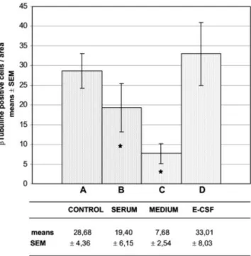
Fig. 3. Quantitative analysis of neuroepithelial cells undergoing neural differentiation measured by the number of3-tubulin-positive cells. Values plotted in the chart show the mean of the3-tubulin-positive cells per area⫾ the standard error. A: Mesencephalic neuroepithelium of chick embryos maintained in vivo until developmental stage HH23. B: Mesence-phalic neuroepithelium explants cultured in vitro with FCS-supplemented medium. C: Mesencephalic neuroepithelium explants cultured in vitro with a chemically defined serum-free medium. D: Mesencephalic neuroepithe-lium explants cultured in vitro with E-CSF-supplemented medium. Note that the number of3-tubulin-positive cells is slightly higher in the explants cultured with E-CSF-supplemented medium (D) than in the controls (A), although this difference is not significant; the explants cultured with serum-free medium (C) show a drastic reduction in3-tubulin-positive cells. Also note that in the explants cultured with FCS-supplemented medium (B), the number of3-tubulin-positive cells is slightly lower than in the controls and the explants cultured with E-CSF-supplemented medium. Asterisks denote values that differ significantly (P⬍0.05) from controls according to the two-tailed Student’st-test.
HH25 chick embryos did not undergo extensive apoptosis (Fig. 1D) as did the explants cultured only with serum-free medium. On the contrary, apoptotic cells were scarce, as detected in controls. These data suggest that the E-CSF promotes the survival of these cells.
Moreover, the addition of E-CSF to the serum-free me-dium in these cultures largely prevents the drastic de-crease in nuclear BrdU incorporation shown by explants cultured with only serum-free medium (compare Fig. 1G and H). The number of BrdU-positive nuclei in the neuro-epithelial explants cultured in the E-CSF-supplemented medium was slightly lower than that in controls. Although this difference was almost double, the number of BrdU-positive nuclei was clearly higher than that of explants cultured in serum-free medium (Fig. 2).
Subsequently, to test whether the role of the E-CSF in cell replication is due to the presence of diffusible factors within it, we implanted heparin acrylic microbeads soaked in E-CSF in neuroepithelial explants cultured in serum-free medium. Abundant BrdU-positive nuclei were located in the basal portion of the areas of the neuroepithelium that were close to the microbead (Fig. 1I). In this way, the number of BrdU-positive cells progressively decreased from the microbead outward, suggesting the presence of diffusible factors within the E-CSF, which may be in-volved in the control of neuroepithelial cell replication.
Finally, the addition of E-CSF to the serum-free me-dium in these organotypic cultures also induced the ex-pression of3-tubulin in a continuous layer of cells located at the basal portion of the mesencephalic neuroepithe-lium, and some of the 3-tubulin-positive cells showed long cytoplasmatic developments from the basal to the apical part of the neuroepithelium (Fig. 1N), as shown in the controls. The number of 3-tubulin-positive cells present in the neuroepithelial explants cultured with E-CSF-supplemented medium was similar to or slightly higher than in the controls, although this difference was not significant (Fig. 3).
Finally, to determine whether the role of the E-CSF in neural differentiation is due to the presence of diffusible factors within it, we also implanted heparin acrylic mi-crobeads soaked in E-CSF in neuroepithelial explants cul-tured in serum-free medium. Abundant 3-tubulin-posi-tive cells were detected in areas of the neuroepithelium close to the microbead. Moreover, the number of 3-tubu-lin-positive cells progressively decreased from the acrylic microbead outward (Fig. 1O), suggesting the presence of diffusible factors within the E-CSF, which may be in-volved in neurogenesis.
Taken together, all these data indicate that the E-CSF contains diffusible factors that regulate the three basic cellular behavioral parameters of the neuroepithelial stem cells analyzed in this study, i.e., survival, replication, and differentiation, suggesting that the E-CSF could play a key role in early brain development in vivo.
DISCUSSION
We have developed an organotypic tissue culture tech-nique for neuroepithelial explants that allows us to study the influence of external factors on cellular behavior. We have shown the validity of this technique, as the cellular behavior of the neuroepithelial cells of mesencephalic ex-plants cultured in FCS-supplemented medium was very similar to that of controls kept in vivo, i.e., the trophic
factors within the FCS are sufficient to ensure an almost normal rate of cell survival, proliferation, and differenti-ation. Despite the fact that the behavior of neuroepithelial cells in vivo differs somewhat from that observed in cul-tures with an FCS-supplemented medium, these differ-ences could be due to the downtime that all tissues take to adjust to the trauma, in vitro conditions, etc. We have also demonstrated that mesencephalic neuroepithelial ex-plants cultured in vitro in serum-free medium cannot maintain the normal rate of cell survival, proliferation, and differentiation by themselves, but that the addition of E-CSF to a serum-free medium is also sufficient to main-tain almost normal cellular behavior compared with con-trols in vivo. These results clearly suggest that E-CSF exerts a trophic effect on mesencephalic neuroepithelial cells in vivo, having a direct influence on the behavior of neuroepithelial cells.
Regulation of Cellular Behavior of
Neuroepithelial Cells Requires Extraneural Factors
Neuroepithelial cells undergo intense cell proliferation and simultaneously begin neurogenic differentiation (Panchision and McKay, 2002). Several growth factors have been involved in the control of these processes, in-cluding FGF2 and EGF (Murphy et al., 1990; Ciccolini and Svendsen, 1998; Tropepe et al., 1999; Vaccarino et al., 1999a, 1999b; Raballo et al., 2000), which are expressed together with their receptors by the neuroepithelial cells from early stages of development and act in an autocrine and/or paracrine fashion (Heuer et al., 1990; Kalcheim and Neufeld, 1990; Ozawa et al., 1996; Wilke et al., 1997; Raballo et al., 2000; Panchision and McKay, 2002).
However, it has also been suggested that there are other behavioral influences different from neuroepithelial self-stimulation (Kalcheim and Neufeld, 1990; Miyan et al., 2003). In this regard, Gato et al. (1998) and Raballo et al. (2000) have suggested that FGF2 is also secreted to the E-CSF, and that it may help regulate neuroepithelial cell behavior, interacting with apical receptors. Moreover, an anti-FGF2 antibody introduced by microinjection into ce-phalic ventricular cavities in vivo impairs cell replication and neurogenesis in the subventricular region (Tao et al., 1997), which is the fetal and adult successor of embryonic neuroepithelial cells (Bruni, 1998; Tramontin et al., 2003). These findings support the hypothesis that the CSF is an alternative fluid way to control the cellular behavior of the cells that are in close contact with brain cavities (Tao et al., 1997; Gato et al., 1998, 2004; Vaccarino et al., 1999a, 199b).
mus), we cannot rule out the possibility of a supplemen-tary extraneural source of growth factors in vivo, which could be extraneural tissues such as the notochorda or the E-CSF itself.
E-CSF Helps Control Cellular Behavior in Neuroepithelial Cells
Classically, E-CSF has been involved in some epigenetic processes during early brain development, e.g., the posi-tive pressure that drives expansion and morphogenesis (Jelinek and Pexieder, 1968, 1970; Desmond and Jacob-son, 1977), which is regulated by apical secretion of os-motically active molecules belonging to the family of pro-teoglycans (Gato et al., 1993; Alonso et al., 1998, 1999).
On the other hand, E-CSF has a complex protein com-position, and the exposure of the apical surface of neuro-epithelial cells to this fluid could affect their behavior (Dziegielewska et al 1980a, 1980b, 1981, 1991, 2000; Che-ciu et al., 1984; Gato et al., 2004). Experimental data demonstrate that the loss or modification of the composi-tion of the E-CSF strongly decreases the neuronal cell population (Desmond and Jacobson, 1977; Desmond, 1985). Moreover, Mashayesky et al. (2002) and Owen-Lynch et al. (2003) have reported that the phenotype of the mutant hydrocephalic Texas rats is due to an alter-ation of the proliferalter-ation of brain primordium stem cells and cortical neurogenesis due to changes in the composi-tion of the CSF. In this regard, the trophic accomposi-tion of CSF from rat fetus on cortex neuroblastic cells cultured in vitro has been demonstrated (Miyan et al., 2003). Here we provide experimental evidence that neuroepithelial cell survival, replication, and differentiation are activated by E-CSF in organotypic cultures, suggesting that the E-CSF contains key factors involved in the control of these cellu-lar processes and, consequently, plays a relevant role in the development of the embryonic brain in vivo. Our re-sults show that cell replication (but not neurogenesis) is slightly but significantly lower in the explants cultured with E-CSF than in the controls developed in vivo, sug-gesting that other factors from neural or extraneural tis-sues could be involved in maintaining the normal rate of neuroepithelial cell replication.
Finally, in the past years it has been suggested that adult CSF plays a key role as a fluid way to deliver diffusible signals and thus influence the cellular behavior of determined brain parenchyma cells (Nicholson, 1999). More recently, Alvarez-Buylla and Garcı´a-Verdugo (2002) and Tramontin et al. (2003) have demonstrated that in the adult brain, the neurogenic stem cells of the subventricu-lar zone have transitory contact with the ventricusubventricu-lar brain cavity, suggesting that adult neurogenesis could be condi-tioned by the CSF.
How Does E-CSF Control Cellular Behavior of Neuroepithelial Pluripotent Stem Cells?
We have reported that the E-CSF could play a relevant role in the early development of the brain, in addition to other morphogenetic mechanisms (expansion of the brain vesicle cavities). As stated above, several studies have demonstrated that the protein composition of the E-CSF is more complex than that of the adult CSF and it has been suggested that there exist proteins involved in the regu-lation of cell behavior, e.g., growth factors and cytokines.
Moreover, it has also been suggested that there exist var-ious growth factors in chick embryo brain cavities (Gato et al., 1993; Ojeda and Piedra, 2000). In this regard, we have previously detected the presence of FGF2 in the E-CSF of chick embryos at early stages of development (Gato et al 1998). We are currently analyzing the presence of several growth factors and cytokines in the E-CSF, as well as their role in neuroepithelial stem cell behavior in vivo.
Regarding the origin of the diffusible factors within the E-CSF during early developmental stages, prior to the formation of the choroids plexus, this fluid may be se-creted at least partially by the neuroepithelial cells them-selves (Miyan et al., 2003). However, the protein compo-sition of the E-CSF is very similar to embryonic serum at the same developmental stages (Gato et al., 2004), which suggests that at least some of the proteins within the E-CSF could gather and be selectively transported from the embryonic serum to the E-CSF.
Perspectives
To conclude, this study supports the idea that early histogenesis of the embryonic brain might not be an in-trinsic process of the neuroepithelium, but one requiring the action of external factors, some of which act from the E-CSF. Our data support the hypothesis that the fluid within the cavity of the embryonic brain is a key pathway for the diffusion of molecular signals in vivo. This implies that a complete understanding of the process of embryonic neurogenesis requires further analysis of the molecules within the E-CSF and their role in neurogenesis. These data may also improve our knowledge of the cellular re-quirements for neuronal culture techniques and allow us to extrapolate them to the regulation of neurogenesis in adults.
ACKNOWLEDGMENTS
The authors thank Dra. Sagrario Callejo for laser con-focal microscopy technical support, Professor David Rix-ham for language translation assistance, and Rufo Mar-tı´n, Pilar MarMar-tı´n, and Isabel Garcı´a for technical assistance. A.G. was supported by Ministerio de Sanidad y Consumo, Instituto de Salud Carlos III, grant number 02/0961 (cofinanced by European Community FEDER), and Junta de Castilla y Leo´n, grant numbers VA049/04 and VA17/03. D.B. was supported by Ministerio de Sanidad y Consumo, Instituto de Salud Carlos III, grant number 02/0915 (cofinanced by European Community FEDER).
LITERATURE CITED
Alonso MI, Gato A, Moro JA, Barbosa E. 1998. Disruption of proteo-glycans in neural tube fluid by-D-xyloside alters brain enlarge-ment in chick embryos. Anat Rec 252:499 –508.
Alonso MI, Gato A, Moro JA, Martı´n P, Barbosa E. 1999. Involvement of sulfated proteoglycans in embryonic brain expansion at earliest stages of development in rat embryos. Cells Tissues Organs 165: 1–9.
Alvarez-Buylla A, Garcı´a-Verdugo JM. 2002. Neurogenesis in adult subventricular zone. J Neurosci 22:629 – 634.
Bally-Cuif L, Bonicelli E. 1997. Transcription factors and head for-mation in vertebrates. Bioessays 19:127–135.
Brunet CL, Sharpe PM, Ferguson MW. 1993. The distribution of epidermal growth factor binding sites in the developing mouse palate. Int J Dev Biol 37:451– 458.
Bruni JE. 1998. Ependymal development, proliferation, and functions: a review. Microsc Res Tech 41:2–13.
Bueno D, Skinner J, Abud H, Heath JK. 1996. Spatial and temporal relationships betweenShh,Fgf4andFgf8gene expression at di-verse signalling centers during mouse development. Dev Dyn 207: 291–299.
Checiu I, Prelipceanu O, Popescu O. 1984. The role of cerebrospinal fluid during embryonic development: a biochemical study. Morphol Embryol (Bucur) 30:243–250.
Ciccolini F, Svendsen CN. 1998. Fibroblast growth factor 2 (FGF-2) promotes acquisition of epidermal growth factor (EGF) responsive-ness in mouse striatal precursor cells: identification of neural pre-cursors responding to both EGF and FGF-2. J Neurosci 18:7869 – 7880.
Crossley PH, Martin GR. 1995. The mouse Fgf8 gene encodes a family of polypeptides and is expressed in regions that direct outgrowth and patterning in the developing embryo. Development 121:439 – 451.
Crossley PH, Martinez S, Martin GR. 1996. Midbrain development induced by FGF8 in the chick embryo. Nature 380:66 – 68. Desmond ME, Jacobson AG. 1977. Embryonic brain enlargement
requires cerebrospinal fluid pressure. Dev Biol 57:188 –198. Desmond ME. 1985. Reduced number of brain cells in so-called neural
overgrowth. Anat Rec 212:195–198.
Desmond ME, Levitan ML. 2002. Brain expansion in the chick embryo initiated by experimentally produced occlusion of the spinal neuro-coel. Anat Rec 268:147–159.
Duboule D. 1994. Temporal colinearity and the phylotypic progression: a basis for the estability of a vertebrate Bauplan and the evolution of morphologies through heterochrony. Development (Suppl):135–142.
Dziegielewska KM, Evans CAN, Fossan G, Lorscheider FL, Mali-nowska DH, Mollgard K, Reynolds ML, Saunders NR, Wilkinson S. 1980a. Proteins in cerebrospinal fluid and plasma of fetal sheep during development. J Physiol 300:441– 455.
Dziegielewska KM, Evans CAN, Malinowska DH, Mollgard K, Reyn-olds ML, Saunders NR. 1980b. Blood-cerebrospinal fluid transfer of plasma proteins during fetal development in the sheep. J Physiol 300:457– 465.
Dziegielewska KM, Evans CAN, Lai PCW, Lorscheider FL, Mali-nowska DH, Mollgard K, Saunders NR. 1981. Proteins in cerebro-spinal fluid and plasma of fetal rats during development. Dev Biol 83:193–200.
Dziegielewska KM, Habgood MD, Mollgard K, Stagaard M, Saunders NR. 1991. Species-specific transfer of plasma albumin from blood into different cerebrospinal fluid compartiments in the fetal sheep. J Physiol 439:215–237.
Dziegielewska KM, Knott GW, Saunders NR. 2000. The nature and composition of the internal environment of developing brain. Cell Mol Neurobiol 20:41–56.
Echelard Y, Epstein DJ, St-Jacques B, Shen L, Mohler J, McMahon JA, McMahon AP. 1993. Sonic hedgehog, a member of a family of putative signaling molecules, is implicated in the regulation of CNS polarity. Cell 75:1417–1430.
Garda AL, Echevarrı´a D, Martı´nez S. 2001. Neuroepithelial co-ex-pression of Gbx2 and Otx2 precedes Fgf8 exco-ex-pression in the isthmic organizer. Mech Dev 101:111–118.
Gato A, Moro JA, Alonso MI, Pastor JF, Represa J, Barbosa E. 1993. Chondroitin sulphate proteoglycan and embryonic brain enlarge-ment in the chick. Anat Embryol 188:101–106.
Gato A, Alonso MI, Moro JA, Martı´n P, Barbosa E. 1998. Presence of FGF-2 in chick embryo neural tube fluid. Eur J Anat 2:185–186. Gato A, Martı´n P, Alonso MI, Martı´n C, Pulgar MA, Moro JA. 2004.
Analysis of cerebro-spinal fluid protein composition in early devel-opmental stages in chick embryos. J Exp Zool 301:280 –289. Gaufo GO, Thomas KR, Capecchi MR. 2003. Hox3 genes coordinate
mechanisms of genetic suppression and activation in the generation of branchial and somatic motoneurons. Development 130:5191– 5201.
Gensburger C, Laburdette G, Sensenbrenner M. 1987. Brain basic fibroblast growth factor stimulates the proliferation of rat neuronal precursor cells in vitro. Natl Lib Med 217:1–5.
Hamburger V, Hamilton HL. 1951. A serie of normal stages in the development of the chick embryo. J Morphol 88:49 –92.
Heuer J, Von Bartheld CS, Kinoshita Y, Evers PC, Bothwell M. 1990. Alternating phases of FGF receptor and NGF receptor expression in the developing chicken nervous system. Neuron 5:283–296. Jelinek R, Pexieder T. 1968. The pressure on cephalic fluid in chick
embryo between the 2nd and 6th day of incubation. Phisiol Bohe-mos 17:297–305.
Jelinek R, Pexieder T. 1970. Presure of the CNS I chick embryo. Fol Morphol 2:102–110.
Kalcheim C, Neufeld G. 1990. Expression of basic fibroblast growth factor in the nervous system of early avian embryos. Development 109:203–215.
Kilpatrick TJ, Bartlett PF. 1993. Cloning and growth of multipoten-tial neural precursors: requirements for proliferation and differen-tiation. Neuron 10:255–265.
Martı´ E, Takada R, Bumcrot DA, Sasaki H, McMahon AP. 1995. Distribution of sonic hedgehog peptides in the developing chick and mouse embryo. Development 121:2537–2547.
Mashayekhi F, Draper CE, Bannister CM, Pourghasem M, Owen-Lynch PJ, Miyan JA. 2002. Deficient cortical development in the hidrocephalic texas (H-Tx) rat: a role for CSF. Brain 125:1859 – 1874.
Miyan JA, Nabiyouni M, Zendah M. 2003. Development of the brain: a vital role for cerebrospinal fluid. Can J Physiol Pharmacol 81:317–328. Murphy M, Drago J, Bartlett PF. 1990. Fibroblast growth factor stimulates the proliferation and differentiation of neural precursor cells in vitro. J Neurosci Res 25:463– 475.
Nicholson C. 1999. Signals that go with the flow. Trends Neurosci 22:143–145.
Ojeda JL, Piedra S. 2000. Evidence of a new transitory extracellular structure within the developing rhombencephalic caviti: an ultra-structural and immunoelectron-microscopic study in the chick. Anat Embryol 202:257–264.
Owen-Lynch PJ, Draper CE, Mashayekhi F, Bannister CM, Miyan JA. 2003. Defective cell cycle control underlies abnormal cortical development in the hydrocephalic texas rat. Brain 126:623–231. Ozawa K, Uruno T, Miyakawa K, Seo M, Imamura T. 1996.
Expres-sion of the fibroblast growth factor family and their receptor family genes during mouse brain development. Mol Brain Res 41:279 –288. Panchision DM, McKay RDG. 2002. The control of neural stem cells
by morphogenic signals. Curr Opin Genet Dev 12:478 – 487. Placzek M, Yamada T, Tessier-Lavigne M, Jessell T, Dodd J. 1991.
Control of dorsoventral pattern in vertebrate neural development: induction and polarizing properties of the floor plate. Development 2:105–122.
Placzek M, Jessell TM, Dodd J. 1993. Induction of floor plate differ-entiation by contact-dependent, homeogenetic signals. Develop-ment 117:205–218.
Raballo R, Rhee J, Lyn-Cook R, Leckman JF, Schwartz ML, Vaccarino FM. 2000. Basic fibroblast growth factor (Fgf2) is necessary for cell proliferation and neurogenesis in the developing cerebral cortex. J Neurosci 20:5012–5023.
Rajan P, Panchision DM, Newell LF, McKay RDG. 2003. BMPs signal alternately through a SMAD or FRAP-STAT pathway to regulate fate choice in CNS stem cells. J Cell Biol 161:911–921.
Roelink H, Augsburger A, Heemskerk J, Korzh V, Norlin S, Ruiz i Altaba A, Tanabe Y, Placzek M, Edlund T, Jessell TM, et al. 1994. Floor plate and motor neuron induction by vhh-1, a vertebrate homolog of hedgehog expressed by the notochord. Cell 76:761–775. Shamim H, Mahmood R, Logan C, Doherty P, Lumsden A, Mason I. 1999. Sequantial roles for Fgf4, En1 and Fgf8 in specification and regionalisation of the midbrain. Development 126:945–959. Tao Y, Black IB, DiCicco-Bloom E. 1997. In vivo neurogenesis is
inhibited by neutralizing antibodies to basic foibroblast growth factor. J Neurobiol 33:289 –296.
Toresson H, Potter SS, Campbell K. 2000. Genetic control of dorso-ventral identity in the telencephalon: opposing roles for Pax6 and Gsh2. Development 127:4361– 4371.
Tramontin AD, Garcı´a-Verdugo JM, Lim DA, Alvarez-Buylla A. 2003. Postnatal development of radial glia and the ventricular zone (VZ): a continuum of the neural stem cell compartment. Cereb Cortex 13:580 –587.
Tropepe V, Sibilia M, Ciruna BG, Rossant J, Wagner EF, Van der Kooy D. 1999. Distinct neural stem cells proliferate in response to EGF and FGF in the developing mouse telencephalon. Dev Biol 208:166 –188. Vaccarino FM, Schwartz ML, Raballo R, Nilsen J, Rhee J, Zhou M,
Doetschman T, Coffin JD, Wyland JJ, Hung YT. 1999a. Changes in
cerebral cortex size are governed by fibroblast growth factor during embryogenesis. Nat Neurosci 2:246 –253.
Vaccarino FM, Schwartz ML, Raballo R, Rhee J, Lyn-CooK R. 1999b. Fibroblast growth factor signaling regulates growth and morphog-nesis at multiple steps during brain development. Dev Biol 44:179 – 201.
Wilke TA, Gubbels S, Schwartz J, Richman JM. 1997. Expression of fibroblast growth factor receptors (FGFR1, FGFR2, FGFR3) in the developing head and face. Dev Dyn 210:41–52.
Yamada T, Placzek M, Tanaka H, Dodd J, Jessell TM. 1991. Control of cell pattern in the developing nervous system: polarizing activity of the floor plate and notochord. Cell 64:635– 647.