Estudio de la función de la familia de Proteínas Quinasas C en el cáncer de mama
326
0
0
Texto completo
(2)
(3) UNIVERITY OF MURCIA. DEPARTMENT OF BIOCHEMISTRY AND MOLECULAR BIOLOGY (A). ROLE OF PROTEIN KINASE C IN BREAST CANCER CELLS. RUBÉN LÓPEZ NICOLAS 2011.
(4)
(5) ÍNDICE.
(6)
(7) Index. i. PRÓLOGO (RESUMEN EN CASTELLANO). 1. I. INTRODUCCIÓN Y OBJETIVOS. I.1. PROTEÍNAS QUINASAS C: Estructura y clasificación. I.2. Proteínas Quinasas C y cáncer. I.3. Objetivos.. 3 3 5 6. II. MATERIALES Y MÉTODOS. II.1. Construcciones de ADN plasmídico. II.2. Cultivos celulares y transfecciones. II.3. Microscopía confocal. II.4. Purificación y medidas de actividad quinasa. II.5. Microarrays. II.6. Análisis estadístico. III. LOCALIZACIÓN ARAQUIDÓNICO.. DE. PKCα. DEPENDIENTE. 7 7 8 8 9 9 10 DE. ÁCIDO 11. IV. EFECTO DEL ÁCIDO OLEICO EN CÉLULAS MODELO DE CÁNCER DE MAMA A TRAVÉS DE PKCα.. 12. V. EFECTO DE LOS ÁCIDOS GRASOS OMEGA-3 Y LA INHIBICIÓN DE LA EXPRESIÓN DE PKCα EN CÉLULAS MODELO DE CÁNCER DE MAMA.. 13. VI. CARACTERIZACIÓN TRAVÉS DE PKCs.. 14. BIOLÓGICA. DE. DAG-LACTONAS. A. VII. PERFIL DE EXPRESIÓN GÉNICA EN CÉLULAS MODELO DE CÁNCER DE MAMA EN AUSENCIA DE PKCα.. 15. VIII. EL TRATAMIENTO DE CÉLULAS MODELO DE CÁNCER DE MAMA CON SALINOMICINA POTENCIA EL EFECTO DE LA INHIBICIÓN DE LA PKCα.. 16. IX. CONCLUSIONES.. 17.
(8) Index. ii. ABBREVIATIONS. 19. CHAPTER I: INTRODUCTION AND OBJECTIVES.. 21. 1. Introduction classification.. to. PROTEIN. KINASE. C.. Structure. and. 23. 2. Conserved regions in PKC. 2.1. Pseudosubstrate domain. 2.2. C1 domain. Structure, function and regulation. 2.3. C2 domain. Structure, function and regulation. 2.4. Catalytic domain. Structure and regulation.. 25 25 25 28 35. 3. Variable regions.. 38. 4. PKC activation mechanism. 4.1. Activation model of classical and novel PKCs.. 39 40. 5. Pathways where PKC is involved.. 43. 6. Other bioactive lipids able to activate PKC. 6.1. Fatty acids. 6.2. DAG-lactones.. 45 45 47. 7. Introduction to PKC and cancer.. 49. 8. PKC isoforms in Breast Cancer.. 54. 9. PKC isoforms as target in cancer therapy.. 57. 10. Objectives.. 60. CHAPTER II: MATERIALS AND METHODS. 1. Construction of expression plasmids. 1.1. Construction in plasmid pEGFP-N3. 1.2. Construction in plasmid pECFP-N3. 1.3. Construction in plasmid pCGN. 1.4. Site-directed Mutagenesis. 1.5. Amplification and purification plasmid DNA.. 61 63 64 65 66 67 68.
(9) Index. iii. 2. Cell culture. 2.1. MCF-7 cells. 2.2. BT-474 cells. 2.3. MDA-MB-231 cells. 2.4. Inhibition of PKCα expression using small interference RNA. 2.5. Migration assays. 2.6. Invasion assays. 2.7. Proliferation assays. 2.8. Apoptosis assays.. 73 73 75 76 77 78 79 80 81. 3. Confocal microscopy. 3.1. Introduction to confocal imaging. 3.2. Microscope used. 3.3. Fluorescent substances used. 3.4. Media used in stimulation of living cells in confocal microscope. 3.5. Image processing and analysis. 3.6. Immunofluorescence.. 83 83 85 87. 4. Electrophoresis techniques. 4.1. Agarose gel preparation. 4.2. Acrylamide gel preparation.. 98 98 99. 5. Western Blot.. 99. 89 89 97. 6. Fluorescence Resonance of Energy Transfer (FRET).. 109. 7. PKCα purification.. 103. 8. Kinase activity assays.. 104. 9. Microarrays. 9.1. Introduction of microarrays. 9.2. Overview of preparation for gene expression array. 9.3. Sample preparation.. 105 105 106 110. 10. Statistical analysis.. 113.
(10) iv. Index. CHAPTER III: MOLECULAR MECHANISM OF PKCα LOCALIZATION BY ARACHIDONIC ACID. THE C2 115 DOMAIN ALSO PLAYS A ROLE. 1. Introduction.. 117. 2. Results. 2.1. Arachidonic acid induces the PKCα translocation to the plasma membrane in a Ca2+-dependent manner. 2.2. The C2 domain of PKCα plays an important role in the AAdependent membrane localization of the enzyme. 2.3. Role of the C1 domain of PKCα in the AA-dependent membrane localization.. 118 118 125 128. CHAPTER IV: EFECT OF OLEIC ACID ON BREAST 131 CANCER CELLS THROUGH PKCα. 1. Introduction.. 133. 2. Results. 2.1. Oleic acid induces PKCα plasma membrane translocation and co-localization with actin filaments in breast cancer cells. 2.2. Characterization of the activation mechanism of PKCα by oleic acid. 2.3. Oleic acid induces the PKCα translocation to plasma membrane in a Ca2+-dependent manner. 2.4. Inhibition of PKCα expression reduces the proliferation rate in breast cancer cell lines and synergizes with oleic acid. 2.5. Effect of oleic acid addition and inhibition of PKCα expression on the migration and invasion capacities of MCF-7 and MDAMB-231 cell lines. 2.6. Oleic acid and inhibition of PKCα induce apoptosis in breast cancer cell lines.. 135 135 139 140 145. 148. 151.
(11) Index. v. CHAPTER V: STUDY OF THE ROLE OF OMEGA-3 FATTY ACIDS AND PKCα DEPLETION IN BREAST 153 CANCER CELLS. 1. Introduction.. 155. 2. Results. 2.1. Omega-3 fatty acids induce PKCα plasma membrane translocation and co-localization with actin filaments in breast cancer cells. 2.2. Characterization of the activation mechanism of PKCα by DHA and EPA. 2.3. Effect of omega-3 fatty acids and inhibition of PKCα expression on the migration and invasion capacities of MCF-7 and MDA-MB-231 cell lines. 2.4. Omega-3 fatty acids and inhibition of PKCα induce apoptosis in breast cancer cell lines.. 157. 157 161. 164 169. CHAPTER VI: CHARACTERIZATION OF BIOLOGICAL ACTIVITY OF DIACYLGLYCEROL173 LACTONES AND ITS EFFECT ON PKCs. 1. Introduction.. 175. 2. Results. 2.1. Translocation of EGFP-tagged PKC isoenzymes in response to DAG-lactone derivatives. 2.1.1. Effect of PMA and 153C-022 on PKC localization. 2.1.2. Effect of 153B-143 on PKC localization. 2.2. Tightness of the plasma membrane binding interactions measured by FRAP. 2.2.1. Effect of PMA and 153C-022 on fluorescence recovery after photobleach PKCα. 2.2.2. Effect of PMA, 153C-022 and 153B-143 on fluorescence recovery after photobleach PKCε. 2.2.3. Effect of PMA, 153C-022 and 153B-143 on fluorescence recovery after photobleach PKCδ.. 180 180 181 186 189 189 190 191.
(12) vi. Index. CHAPTER VII: GENE EXPRESSION PROFILE OF 193 BREAST CANCER CELLS IN ABSENCE OF PKCα. 1. Introduction.. 195. 2. Results. 2.1. Inhibition of PKCα expression in MCF-7 cells. 2.1.1. Genes down-regulated after PKCα inhibition. 2.1.2. Genes up-regulated after PKCα inhibition. 2.1.3. Validation of arrays data. 2.2. Inhibition of PKCα expression in MDA-MB-231 cells. 2.2.1. Genes down-regulated after PKCα inhibition. 2.2.2. Genes up-regulated after PKCα inhibition. 2.2.3. Validation of arrays data.. 196 196 197 199 201 208 208 210 212. CHAPTER VIII: CHARACTERIZATION OF SALINOMYCIN EFFECT ON BREAST CANCER 219 CELLS THROUGH PKCα. 1. Introduction. 2. Results. 2.1. Effect of salinomycin on the migration and invasion capacities of breast cancer cell lines with down-regulated PKCα. 2.2. Salinomycin induces apoptosis in breast cancer cell lines with down-regulated PKCα.. CHAPTER IX: DISCUSSION AND CONCLUSIONS. 1. Discussion. 1.1. Role of the C2 domain in the arachidonic acid-dependent localization and activation of PKCα. 1.2. Role of the C1 domain in the arachidonic acid-dependent localization and activation of PKCα. 1.3. Role of C1 and C2 domains in the localization and activation of PKCα induced by oleic acid. 1.4. Effect of PKCα depletion and oleic acid on breast cancer cell lines. 1.5. Localization and activation mechanism of PKCα induced by omega-3 fatty acids.. 221 223 223 227. 231 233 233 235 237 239 241.
(13) Index. 1.6. Omega-3 fatty acids reduce the malignancy of breast cancer cell lines through PKCα. 1.7. Synthetic charged DAG-lactones induce localization of several PKC isoform to the plasma membrane. 1.8. DAG-lactones anchorage PKC isoform to the plasma membrane in a more subtle manner than phorbol ester. 1.9. Studies on gene expression arrays reveal that PKCα depletion decreases the malignancy of MCF-7 and MDA-MB-231 breast cancer cells. 1.10. The effects of salinomycin on breast cancer cells are enhanced by PKCα depletion. 2. Conclusions.. vii. 243 244 247. 248 249 251. ANNEX I (MCF-7 DEREGULATED GENES). 255. ANNEX II (MDA-MB-231 DEREGULATED GENES). 263. BIBLIOGRAPHY. 269.
(14)
(15) PRÓLOGO RESUMEN EN CASTELLANO.
(16)
(17) Resumen en castellano. 3. I. INTRODUCCIÓN. I.1. PROTEÍNAS clasificación.. QUINASAS. C:. Estructura. y. Las Proteínas Quinasas C (PKC) constituyen un grupo de enzimas con actividad fosfotransferasa que fosforilan específicamente residuos de Ser y Thr de sus proteínas sustrato. Están involucradas en diversas vías de transducción de señales en las células, participando en multitud de funciones fisiológicas, así como procesos patológicos come el cáncer y determinadas cardiopatías entre otros (Nishizuka, 1995; Newton, 2001; Ohno y Nisizuka, 2002; Corbalán-García y Gómez-Fernández, 2006). Su ubicuidad y diversidad funcional, las hacen una familia muy importante de proteínas, cuyo objeto de estudio es de gran relevancia. Esta familia de enzimas se compone de 10 isoenzimas, que están codificados por 9 genes en mamíferos. Cada isoenzima se expresa en una amplia variedad de células y además cada célula expresa un gran número de estas proteínas, siendo así una familia muy ubicua, además del gran número de funciones celulares que desempeña. Las isoenzimas de la PKC se han clasificado en tres subfamilias en base a su estructura primaria y dependencia de distintos cofactores para obtener un incremento de su actividad enzimática (Figura I.1): a) PKC clásicas o convencionales: incluye las isoenzimas PKCα, βI, βII y γ (Inoue y col., 1977; Coussens y col., 1986). Presentan un dominio pseudosubstrato, el dominio C1 capaz de interaccionar con DAG y ésteres de forbol, y un dominio C2 capaz de unir Ca2+ y fosfolípidos aniónicos. b) PKC nuevas: en esta subfamilia se agrupan las isoenzimas ε, δ (Ono y col., 1987a), η (Osada y col., 1990) y θ (Osada y col., 1992). Este grupo posee los mismos dominios reguladores que las PKC clásicas, pero el dominio C2 es insensible a Ca2+, por eso este grupo de enzimas solo necesita de fosfolípidos aniónicos y DAG o ésteres de forbol para activarse totalmente. c) PKC atípicas: es una subfamilia compuesta por dos miembros, la PKCζ y la PKC ι/λ [PKCι (isoforma en humanos) (Selbie y col., 1993)/ PKCλ (isoforma en ratón) (Akimoto y col., 1994)]. Este grupo solo posee el dominio pseudosubstrato, un dominio C1 incapaz de interaccionar con DAG o ésteres de forbol y otro llamado PB1 encargado de interaccionar con otras proteínas..
(18) 4. Prologo. Figura I.1. Representación esquemática de la estructura primaria de los isoenzimas de la PKC. Se muestran los distintos dominios que lo forman, así como los requerimientos de cofactores para su regulación. En el extremo amino terminal se encuentra la región reguladora, con el dominio pseudosustrato, y los dominios C1 y C2, que unen diferentes cofactores según el tipo de isoenzima. El extremo carboxilo terminal, contiene la región catalítica del enzima con el dominio quinasa de unión al ATP y al sustrato. Las regiones variables se muestran nombradas como V1, V2, V3, V4 y V5. La región en azul presente en el dominio PB1 de las atípicas. (Tomado de Corbalán García y Gómez Fernández, 2006).. El dominio C1 suele presentarse en tandem formando 2 subdominios, cada uno de los que posee una secuencia consenso del tipo HX12CX2CX13/14CX2CX4H X2CX2CX7C siendo imprescindible para la coordinación de dos iones de zinc. Presentan una estructura globular, donde los aminoácidos se agrupan en una distribución polarizada; en la parte superior, por donde se unen al DAG o ésteres de forbol, se encuentran residuos aromáticos, mientras que en la parte media se encuentras aminoácidos catiónicos. El otro dominio de la región reguladora es el dominio C2, cuya estructura es un β-sandwich compuesto por ocho cadenas β dispuestas de forma antiparalela y que están conectadas por medio de lazos de estructura flexible tanto en la parte alta como baja del dominio (Figura I.2) (Cho y Stahelin, 2006). En las PKCs clásicas el principal papel de este dominio es actuar como motivo de anclaje a la membrana activado por Ca2+ (CorbalánGarcía y Gómez-Fernández, 2006; Cho y Stahelin, 2006). El dominio presenta dos regiones funcionales importantes; la primera es la región de unión a calcio localizada en la parte superior y encargada de unir dos o tres iones Ca2+ dependiendo de la isoenzima, además de PtdSer (Conesa-Zamora y col., 2000; Ochoa y col., 2002). La otra región importante es la región polibásica o rica en lisinas, la cuál ha sido sugerida como la que une específicamente PtdIns(4,5)P2 (Sánchez-Bautista y col., 2006)..
(19) Resumen en castellano. A. 5. B. Figura I.2. Representación del dominio C2. (A) Se observa la unión a la PtdSer por la región de unión de Ca2+ en la parte superior del esquema, y a una molécula de PtdIns(4,5)P2 (o PIP2) por la región polibásica en el centro del esquema. (B) Se representa amplificado un detalle de la región polibásica del dominio C2, concretamente los residuos de las cadenas β3 y β4 encargados de la interacción con el PtdIns(4,5)P2 (Tomado de Guerrero-Valero y col., 2009).. Para la activación de la PKC, antes debe localizarse en membranas biológicas donde se encuentren sus cofactores. En el caso de las PKCs clásicas, la translocación es mediada por los dominios C1 y C2 siendo primeramente la interacción del dominio C2 con la PtdSer de la membrana a través de Ca2+, lo que favorece que el dominio C1 interaccione con DAG, permitiendo un mejor anclaje de la PKC a la membrana (Corbalán-García y Gómez Fernández, 2006). La PtdSer y el DAG son los cofactores clásicos, pero hoy en día se ha visto que otros lípidos también son capaces de activar diferentes isoenzimas de PKC (López-Andreo y col., 2005).. I.2. Proteínas Quinasas C y cáncer. La familia de PKC, además de participar en multitud de funciones biológicas, fue involucrada en el cáncer inicialmente por ser el receptor celular de los ésteres de forbol. Más tarde se vio su relación con otros oncogenes, confirmando el papel de la PKC en el cáncer. A diferencia de otras proteínas, su influencia en el desarrollo de cáncer no es por mutación, sino por una desregulación de sus niveles en células tumorales, pudiendo estar más o menos sobre- o sub-expresada en función de la isoenzima y tipo de cáncer. En el caso concreto del cáncer de mama, se ha visto una sobre-expresión de la PKCα siendo la causante de la generación de resistencia frente a diversos agentes terapéuticos (Gill y col., 2001)..
(20) Prologo. 6. Por todo ello las diferentes isoenzimas de PKC han sido elegidas como posibles dianas en el tratamiento de varios tipos de cáncer. Hoy en día se han rechazado muchos de los compuestos propuestos, principalmente por la falta de especificidad contra un determinada isoenzima de PKC. Dado que el cáncer es una afección multifactorial donde están implicadas un gran número de proteínas, la estrategia seguida en la actualidad se basa en el tratamiento con varios compuestos simultáneamente con el fin de inhibir al máximo número de rutas posibles (entre ellas las conducidas por PKC), mejorando así la eficacia del tratamiento.. I.3. OBJETIVOS. Los objetivos concretos de esta Tesis Doctoral son los siguientes: Caracterización del mecanismo molecular por el cuál el ácido araquidónico induce la localización y posterior activación de PKCα. Determinación de la función de los dominios reguladores C1 y C2 de la PKCα en la localización celular y activación provocada por el ácido araquidónico, oleico, eicosapentaenoico y docosahexaenoico. Determinación de los efectos que tienen el ácido oleico y algunos ácidos grasos omega-3 como el EPA y DHA en cultivos celulares provenientes de cáncer de mama. Caracterización de las funciones desempeñadas por la PKCα en células MCF-7 y MDA-MB-231 estimuladas con ácido oleico, eicosapentaenoico y docosahexaenoico. Caracterización de los efectos que tienen las DAG-lactonas con carga positiva en la localización de diferentes isoformas de PKC en células MCF-7. Determinación del grado de anclaje de diferentes isoformas de PKC en la membrana plasmática de células MCF-7 en presencia de DAG-lactonas con carga positiva. Determinación de las funciones de PKCα en líneas celulares invasivas (MDA-MB-231) y no invasivas (MCF-7). Caracterización de los efectos de la salinomicina en líneas celulares de cáncer de mama y la función que PKCα desempeña en este proceso..
(21) Resumen en castellano. 7. II. Materiales y Métodos. II.1. Construcciones de ADN plasmídico. Los ADN correspondientes a diferentes isoenzimas de PKC fueron cedidos por los Drs. Y Nishizuka y S Ono. Éstos fueron amplificados mediante PCR y expresados en Escherichia coli siguiendo el protocolo propuesto por Cohen y col., 1973, con el fin de producir grandes cantidades de plásmidos. Las construcciones que se generaron fueron las siguientes:. CONSTRUCCIONES. OLIGONUCLEOTIDOS USADOS. ENZIMAS RESTRICCION. PKCα-WT-EGFP (PKCα-EGFP). 5´ATTCTCGAGCTATGGCTGACGT 3´CCGCCTACCTACGCACTTTGCAAGAT. KpnI/XhoI. PKCαD246N/D248N-EGFP. 5´ATTCTCGAGCTATGGCTGACGT 3´CCGCCTACCTACGCACTTTGCAAGAT. KpnI/XhoI. PKCαK197A/199A-EGFP. 5´ATTCTCGAGCTATGGCTGACGT 3´CCGCCTACCTACGCACTTTGCAAGAT. KpnI/XhoI. PKCαK209A/K211A-EGFP. 5´ATTCTCGAGCTATGGCTGACGT 3´CCGCCTACCTACGCACTTTGCAAGAT. KpnI/XhoI. PKCαW58G-EGFP. 5´ATTCTCGAGCTATGGCTGACGT 3´CCGCCTACCTACGCACTTTGCAAGAT. KpnI/XhoI. PKCαF60G-EGFP. 5´ATTCTCGAGCTATGGCTGACGT 3´CCGCCTACCTACGCACTTTGCAAGAT. KpnI/XhoI. PKCαY123G-EGFP. 5´ATTCTCGAGCTATGGCTGACGT 3´CCGCCTACCTACGCACTTTGCAAGAT. KpnI/XhoI. PKCαL125G-EGFP. 5´ATTCTCGAGCTATGGCTGACGT 3´CCGCCTACCTACGCACTTTGCAAGAT. KpnI/XhoI. PKCδ-WT-ECFP (PKCδ-ECFP). 5´AAGGTACCGGGCGGCGCACCGTTCCTGCGCATC 3´CGGGATCCTCACTATTCCAGGAATTGCTCATA. PKCα-WT-HA (PKCα-HA). 5´ATTCTCGAGCTATGGCTGACGT 3´CCGCCTACCTACGCACTTTGCAAGAT. XbaI/KpnI. PKCαD246N/D248N-HA. 5´ATTCTCGAGCTATGGCTGACGT 3´CCGCCTACCTACGCACTTTGCAAGAT. XbaI/KpnI. PKCαK197A/199A-HA. 5´ATTCTCGAGCTATGGCTGACGT 3´CCGCCTACCTACGCACTTTGCAAGAT. XbaI/KpnI. PKCαK209A/K211A-HA. 5´ATTCTCGAGCTATGGCTGACGT 3´CCGCCTACCTACGCACTTTGCAAGAT. XbaI/KpnI. PKCαW58G-HA. 5´ATTCTCGAGCTATGGCTGACGT 3´CCGCCTACCTACGCACTTTGCAAGAT. XbaI/KpnI. BamHI/KpnI.
(22) 8. Prologo. II.2. Cultivos celulares y transfecciones. Para esta Tesis Doctoral se usaron tres líneas celulares provenientes de cáncer de mama, concretamente BT-474, MCF-7 y MDA-MB-231. Todas se crecieron en medio de cultivo DMEM 4,5 g/l glucosa suplementado con 10% suero bovino fetal, antibiótico, glutamina y piruvato; además de ello la línea MDA-MB-231 también se suplementó con aminoácidos no esenciales. Las transfecciones con ADN plasmídico se realizaron de forma distinta según la línea celular, para MCF-7 se utilizó Lipofectamina 2000 siguiendo las recomendaciones del fabricante, mientras que para BT-474 se usó la electroporación con los siguientes parámetros: Protocolo Square wave: 2 pulsos de 200 V de 8 ms cada uno, con descanso de 5 s entre ambos. Para la inhibición de la expresión de PKCα se utilizaron diferentes oligos de ARN de interferencia según la línea celular y se electroporó siguiendo las condiciones siguientes para las tres líneas celulares: Protocolo Square wave: 2 pulsos de 800 V de 0,2 ms cada uno, con descanso de 5 s entre ambos.. II.3. Microscopía confocal. Se usó el microscopio confocal Laica TCS SP2 AOBS (Leica, Heidelberg, Germany) con el objetivo Nikon PLAN APO-CS 63x 1.4 NA (aceite de inmersión) para visualizar las células que expresan las construcciones fluorescentes y que han incorporado el indicador de Ca2+ Fluor-3. Las células cargadas con Fluo-3 o transfectadas con construcciones de EGFP, fueron excitadas con el láser Ar/ArKr a 488 nm y la emisión se recogió entre 500-520 nm. En el caso de construcciones ECFP se utilizó el láser diodo azul a 405 nm para la excitación y 470-490 nm para la detección. Se recogieron series de imágenes respecto al tiempo y los datos de localización y flujos de Ca2+ se analizaron con el programa Imagen NIH (http://rsb.info.nih.gov/ij/ 1997-2010)..
(23) Resumen en castellano. 9. II.4. Purificación y medidas de actividad quinasa. Para la obtención de proteínas parcialmente purificadas (concretamente los dos mutantes de la PKCα de la región polibásica y un mutante del dominio C1A) se transfectaron células HEK-293 mediante la técnica de fosfato de calcio. Después de expresar la proteína de interés, se lisaron las células y se introducía el sobrenadante en una columna de DEAE-Sephacel. Para la elución de la proteína de interés se utilizó el sistema EconoSystem por medio de un gradiente salino. Con estas enzimas parcialmente purificadas se llevaron a cabo experimentos de actividad enzimática utilizando el isótopo radiactivo 32P. El proceso consiste en simular las condiciones óptimas para la activación de la enzima, es decir, presencia de vesículas lipídicas, Ca2+, ATP (una parte marcado con radiactividad) y como sustrato se utilizó la histona III-S. Tras 10 minutos de actuación de la PKC, se para la reacción con BSA y tricloroacético para finalmente medir, mediante un contador de centelleo, el grado de marcaje de la histona con el isótopo radiactivo.. II.5. Microarrays. Después de crecer las células en las condiciones adecuadas, se les extrae el ARN total utilizando el RNeasy Plus kit para posteriormente marcarlo con biotina. Este proceso de marcaje conlleva una serie de pasos como la transcripción reversa, purificación del cADN obtenido para después llevar a cabo la transcripción in vitro donde el nuevo ARN sintetizado es marcado con biotina. Para todo esto se utilizó el MessageAmp™ II-Biotin Enhanced Kit siguiendo las condiciones del fabricante. A continuación ese ARN marcado se fragmenta para hibridarlo con el GeneChip® Probe Array (Affymetrix) durante 16 horas a 45ºC. Por último se introduce el chip en las “Fluidics Stations” antes de escanearlo en el Affymetrix® GeneChip® Scanner 3000. Los resultados obtenidos tras el escaneo, se analizan usando el programa R con el fin de extraer una lista con los genes expresados con diferencias significativas entre las células control y aquellas donde se ha inhibido la expresión de PKCα. Por último esa lista de genes se introduce en la base de datos “GENECODIS” para hacer una clasificación según Gene Ontology y KEGG pathways..
(24) 10. Prologo. II.6. Análisis estadístico. Los análisis estadísticos se realizaron por medio de los test de la chicuadrado, Kruskal-Wallis, Man-Whitney y de la varianza según cada caso. Se utilizó el paquete estadístico SPSS y se consideró un diferencia significativa cuando p<0.05..
(25) Resumen en castellano. 11. III. Localización de PKCα dependiente de ácido araquidónico. El ácido araquidónico es un ácido graso del tipo omega-6 muy frecuente en la grasa animal, así como en las membranas biológicas. Hay evidencias que sugieren que estos tipos de lípidos poseen capacidad tumoral y de metástasis (Rose y Connolly, 2000), de ahí el interés de estudiar el efecto de este ácido graso en células de cáncer de mama a través de la localización y posterior activación de la PKCα. Los resultados nos confirman la capacidad del ácido araquidónico de localizar la PKCα, llevándolo a cabo de una forma dependiente de Ca2+. Además se observó que aunque el propio ácido graso no producía el aumento de calcio citosólico, cooperaba con la ionomicina (un ionóforo para el Ca2+) a la hora de incrementar la concentración de dicho ión en el citoplasma. Estudios posteriores han determinado que el dominio C2, principalmente la región de unión a calcio, de la PKCα es muy importante en el proceso de localización. En relación al dominio C1 se observó que el realmente importante es el subdominio C1A, mientras que el C1B no juega un papel tan importante en el proceso de localización de la PKCα mediado por ácido araquidónico. De los resultados obtenidos se pudo concluir el mecanismo molecular por el que el ácido araquidónico induce la localización y posterior activación de la PKCα. Básicamente consiste en que tras una elevación en los niveles de Ca2+ en el citoplasma la PKCα interacciona inicialmente con el ácido araquidónico a través de la región de unión a Ca2+ del dominio C2 utilizando dicho ión como puente. Esta inicial interacción favorece que el subdominio C1A se una al ácido araquidónico disponible en la membrana plasmática, anclando así más firmemente la PKCα y permitiendo su posterior activación..
(26) 12. Prologo. IV. Efecto del ácido oleico en células modelo de cáncer de mama a través de PKCα. Desde la antigüedad hasta nuestros días se le ha asignado un efecto protector del aceite de oliva frente al cáncer (y concretamente al de mama) y otras enfermedades cardiovasculares. Básicamente ese efecto es debido al ácido oleico y otras sustancias minoritarias como los compuestos fenólicos. El ácido oleico, al igual que el araquidónico, es capaz de localizar la PKCα de una manera dependiente de Ca2+, interaccionando a través del la región de unión a Ca2+ del dominio C2, jugando está un papel esencial en la localización y posterior activación de PKCα. En dichos procesos, tanto la región rica en lisinas como el dominio C1 no muestran un papel muy relevante aunque también son necesarios para una localización total y estable en la membrana plasmática para una posterior activación enzimática. En relación a las funciones celulares estudiadas en los distintos modelos de cáncer de mama, se observó que el ácido oleico es capaz de inhibir levemente la capacidad proliferativa, migrativa e invasiva de las células estudiadas, así como activar la apoptosis también de una forma leve. Los mayores efectos en estos aspectos se encontraron cuando la expresión de la PKCα fue inhibida. El mayor efecto mostrado por el ácido oleico en las células de cáncer de mama fue la desorganización del citoesqueleto provocando que las células se agranden y no lleguen a dividirse (de ahí la inhibición de la proliferación celular). Este efecto se vio potenciado con la inhibición de la expresión de PKCα en dichas células mediante el uso de ARN de interferencia..
(27) Resumen en castellano. 13. V. Efecto de los ácidos grasos omega-3 y la inhibición de la expresión de PKCα en células modelo de cáncer de mama. A lo largo de la historia se ha considerado una relación entre la mayor ingesta de grasa en la dieta y una mayor incidencia en cáncer, pero estudios recientes revelan que es más importante el tipo de grasas ingeridas y no tanto la cantidad. De estos estudios se deduce que los ácidos grasos poliinsaturados, entre los que se incluyen los omega-3, ejercen un efecto protector y también mejoran el tratamiento con otros compuestos quimioterápicos. En este estudio se valoró el efecto de dos omega-3 (EPA y DHA), así como la inhibición de la expresión de PKCα, en distintos aspectos del metabolismo en diversas líneas celulares provenientes de cáncer de mama. Ambos ácidos grasos fueron capaces de localizar la PKCα en la membrana plasmática de las células estudiadas aunque solo el DHA fue capaz de conseguir la activación de la enzima estudiada. En este proceso se observó el papel esencial de la región de unión de Ca2+ y la región rica en lisinas, ambas presentes en el dominio C2 de la proteína, mientras que el dominio C1 no era tan importante. Después de estudiar la capacidad migratoria e invasiva de las células de cáncer de mama, observamos que ambos lípidos las inhiben, siendo más efectivo el DHA. Además, en función de la línea estudiada, se aprecia o no una gran sinergia en estos efectos cuando además se inhibe la expresión de la PKCα, llegando incluso a abolir la migración en células MCF-7. Respecto a la apoptosis, ambos ácidos grasos aumentan la muerte celular programada, siendo mayor el efecto del DHA. Además, se ha diferenciado entre apoptosis temprana y tardía, poniéndose de manifiesto que estos ácidos grasos poli-insaturados actúan de una forma moderadamente rápida, ya que en tan solo 4 días de tratamiento, ya existe apoptosis temprana. En este aspecto, también existe una sinergia entre el tratamiento con estos omega-3 y la inhibición de la expresión de PKCα, pero esta vez en las células MDA-MB-231, induciéndoles un 70% de apoptosis cuando inhibimos la PKCα y tratamos con DHA al mismo tiempo..
(28) Prologo. 14. VI. Caracterización biológica lactonas a través de PKCs.. de. DAG-. Las DAG-lactonas son compuestos sintetizados químicamente y formadas por un anillo de cinco miembros al que se le unen diferentes cadenas pudiendo tener cientos de compuestos. Estas sustancias surgieron en un pasado reciente con el fin de modular específicamente la actividad de distintas enzimas que posean dominios C1 (entre ellas, la familia de PKC) ya que es dicho dominio la diana de estos compuestos. De todas las DAG-lactonas probadas en este estudio, solo dos (153C022 y 153B-143) mostraron efecto sobre alguna isoforma de PKC. Además también se demostró que ambos compuestos producían una más rápida localización que el éster de forbol en todos los casos. El compuesto 153C-022 consiguió localizar tanto a isoenzimas clásicas (PKCα) como nuevas (PKCε y PKCδ) a la membrana plasmática de células MCF-7, mientras que la otra DAG-lactona solo consiguió dicho efecto en las isoenzimas nuevas, sugiriendo que la estructura ramificada de 153B-143 muestra especificidad por los dominios C1 de esas PKCs. También se estudió el grado de anclaje que ejercían las DAG-lactonas en las diferentes isoenzimas de PKC en la membrana plasmática tras su localización. Se observó que la DAG-lactona 153C-022 ancla de una manera más ligera la PKCα a la membrana que la PKCε, permitiendo una mayor movilidad lateral por la membrana plasmática a la PKC clásica que a la nueva. Además se comprobó que esta mayor movilidad de la PKCα unida a la DAGlactona también es significativamente mayor que la producida por dicha isoenzima unida a ésteres de forbol, lo que explicaría el efecto beneficioso del tratamiento con 153C-022 ya que la PKCα activaría una gran cantidad de rutas de señalización (tanto de proliferación como apoptosis), mientras que con ésteres de forbol se activaría pocas rutas (principalmente de crecimiento y proliferación) provocando el efecto tumorogénico típico de este compuesto..
(29) Resumen en castellano. 15. VII. Perfil de expresión génica en células modelo de cáncer de mama en ausencia de PKCα. Una vez comprobados los efectos en la disminución de la capacidad proliferativa, migrativa e invasiva, así como la inducción de la apoptosis en diferentes modelos de cáncer de mama después de eliminar la PKCα en dichas líneas celulares, decidimos estudiar los genes que se veían afectados en estos eventos. Para ello, se inhibió la expresión de PKCα en una línea celular invasiva (MDA-MB-231) y otra no invasiva, se les extrajo el ARNm y se realizaron unos microarrays de expresión diferencial de genes de la plataforma Affymetrix. Después de obtener la lista de genes con diferente expresión significativa, se clasificaron según el criterio de KEGG pathways presente en la base de datos GeneCodis disponible en la web. En ambas líneas celulares los grupos generados sugerían la hipótesis de que tras inhibir una enzima implicada en el desarrollo de cáncer (como es la PKCα), muchos otros genes de supervivencia y multiplicación celular también se verían con una expresión reducida, mientras que aquellos genes implicados en apoptosis y parada del ciclo celular estarían sobre-expresados. Esto nos sugiere que la PKCα controla diversas rutas para la estimulación del crecimiento y la inhibición de apoptosis. Los genes significativamente sobre- y sub-expresados en ambas líneas celulares son diferentes, aunque globalmente las rutas de señalización afectadas son similares y proponen efectos fenotípicos parecidos. Los resultados nos sugerían la hipótesis de que tras inhibir la expresión de PKCα, las células tumorales tratan de suplir esa ausencia sobreexpresando otros genes, por eso decidimos hacer tratamientos conjuntos con la inhibición de la expresión de PKCα a la vez que inhibíamos específicamente las proteínas cuyos genes tenían una mayor expresión. En el caso de MCF-7, las proteínas inhibidas fueron PLC, PKA, HER y PDGF, obteniendo resultados satisfactorios en la inhibición de la migración, llegando en algunos casos a la eliminación de la capacidad migratoria celular, así como en la estimulación de la apoptosis. Sin embargo en el caso de las MDA-MB-231, apenas se observó efecto en la migración, invasión y apoptosis tras inhibir específicamente las proteínas GGTaseI, FTase y MMP a la vez que se inhibía la expresión de PKCα..
(30) 16. Prologo. VIII. El tratamiento de las células modelo de cáncer de mama con salinomicina potencia el efecto de la inhibición de PKCα. La salinomicina es un antibiótico que fue muy utilizado en el pasado para el tratamiento de coccidiosis en aves de corral, así como para un mayor engorde del ganado. Sin embargo hoy en día se ha visto el efecto beneficioso en el tratamiento del cáncer ya que es capaz de neutralizar las múltiples resistencias que generan las células cancerosas frente a numerosos compuestos, así como inducirles apoptosis. En nuestro trabajo se planteó el estudio del mecanismo, a través de PKCα, por el cuál este antibiótico ejerce su acción en dos líneas celulares de cáncer de mama. En MCF-7 observamos una gran inhibición de la migración, incluso fue eliminada cuando tratamos conjuntamente las células con este antibiótico e inhibimos la expresión de PKCα. También se obtuvieron resultados satisfactorios en apoptosis, ya que cuadruplicaba el porcentaje de células apoptóticas respecto a las células sin tratar. Estos resultados nos sugerían que la inhibición de la migración ejercida por la salinomicina la hacía implicando a la PKCα, mientras que la inducción de la apoptosis es independiente de esta enzima. En el caso de MDA-MB-231, se obtuvieron resultados similares resultados en cuanto a la inhibición de la migración e invasión, a la vez que estimulaba la apoptosis. Sin embargo en esta línea celular las rutas implicadas en estos aspectos celulares fueron distintas ya que la PKCα no intervenía en la inhibición de la migración inducida por la salinomicina, mientras que sí juega un papel importante en la inhibición de la invasión y la estimulación de la apoptosis en dicha línea celular..
(31) Resumen en castellano. 17. IX. CONCLUSIONES. Las conclusiones más importantes de esta Tesis Doctoral son: 1) La localización de PKCα en la membrana plasmática de células MCF-7 inducida por ácido araquidónico es un proceso dependiente de Ca2+. Este proceso empieza con la interacción del dominio C2 con el ácido araquidónico para finalizar con el anclaje estable a través del dominio C1A. 2) En la localización y activación de la PKCα por ácido oleico la región de unión a calcio juega un papel esencial, mientras que la región rica en lisinas y el dominio C1 tienen un papel menos relevante. 3) Los ácidos grasos omega-3 eicosapentaenoico y docosahexaenoico reducen la migración e invasión de células MDA-MB-231 de forma independiente a PKCα, mientras que en células MCF-7 solo el docosahexaenoico muestra efecto en la inhibición de la migración, aunque lo hace de una forma dependiente de PKCα. En la apoptosis, ambos omega-3 aumentan la muerte celular programada en ambas líneas celulares. Lo hacen de una forma dependiente de PKCα en el caso de MDA-MB-231, e independientemente en MCF-7. 4) La DAG-lactona 153B-143 isoenzimas de PKC nuevas.. interacciona. específicamente. con. las. 5) Las diferentes isoenzimas de PKC muestran más afinidad por las DAGlactonas que por el PMA, localizándose más rápido las nuevas que las clásicas. 6) La inhibición de la PKCα usando ARN de interferencia reduce la agresividad de las líneas celulares de cáncer de mama, frenándoles la tasa de crecimiento, migración e invasión, así como aumentando el nivel de apoptosis. 7) Tanto las células MCF-7 como MDA-MB-231 muestran un perfil de expresión génica tras inhibirles la expresión de PKCα, donde la mayoría de genes se expresan menos que en las células control. Esto indica el papel que tiene la PKCα en dichas líneas celulares. 8) La salinomicina inhibe la migración en MCF-7 de un modo dependiente de PKCα, mientras que la inducción de apoptosis lo hace independientemente de dicha enzima. En el caso de las células MDA-MB231, la salinomicina inhibe la invasión y estimula la apoptosis involucrando la PKCα, mientras que la inhibición de la migración en dicha línea celular lo hace independientemente de la mencionada isoenzima..
(32)
(33) COMMON ABBREVIATIONS USED AA ADP ALA AOBS AOTF ATP BME BSA CDK CBR CFP CMV CSC DAG DAPS DGK DHA DMBA DMEM PH domain dsDNA DTT ECFP EDTA EGFP EGFR EGTA EPA ER ERK FCS FRAP FRET FTase GAP43 GGTase I GLA GTPasa HA-tag HBS HSP90 IGFBPs Ins(1,4,5)P3 IVT JNK Kanr. Arachidonic acid Adenosine diphosphate Alpha linolenic acid Acousto optical beam splitter Acousto optical tunable filter Adenosine-5’-triphosphate Basement membrane extract Bovine serum albumin Cyclin-dependent kinase Calcium Binding Region Cyan fluorescent protein Cytomegalovirus promoter Cancer stem cells Diacylglycerol 1,2-diacetyl-sn-phosphatidyl-L-serine Diacylglycerol kinase Docosahexaenoic acid 7,12-dimethylbenz(a)anthracene Dulbecco´s modified Eagle´s medium Pleckstrin homology domain Double strand DNA Dithiothreitol Enhanced cyan fluorescent protein Ethylenediaminetetraacetic acid Enhanced green fluorescent protein Epidermal growth factor receptor Ethylene glycol tetraacetic acid Eicosapentaenoic acid Estrogen receptor Extracellular signal-regulated kinase Fetal calf serum Fluorescence recovery after photobleaching Fluorescence resonance energy transfer Farnesyltransferase Growth associated protein 43 Geranylgeranyltransferase I Gamma linolenic acid Guanosine triphosphate hydrolase Hemagglutinin-tag Hepes buffer salinum Heat shock protein-90 Like-insulin growth factor binding proteins Inositol-1,4,5-trisphosfate In vitro transcription c-Jun N-terminal kinase Kanamycin resistance.
(34) LA LB MAPK MARCKS MCS MDR MMP MUFA NSCLC OA PC-PLC PCR PDK-1 PI PIP kinases PI3K PKA PKB PKC PKD PLC PLC-PI PLD PMA PSA PtdCho PtdIns(4,5)P2 PtdSer PUFA rasGRP RACK RB RNA ROS RT RTK SDS-PAGE SFA siRNA siRNAα STAT STICKs TCA TEMED TGFαR TNF-α VEGF. Linoleic acid Lysogeny broth or Luria Bertani Mitogen-activated protein kinase Myristoylated alanine-rich C-kinase substrate Multiple cloning site Multidrug resistance Matrix metalloproteinase Monounsaturated fatty acid Non-small cell lung cancer Oleic acid Phosphatidylcholine-dependent phospholipase C Polymerase chain reaction Phosphinositide-dependent protein kinasePropidium iodure Phosphatidylinositol phosphate kinases Phosphoinositide 3-kinase Protein kinase A Protein kinase B Protein Kinase C Protein Kinase D Phospholipase C Phosphoinositide phospholipase C Phospholipase D phorbol 12-myristate 13-acetate Ammonium persulfate Phosphatidylcholine Phosphatidylinositol-4,5-bisphosphate Phosphatidylserine Polyunsaturated fatty acid Ras guanyl-releasing protein Receptor for activated C-kinase Retinoblastoma protein Ribonucleic acid Reactive oxygen species Room temperature Receptor tyrosine kinase Sodium dodecil sulphate-polyacrylamide gel electrophoresis Saturated fatty acid Small interference RNA Small interference RNA to inhibited PKCα expression Signal transducer and activator of transcription Substrate that interact with C-kinases Trichloroacetic Tetramethylethylenediamine Transforming growth factor α receptor Tumour necrosis factor α Vascular endothelial growth factor.
(35) CHAPTER I INTRODUCTION AND OBJECTIVES.
(36)
(37) Introduction and objectives. 23. 1. Introduction to PROTEIN KINASE C. Structure and classification. There are many signalling enzymes in living cells which function by transmitting information from the plasma membrane to the nucleus. Protein kinases C (PKCs) are included in a group of enzymes with phosphotransferase activity. They specifically phosphorylate Ser/Thr in their target protein and they have a crucial role in signalling transmission since they are involved in several pathways, taking part in an enormous variety of physiological functions, including mitogenesis and cell proliferation, metabolism regulation, apoptosis, platelet activation, reorganization of actin cytoeskeleton, ion channel modulation, secretion and neural differentiation, and also in many illnesses like cancer, lung and heart diseases (Nishizuka, 1986; Nishizuka, 1992; Coleman and Wooten, 1994; Mangoura et al., 1995; Dekker and Parker, 1997; Mellor and Parker, 1998; Ron and Kazanietz, 1999; Dempsey et al., 2000; Griner and Kazanietz, 2007). There are several isoenzymes, which are expressed in a large variety of tissues, and every cell expresses a large number of these proteins which are activated in response to different metabolites. Bearing all this in mind, PKC is seen to be an ubiquitous enzyme and to possess wide functional diversity, which underlines the enormous importance of their study (Bell et al., 1986; Nishizuka, 1992; Newton, 1995; Mellor and Parker, 1998). Classification of the ten PKC isoenzymes that exist is based on their primary structure and their dependence on different cofactors to attain the maximum enzymatic activity. These ten isoenzymes can be distributed in three groups: . Classical or conventional PKCs (cPKC): these include PKCα, βI, βII and γ. Two isoforms of PKCβ result from differential RNA processing. The difference lies in their carboxyl-terminal region, more specifically the V5 region, varying the localization in the active or inactive state (Ono et al., 1986; Ono et al., 1987a; Disatnik et al., 1994; Luria et al., 2000). Isoenzymes included in this family need DAG or phorbol ester, anionic phospholipid and Ca2+ ions to reach their maximum activity.. . Novel PKCs (nPKC): this sub-family brings together isoenzymes ε, δ (Ono et al, 1987a), η (Osada et al, 1990) and θ (Osada et al, 1992). These isoforms require anionic phospholipids and DAG or phorbol esters to attain the highest activity, and they are Ca2+-independent.. . Atypical PKCs (aPKC): this sub-family is composed by only two members, PKCζ and PKC ι/λ [PKCι for human (Selbie et al, 1993)/ PKC λ for mouse (Akimoto et al, 1994)]. Enzymes in this group only require acidic.
(38) 24. Chapter I. phospholipids to be activated, although they can be regulated by ceramides and protein-protein interactions.. Figure I.1. Scheme of primary structures of the members of Protein Kinase C family showing domain composition. The regulatory domain is located in the amino terminal region containing different domains, depending on the isoenzyme. Pseudosubstrate (yellow); the C1 domain (green); the C2 domain (purple). The PB1 domain (violet), present in atypical PKC, is represented by a blue box [OPCA (Octicosapeptide repeat domain) is the motif where aPKC interact] (Taken from Corbalán García and Gómez Fernández, 2006).. As regards the primary structure of these mammalian kinases, it is possible to differentiate between a regulatory domain in the amino-terminal and a catalytic domain in the carboxyl-terminal region (Coussens et al., 1986). Of the five variable regions, the most important is V3, which separates both domains. The main differences between sub-families concern the regulatory region, while the catalytic domain is well preserved (Fig I.1). Three domains can be distinguished in the regulatory region of cPKCs: a pseudosubstrate sequence, and the C1 and C2 domains. The pseudosubstrate sequence is able to block the catalytic centre and so inhibit enzymatic activity (House and Kemp, 1987). The C1 domain has a cysteine-rich region, appears in tandem (C1A and C1B) and it is responsible for binding diacylglycerol (DAG) and phorbol esters (Bell and Burns, 1991). The C1 domain is followed by the C2 domain, which is of type I and has binding sites for acidic phospholipids, Ca2+ and also for PtdIns(4,5)P2 (Medkova and Cho, 1998; Corbalán-García et al., 1999; Verdaguer et al., 1999; Conesa-Zamora et al., 2000; Sánchez-Bautista et al., 2006; Marín-Vicente el al., 2007). Structurally, the nPKC group is very similar to cPKC, since novel isoforms have two C1 domains in tandem, but the C2 domain is type II and is nearer the amino-terminal region than the C1 domain (Nalesfki and Falke,.
(39) Introduction and objectives. 25. 1996); it does not present a Ca2+ binding site and interacts with anionic phospholipids (García-García et al., 2001; Ochoa et al., 2001). The regulatory region in the aPKC group is totally different from the others because they do not have a C2 domain, but only contain one C1 domain and a specific domain called PB1, which is located in amino-terminal region. The catalytic domain, which is conserved in all subfamilies, contains the ATP binding site and the place where target proteins susceptible to be phosphorylated by PKC, are bound (Hanks et al., 1988; Kemp and Pearson, 1990).. 2. Conserved regions in PKC. 2.1 Pseudosubstrate domain. Every PKC isoform has an auto-inhibitory pseudosubstrate domain, which is a small polybasic sequence formed by 20 amino acids and which maintains PKC in an inactive state by sterically blocking the active site of the kinase (House and Kemp, 1987; Makowske and Rosen, 1989; Soderling, 1990; Orr et al., 1992). The sequence of this domain is highly conserved in all isoforms, the only difference being the location along the PKC primary structure. It is also very similar to target sequences in proteins that are susceptible to phosphorylation by PKC. In this case, the difference is the substitution of one amino acid: while the target proteins contain a Ser/Thr residue, the pseudosubstrate contains an alanine (House and Kemp, 1987; Nishikawa et al., 1997). Several hypotheses have been proposed to explain the mechanism of PKC activation, and in all of them the pseudosubstrate domain must be released from the active site.. 2.2. C1 domain. Structure, function and regulation. The C1 domain is in the regulatory region of each PKC isoform and is a member of a superfamily of Cys-rich domains that it is composed by approximately 50 amino acids. In general, these domains are classified in two groups, depending on their capacity to bind DAG or phorbol esters..
(40) 26. . . Chapter I. Typical C1 domains can bind effectors and are present in cPKC and nPKC (Fig I.2) (Ono et al., 1989a; Burns and Bell, 1991; Quest et al., 1994; Kazanietz et al., 1994; Wender et al., 1995; Bittova et al., 2001). The C1 domain is also found in other proteins like Protein Kinase D (PKD), Diacylglycerol Kinase (DGK) (isoforms β and γ), Chimerins, RasGRP and Munc 13, among others (Yang and Kazanietz, 2003). Atypical C1 domains can not bind DAG or phorbol esters. They are present in aPKC (Mellor and Parker, 1998; Newton and Johnson, 1998). Proteins other than PKC also possess this kind of C1 domain, for example DGK (isoforms α, δ, η, κ, ε, ζ, ι and θ), Raf proteins, Vav proteins, Ras suppressor kinase and Rho kinase (Van Blitterswijk and Houssa, 2000; Kanoh et al., 2002; Zhou et al., 2002).. In cPKC and nPKC, the C1 domain is composed of a tandem of two subdomains, called C1A and C1B according to their position in aminoterminal end (Hurley et al., 1997). Each of these subdomains shows a conserved sequence (HX12CX2CX13/14CX2CX4H X2CX2CX7C, where “H” is His, “X” is any amino acid and “C” is Cys) which is essential to coordinate two Zn2+ ions (Ahmed et al., 1991; Burns and Bell, 1991; Quest et al., 1992). Both of these subdomains can bind DAG or phorbol esters, although some studies have demonstrated that each subdomain does so with a different affinity (Ananthamarayanan et al., 2003), while the presence of only one is sufficient to ensure the smooth running of this part of the protein (Burns and Bell, 1991; Quest et al., 1994; Kazanietz et al., 1994; Wender et al., 1995; Bittova et al., 2001).. Figure I.2. Overall structure of the C1B domains of PKCγ and PKCδ. (A) Overall structure of the C1B domain of PKCγ. The zinc atoms are represented by big purple balls and the residues involved in coordinating these two zinc ions are represented by stick models with carbon in grey, nitrogen in blue and sulphur in yellow. (B) Overall structure of the C1B domain of PKCδ in complex with phorbol 13-acetate. The phorbol ester is represented as a stick model with carbon in yellow and oxygen in red (Taken from Corbalán-García and Gómez-Fernández, 2006)..
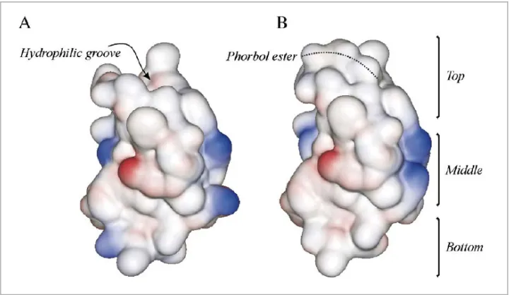
(41) Introduction and objectives. 27. Both C1 subdomains of this cPKC present a polarized distribution of hydrophobic and ionic amino acids. The top part of the molecule, where effectors like DAG/phorbol esters are bound, contains aromatic and aliphatic residues, whereas the middle part contains cationic residues (Fig I.3). The results of many studies in which the role of most important C1 domain amino acids has been resolved, suggest that typical C1 domains present a polarized amino acid distribution, being hydrophobic at the top and bottom, and containing mainly cationic residues in the middle. The hydrophilic groove in the top is the site where ligands like DAG/phorbol esters bind and lead the penetration of this part of the domain into the membrane because of the hydrophobic amino acids in this part and the cationic residues in the middle part (Zhang et al., 1995; Xu et al., 1997; Pak et al., 2001; Hurley and Meyer, 2001).. Figure I.3. Molecular surface drawing of the C1B domain of PKCδ in the absence (A) or presence (B) of phorbol ester. Positively and negatively charged regions are shown in blue and red, respectively, while the hydrophobic surface is depicted in grey. Note how the surface of the top third of the molecule is highly hydrophobic when the phorbol ester fits into the hydrophilic groove, facilitating membrane insertion of the domain under these conditions. The area occupied by the phorbol ester has been marked with a dotted line to facilitate interpretation of the figure (Taken from Corbalán García and Gómez Fernández, 2006).. The C1 domain plays a role in PKC binding to different membranes along the cell. As mentioned above, this domain is found in a large number of proteins which have several functions, and it is regulated by DAG/phorbol ester interactions (Yang and Kazanietz, 2003). A common characteristic of classical and novel PKC is that both subfamilies have two C1 domains in tandem. Several studies have demonstrated that C1 subdomains, whether inside the same or in different.
(42) 28. Chapter I. isoenzymes, are not equivalent and have different affinities for DAG and phorbol esters. Based on our knowledge to date, it can be concluded that C1A and C1B subdomains of PKC respond in different ways to DAG and phorbol esters, so that the physiological results obtained using phorbol esters are not extrapolatable to those using DAG. Likewise, different isoenzymes show different responses, so that the results for one isoenzyme will not necessarily be the same for another even if they are from the same family (Corbalán-García and Gómez-Fernández, 2006). Biologically, these differences reflect the huge variety of situations in which different PKC isoenzymes may participate. Besides DAG and phorbol esters, C1 domains can bind other lipids, such as phospholipids, ceramides and fatty acids. This part of the protein shows different affinities for these compounds, since during the interaction between them, PKC is translocated to different subcellular compartments for subsequent activation (Kashiwagi et al., 2002; Becker and Hannun, 2003; Yagi et al., 2004; Sánchez-Bautista et al., 2009). One example is the case of C1B, but not C1A domain, of PKCε which binds ceramides and arachidonic acid, although every ligand leads to a different location pattern due to the different binding mechanisms involved (Kashiwagi et al., 2002).. 2.3. C2 domain. Structure, function and regulation. The whole structure of several C2 domains from several proteins, including Synaptotagmins and PKC, has been elucidated. In PKCs, the C2 domains were initially discovered as a Ca2+ binding site of cPKC (Coussens et al., 1986; Knopf et al., 1986; Ono et al., 1986). C2 domain is present in numerous proteins involved in cellular signalling, small GTPase regulation or vessel transport (Nalefski and Falke, 1996; Rizo and Südhof, 1998), for example phospholipase C (PLC) (Rebecchi and Pentyala, 2000) or phosphoinositide 3-kinases (PI3Ks) (Walker et al., 1999). It contains approximately 130 amino acids and it, together with C1 domain, takes charge of binding PKC to membrane in a correct position. The structure of all C2-domains has a common overall fold: a single compact Greek-key motif organized as an eight-stranded anti-parallel βsandwich consisting of a pair of four-stranded β-sheets (Shao et al., 1996; Rizo and Südhof, 1998; Sutton and Sprang, 1998). Unlike C1 domain, C2 domains have a high degree of variability among themselves (Nalefski and Falke, 1996; Rizo and Südhof, 1998). The sequence of.
(43) Introduction and objectives. 29. amino acids is much conserved in β-strands and in some residues that have an essential role in this domain, like the amino acids involved in the coordination of Ca2+ ions (in cPKCs), in particular five aspartates (Fig I.4). There is great variability in the binding regions between β-strands, where residues like Pro and His are very common in these zones, which confer flexibility to them. As a result, a structural role is attributed to β-strands, while the binding regions refect the specific function of the domain (Nalefski and Falke, 1996; Rizo and Südhof, 1998).. Figure I.4. Sequence alignment of the seven C2 domains of conventional and novel PKCs. Dashes indicate gaps. The coloured residues represent those exhibiting more than 50% homology. Aromatic residues are labelled in green, hydrophobic residues are labelled in yellow, positively charged residues are labelled in dark blue, negatively charged residues are labelled in red and charged residues in light blue. Black stars indicate the critical Asp residues involved in Ca2+ coordination and green stars indicate the Lys residues located in the Lys-rich cluster in cPKCs. Purple stars indicate the residues involved in the pTyr binding domain of PKCδ. C2 domains with topology I are grouped in the top part of the alignment (PKCα, β and γ) and C2 domains with topology II are grouped in the bottom part. Note that alignment of C2 domains with topology II contains two subgroups that have been denoted due to the important structural differences observed between PKCε and PKCδ (Taken from Corbalán García and Gómez Fernández, 2006).. C2 domains can be classified in two groups (Pappa et al., 1998; Sutton and Sprang, 1998; Verdaguer et al., 1999; Ochoa et al., 2001): . The synaptotagmin-like variants, also referred to as the S-family or type I topology, which include the C2 domains of conventional PKCs..
(44) 30. . Chapter I. The PLC-like variants, also known as the P-family or type II topology, which include the C2 domains of novel PKCs.. The main difference between both topologies is that the first strand in the C2 domain with topology I occupies the structural position of the eighth βstrand in the C2-domain with topology II (Fig I.5). As a result, domains with topology II represent a circular permutation of those with topology I, and both are inter-convertible, since topology I becomes II when its amino- and carboxyl-terminal are linked and new termini are generated, cutting the conection between β1 and β2-strands (Nalefski and Falke, 1996; Rizo and Südhof, 1998).. Figure I.5. Ribbon diagrams of the C2 domain structures of PKCα (A) and PKCε (B) as representative members of topology I and II, respectively. The two different topologies result from a circular permutation of the β-strands that leaves the N- and C-termini either at the top or at the bottom of the β-sandwich, respectively. It is important to note that the membrane interaction area (CBR1 to 3 in topology I, and loops 1 to 3 in topology II) is located at the top of each domain independent of the topology exhibited (Taken from Corbalán García and Gómez Fernández, 2006).. C2 domains in classical and novel PKCs play an essential role in the activation of their proteins, since, together with C1 domain, they anchor the enzyme to the membrane due to their interaction with phospholipids. Besides anchoring PKC to the membrane, this domain has other functions, including protein-protein interactions. The classical ligands to C2 domains are Ca2+ and anionic phospholipids like PtdSer (Newton and Johnson, 1998; Verdaguer et al., 1999). Besides PtdSer, other anionic phospholipids can activate PKC in the presence of Ca2+,.
(45) Introduction and objectives. 31. such as phosphatidylglycerol, phosphatidic acid and phosphatidylinositol-4,5bisphosphate (PtdIns(4,5)P2) (Lee and Bell, 1991; Newton, 1993; Marín-Vicente, et al., 2008). Others lipids also intervene in the regulation of PKC: for instance, ceramides (Kashiwagi et al., 2002; Yakushiji et al., 2003), unsaturated fatty acids like arachidonic acid (O’Flaherty et al., 2001; López-Nicolás et al., 2006) and retinoid compounds (Randominska-Pandya et al., 2000; Boskovic et al., 2002; López-Andreo et al., 2005). In PKCα, a classical PKC isoform, the exact binding site for different lipids has been identified; for example, PtdSer interacts in the so-called Ca2+ binding site and PtdIns(4,5)P2 binds in the so-called lysine rich cluster (GarcíaGarcía et al., 2001; Ochoa et al., 2002; Corbalán-García et al., 2003; GuerreroValero et al., 2009). The C2 domain is responsible for binding Ca2+, and three different binding sites have been characterized (Ca1, Ca2 and Ca3) within the Ca2+ Binding Region. All of them are located at the top of the β-sandwich where Ca2+ ions coordination occurs, specifically in the connection loops between βstrands (Verdaguer et al., 1999; Ochoa et al., 2001). For this reason, the loops located in this part of the C2 domain are called CBR1 (Calcium binding region 1), CBR2 and CBR3, in order of the amino-terminal of the protein (Fig I.5). CBRs provide all the necessary residues involved in Ca2+ coordination: five aspartates (Asp187, 193, 246, 248 and 254), which are highly conserved among all classical PKCs (Fig I.4). Asp187 and Asp193 are located in the CBR1 (loop between β2- and β3-strands), where one of the calcium ions (Ca1) is lodged, whereas CBR3 (loop between β6- and β7-strands) includes Asp246, Asp248 and Asp254, and it is the site where Ca2 is lodged (Sutton and Sprang, 1998; Verdaguer et al., 1999) (Fig I.6). Further mutagenesis studies at the Ca2+-binding site have demonstrated that individual Ca2+ ions and their ligands play different roles in membrane binding and PKCα activation. The model suggested that Ca1 is involved in initial membrane anchoring, whereas Ca2 and Ca3 are involved in conformational changes (Edwards and Newton, 1997; Medkova and Cho, 1998; Corbalán-García et al., 1999; García-García et al., 1999; Conesa-Zamora et al., 2000; Bolsover et al., 2003). The results of all biochemical and cellular studies carry out to date suggest a sequential model for classical PKCs membrane binding and activation. In the first step, an increase in intracellular Ca2+ would result in binding of Ca1 and Ca2 when the protein is still in the cytosol, leading to membrane targeting of the enzyme through the C2 domain. There, PKC penetrates the membrane for a longer period of time, allowing the C1 domain to find the diacylglycerol generated upon receptor stimulation, finally leading.
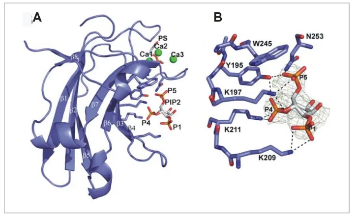
(46) 32. Chapter I. to complete activation of the enzyme (Feng et al., 2000; Conesa-Zamora et al., 2001; Nalefski and Newton, 2001; Bolsover et al., 2003).. Figure I.6. Ca2+ and phosphatidylserine-binding region of the PKCα–C2 domain. (A) Coordination scheme of the calcium ions in the structure determined for PKCα in complex with Ca2+ and DCPS. Dotted lines represent the coordination established between the different carboxylate and oxygen groups, and Ca1, Ca2 and Ca3. (B) Lateral view of the surface model of the Ca2+ binding-region, the Asp residues involved in Ca2+ coordination have been represented as thin-stick models with carbon in grey and oxygen in red. Calcium ions are represented as yellow balls. The residues of the C2 domain directly involved in phosphatidylserine binding are shown as thick-stick models with carbon in grey, nitrogen in blue and oxygen in red. Additionally, DCPS is represented as a stick model and carbons have been colored in yellow. Note how the residues coordinating Ca2+ occupy the bottom of the crevice formed by CBR1, 2 and 3. The DCPS molecule is located on top of this area contributing to coordinate Ca1 through its phosphate moiety and acts as a cap held by residues of the C2 domain that directly interact with it (N189, R216, R249 and T251) (Taken from Corbalán García and Gómez Fernández, 2006).. Further crystallization of the C2 domain of PKCα in complex with Ca2+ and 1,2-diacetyl-sn-phosphatidyl-L-serine (DAPS) demonstrated the presence of an additional binding site for anionic phospholipids in the vicinity of the conserved lysine-rich cluster in cPKCs (Ochoa et al., 2002). More specifically, it was found that Lys197 and Lys199, located in β3 strand, and Lys209 and Lys211, in β4 strand, establish a series of electrostatic interactions with a second DAPS molecule, suggesting that this site participates in the interaction of the C2 domain with the membrane (Fig.
(47) Introduction and objectives. 33. I.7). Other studies demonstrated that this area could bind other negatively charged molecules such as phosphate (Verdaguer et al., 1999), phosphatidic acid (Ochoa et al., 2002), all-trans-retinoic acid (Ochoa et al., 2003; LópezAndreo et al., 2005) or phosphoinositides like PtdIns(4,5)P2 (Marín-Vicente et al., 2008). A more extensive biochemical study using different acidic phospholipids demonstrated that the C2 domain of PKCα bind PtdIns(4,5)P2, preferentially through the β4 strand of the lysine-rich cluster (Lys209/Lys211) (Fig I.7) (Corbalán-García et al., 2003), leading to PKCα activation by means of a mechanism other than the classical one. Recent studies have demonstrated that the C2 domains of three classical PKCs exhibit different affinity for the PtdIns(4,5)P2, which is bound through the lysine-rich cluster (Sánchez-Bautista et al., 2006; Guerrero-Valero et al., 2007). PKCγ-C2 domain shows the lowest affinity for this lipid, while PKCα-C2 domain needs a lower amount of this lipid to localize in membranes (Guerrero-Valero et al., 2007). Furthermore, two aromatic amino acids (Tyr195 and Trp245) have been implicated in the specific interaction between PKCα-C2 domain and the phosphate of the inositol ring from PtdIns(4,5)P2. These residues, similar to other cationic amino acids, like Lys197, Lys209, Lys 211 and Asn253, are highly conserved among all C2 domains with topology I, while the C2 domains of topology II do not preserve most of the residues responsible for the PtdIns(4,5)P2 interaction (Guerrero-Valero et al., 2009).. Figure I.7. Localization of the Lysine-rich cluster in the C2 domain of PKCα. Overall structure of the C2 domain of PKCα represented as a cartoon scheme over its molecular surface. The lysine residues involved in the cluster, which are located in β3 and β4 strands, are represented as stick models with carbon in grey and nitrogen in blue. Acidic surfaces are represented in red and basic in blue. Note how the Ca2+-binding region is an area rich in negatively charged amino acids, while the lysine-rich cluster forms a basic surface. The inset shows an amplification of β3 and β4 strands of PKCα with the Lys residues forming the cluster represented by stick models (Taken from Corbalán García and Gómez Fernández, 2006)..
(48) 34. Chapter I. Besides anchoring PKC in membranes, C2 domain seems to be involved in protein-protein interactions, mainly through the lysine-rich cluster (MochlyRosen et al., 1992; Ron et al., 1995). It can be concluded that both PtdSer and PtdIns(4,5)P2 are very important for regulating the localization and activation of classical PKCs through C2 domain interaction, specifically through the Ca2+-binding site and the lysine-rich cluster (Bolsover et al., 2003; Landgraf et al., 2008; MarinVicente et al., 2008). In novel PKCs, the C2 domain shows some differences from classical isoforms; for example, it is the first conserved domain of amino-terminal; it presents a type II topology and it anchors to membranes in a Ca2+independent manner (García-García et al., 2001; Corbalán-García et al., 2003). This subfamily also contains an important region at the top of βsandwich, although now, the connection chains between β-strands are called loops and no Calcium Binding Regions since these isoforms do not bind Ca2+ ions. Loop 1 and loop 3 are located in this region and are formed by β-1 and β2, and β-5 and β-6 connections, respectively. Unlike the Calcium Binding Region (in cPKC), this region exhibits significant differences among novel isoforms (Fig I.4). The binding mechanisms of nPKCs differ from those of cPKCs, not only due to the absence of appropriate residues to form the Ca2+ Binding Region, but also due to the structure of the lipid docking motif formed by loops 1 and 3 (Corbalán-García et al, 2003) (Fig I.8).. Figure I.8. Residues involved in lipid binding of PKCε–C2 domain. The cartoon shows the 3D structure of PKCε. The critical residues involved in phospholipid binding have been represented as stick models with carbon in grey, nitrogen in blue and oxygen in red (Trp23, Arg26 and Arg32 in loop1 and Ile89 in loop 3). Additional residues not involved in lipid binding are also shown: side chains of Asp86 and Asp92 are represented as stick models; observe how these chains point to the surface of the molecule, in the opposite direction to the side chains of the residues involved in phospholipid binding (Taken from Corbalán García and Gómez Fernández, 2006)..
(49) Introduction and objectives. 35. 2.4. Catalytic domain. Structure and regulation. The members of the ABC kinases family, PKA, PKB/Akt and PKC, show 40% sequence homology in their catalytic domain. Among PKC this percentage increases to 60% (Fig I.9) (Newton, 2003; Corbalán-García and GómezFernández, 2006).. Figure I.9. Alignment of the activation loop, turn motif and hydrophobic motif phosphorylation sequences for the PKC isoenzymes, PKBα/Akt1, p70S6 kinase, PRK2 and PKAα. Sequences shown are for human PKC isoenzymes α, ε, ζ, η/L θ and ι/λ and rat PKC γ and δ, rat PKC βI and βII, murine PKB α/Akt1, rat p70S6 kinase and murine PKA α. Amino acid residue numbers are indicated to the left of the sequences (Taken from Newton, 2003).. To date, the 3D structures of catalytic domain of some PKC have been solved: novel PKCθ (Xu et al., 2004), atypical PKCι (Messercschmidt et al., 2005; Takimura et al., 2010), classical PKCβII (Grodsky et al., 2006) and PKCα (Wagner et al., 2009). The catalytic domain of PKCs contains both the ATP binding and the consensus phosphorylation sites; it interacts with substrates and is responsible for phosphotransfer activity. The kinase domains of PKC isoenzymes are closely related, as illustrated in the dendrogram (Fig I.10). Protein kinase fold is separated into two subdomains or lobes. The smaller N-terminal lobe, or N lobe, is composed of a five-stranded β sheet and one prominent α helix, called helix αC. The larger lobe is called the C lobe and is predominantly helical (Fig I.10) (Taylor and Radzio-Andzelm, 1994; Johnson and Lewis, 2001). Between them, there is a cleft where the ATP- and substrate-binding sites are located. N lobe has a preserved region in all kinases, called the glycine-rich loop, whose consensus sequence is XGXGX2GX16KX, where X is any amino acid and G is glycine. This region is located between β1 and β2 strands and it is.
Figure


+7


Documento similar
ΔCm, mitochondrial membrane potential; AMPK, AMP-activated protein kinase; ERK 1/2, extracellular signal-regulated kinase 1/2; IF1, ATPase inhibitory factor 1; IF1 KO , IF1
La campaña ha consistido en la revisión del etiquetado e instrucciones de uso de todos los ter- mómetros digitales comunicados, así como de la documentación técnica adicional de
You may wish to take a note of your Organisation ID, which, in addition to the organisation name, can be used to search for an organisation you will need to affiliate with when you
Where possible, the EU IG and more specifically the data fields and associated business rules present in Chapter 2 –Data elements for the electronic submission of information
The 'On-boarding of users to Substance, Product, Organisation and Referentials (SPOR) data services' document must be considered the reference guidance, as this document includes the
In medicinal products containing more than one manufactured item (e.g., contraceptive having different strengths and fixed dose combination as part of the same medicinal
Products Management Services (PMS) - Implementation of International Organization for Standardization (ISO) standards for the identification of medicinal products (IDMP) in
Products Management Services (PMS) - Implementation of International Organization for Standardization (ISO) standards for the identification of medicinal products (IDMP) in
