Association between Low
Testosterone Levels and Sarcopenia in Cirrhosis:
A Cross-sectional Study
Carlos Moctezuma-Velázquez,* Gavin Low,† Marina Mourtzakis,‡ Mang Ma,* Kelly W. Burak,§ Puneeta Tandon,*,|| Aldo J. Montano-Loza*
* Liver Unit, University of Alberta Hospital, Edmonton, Alberta, Canada.
† Department of Radiology, University of Alberta Hospital, Edmonton, Alberta, Canada. ‡ Department of Rehabilitation Medicine, University of Waterloo, Ontario, Canada. § Liver Unit, University of Calgary, Calgary, Canada. || Cirrhosis Care Clinic, University of Alberta Hospital, Edmonton, Alberta, Canada.
July-August, Vol. 17 No. 4, 2018: 615-623
INTRODUCTION
Sarcopenia is one well-known complication of cirrho-sis, it can be present in up to 45% of patients, and its preva-lence increases as liver disease progresses.1 Myosteatosis,
on the other hand, implies an increase in fatty infiltration within the skeletal muscle and is related to muscle quality. Both sarcopenia and myosteatosis, are independent pre-dictors of mortality in this population.2,3
Some of the most important contributors to muscle wasting in cirrhosis are metabolic abnormalities, insuffi-cient intake, malabsorption, impaired liver function to metabolize and endocrine abnormalities.4 Of the latter
The Official Journal of the Mexican Association of Hepatology, the Latin-American Association for Study of the Liver and
the Canadian Association for the Study of the Liver
Manuscript received: Manuscript received:Manuscript received:
Manuscript received:Manuscript received: August 02, 2017. Manuscript accepted:Manuscript accepted:Manuscript accepted:Manuscript accepted:Manuscript accepted: October 16, 2017.
DOI: 10.5604/01.3001.0012.0930
A B S T R A C T A B S T R A C T A B S T R A C T A B S T R A C T A B S T R A C T
Introduction and aim. Introduction and aim.Introduction and aim. Introduction and aim.
Introduction and aim. Sarcopenia is an independent predictor of mortality in cirrhosis. Hypogonadism is common in cirrhosis and has been associated with sarcopenia in non-cirrhotic chronic liver disease populations. The aim of this study is to investigate if sar-copenia is associated with low testosterone levels in patients with cirrhosis. Material and methods. Material and methods. Material and methods. Material and methods. Material and methods. This is a retrospective anal-ysis of prospectively collected data of 211 cirrhotic patients undergoing evaluation for liver transplantation. Sarcopenia was defined by computed tomography (CT) scan using specific cutoffs of the 3rd lumbar vertebra skeletal muscle index (L3 SMI). Morning testo-sterone levels were obtained in all patients. Results.Results.Results.Results.Results. Of the 211 patients, sarcopenia was noted in 94 (45%). Testosterone levels were lower in sarcopenic patients (10.7 ± 1.1 vs. 13.7 ± 1.4 nmol/L, p = 0.03) and hypotestosteronemia was more frequent in them too (34 vs. 16%, p = 0.004). In males, those with sarcopenia had lower testosterone levels (14.6 ± 1.4 vs. 21.9 ± 1.8, p = 0.002), and the corresponding frequency of hypotestosteronemia (42 vs. 19%, p = 0.006) was also higher. There were no significant differ-ences in female patients. There was a weak correlation between L3 SMI and testosterone levels (r 0.37, p < 0.001). On multivariable regression analysis including sex, body mass index (BMI), hypotestosteronemia, MELD and etiology of cirrhosis, only hypotestoster-onemia (RR 2.76, p = 0.005) and BMI (RR 0.88, p < 0.001) were independently associated with sarcopenia. Conclusion.Conclusion.Conclusion.Conclusion.Conclusion. Low tes-tosterone levels are associated with sarcopenia in male cirrhotic patients. The potential therapeutic effect of testes-tosterone to reverse sarcopenia in these patients warrants evaluation in future trials.
Key words. Key words.Key words. Key words.
Key words. (MeSH). Malnutrition. End-stage liver disease. Hypogonadism. Muscular atrophy.
factors, low testosterone levels have been associated with sarcopenia in several chronic disease populations, includ-ing renal failure, heart failure, and obstructive pulmonary disease. The pharmacologic supplementation of this hor-mone has been shown to have anabolic effects on muscle mass in different populations.5,6 The relation between
tes-tosterone and myosteatosis has not been studied as thor-oughly as that with sarcopenia, but in patients with prostate cancer, androgen deprivation therapy results in muscle fatty infiltration.7
androgens and gonadal failure.8,9 In female patients with
cirrhosis, fat depletion is likely to occur before a signifi-cant loss of muscle mass.4 Low testosterone levels are an
independent predictor of mortality in cirrhosis.10,11 To the
best of our knowledge, there have been no studies in pa-tients with cirrhosis which have addressed the association between testosterone levels, sarcopenia, and myosteatosis, as determined by cross-sectional imaging, the current gold standard for estimating muscle mass.12
A clear association between these parameters would add to the evidence to consider testosterone as a therapeutic option for muscle wasting in cirrhosis. Accordingly, we aimed to investigate if sarcopenia and/or myosteatosis were associated with low testosterone levels in patients with cirrhosis.
MATERIAL AND METHODS
Study population
Two hundred and eleven patients were included in our retrospective analysis of prospectively collected data. Eligible subjects were cirrhotic patients who were seen in the liver units of the University of Alberta Hospital and the Foothills Medical Centre to assess their candida-cy for liver transplantation between February 2011 and August 2012. The variables of interest were prospectively collected. Inclusion criteria were age between 18 to 80 years old and a diagnosis of cirrhosis confirmed by either histological (defined as presence of nodules of regenera-tion), radiological (ultrasound or cross-sectional imaging showing a lobulated liver and/or unequivocal signs of portal hypertension) or transient elastography (defined as liver stiffness ≥ 14 kPa) assessment. We excluded pa-tients on any kind of hormonal replacement therapy, with extra-hepatic malignancy, end-stage renal disease on dialysis, chronic pulmonary disease requiring oxygen supplementation, or congestive heart failure with an ejection fraction < 40%. The study protocol was ap-proved a priori by the institutional review board prior to data collection.
Muscularity, sarcopenia, and myosteatosis assessment
Computed tomography (CT) scans used for analysis were performed as part of the liver transplant evaluation. A transverse CT image from the third lumbar vertebra (L3) in the inferior direction was assessed from each scan. Images were analyzed with SliceOmatic V4.3 soft-ware (Tomovision, Montreal, Quebec, Canada), which enables specific tissue demarcation by using previously reported Hounsfield unit (HU) thresholds. Using this
technique, skeletal muscle is identified and quantified by HU thresholds of -29 to +150. Muscles in the L3 region encompass psoas, erector spinae, quadratus lumborum, transversus abdominis, external and internal obliques, and rectus abdominis. The following HU thresholds were used for adipose tis-sues: -190 to -30 for subcutaneous and intermuscular adi-pose tissues, and -150 to -50 for visceral adiadi-pose tissues. Cross-sectional areas (cm2) were automatically
comput-ed by summing tissue pixels and multiplying by pixel surface area. All CT images were analyzed by 2 trained observers. Cross-sectional area of muscle and adipose tissue were normalized for stature (cm2/m2). The
L3-SMI was expressed as cross-sectional muscle area/height. Sarcopenia was defined according to the following cut-offs: L3 Skeletal muscle index (L3 SMI): ≤ 41 cm2/m2 for
women and ≤ 53 cm2/m2 for men with body mass index
(BMI) ≥ 25 kg/m2 and ≤ 43 cm2/m2 in all patients with a
BMI < 25 kg/m2.3 Muscle attenuation analysis was
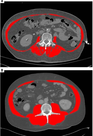
per-Figure 1. Figure 1.Figure 1.
Figure 1.Figure 1. Sarcopenia determined by cross-sectional imaging. Patient of fig-ure 1A had sarcopenia, with a BMI 32 kg/m2 and L3 SMI of 50 cm2/m2.
Pa-tient of figure 2B did not have sarcopenia, with the same BMI of 32 kg/m2
but a L3 SMI of 70 cm2/m2.
Statistical analyses
Continuous data were described using means and standard deviations; for categorical data we used absolute and relative frequencies. For bivariate analysis, for contin-uous and categorical variables we used the χ2 test and the
unpaired t test respectively. The Pearson’s correlation co-efficient was used to evaluate the correlation between tes-tosterone levels and L3 SMI. For multiple logistic regression analysis, we considered sarcopenia as the de-pendent variable. Biochemical parameters (sodium, creat-inine, albumin, INR, testosterone levels), age, etiology of cirrhosis, and sex, were entered as independent variables. Variables of interest plus variables with a p-value < 0.1 in univariate analysis were included in multivariable regres-sion analysis. We also used logistic regresregres-sion to evaluate the association between myosteatosis, as a dependent vari-able, testosterone, and sarcopenia. Statistical analysis was performed using SPSS 22 (SPSS, Inc., Chicago, IL).
RESULTS
Clinical and Biochemical Features of Patients Evaluated
Of the 211 patients, 128 patients were male (61%), and the mean age was 57 ± 1 years. Cirrhosis was caused by hepatitis C virus infection (HCV) in 74 (35%), alcohol in 54 (26%), non-alcoholic fatty liver disease (NAFLD) / cryptogenic in 53 (25%), autoimmune liver disease in 18 (9%), and other etiology in 11 patients (5%).
formed in 87 patients (41%). Myosteatosis was defined according to cut-off values for muscle attenuation that have been associated with mortality, specifically: < 41 HU in patients with a BMI up to 24.9, and < 33 in those with a BMI ≥ 25.13
As an example of the L3 SMI assessment, figures 1A and 1B illustrate two cirrhotic patients with similar BMI (26 kg/m2). Abdominal CT images in horizontal plane
are images taken at L3, the red color indicates skeletal muscles. CT at the left (1A) is from a male patient with sarcopenia with L3 SMI 38 cm2/m2, and the CT at the
right (1B) is from a non-sarcopenic male with a L3 SMI of 56 cm2/m2.
Clinical and laboratory assessment
We collected the following information: morning tes-tosterone levels, age, gender, etiology of cirrhosis, and liv-er tests. Testostliv-erone levels wliv-ere reported in nmol/L (normal males: 10.3-29.5; females: 0.6-2.0) with values be-low the be-lower limit of normally, considered as hypotesto-steronemia. Testosterone in all samples was measured with an electrochemiluminescence immunoassay (ECLIA). Standard commercial kits (Elecsys® Testoster-one II, Roche Diagnostics, Mannheim, Germany) were used and the mean ± SD of the testosterone levels (ng mL-1) were reported. The sensitivity of the assay was 0.04
ng mL-1 for testosterone. The clinical and biochemical
pa-rameters were obtained within 1 week from the index CT scan used to determine the L3 SMI.
Table 1. Features associated with sarcopenia in patients with cirrhosis.
Features All patients No sarcopenia Sarcopenia P-value
(n = 210) (n = 116) (n = 94)
Age (years) 57±1 57± 1 58± 1 0.5
Gender (M: F) 128 : 82 6 : 47 59 : 35 0.7
BMI (kg / m2) 27±0.5 29± 1 25± 0.5 < 0.001
L3 SMI (cm2 / m2) 48±1 54± 1 41± 1 < 0.001
Muscle attenuation (HU, n = 87) 32±1 30± 1 33± 1 0.02
Myosteatosis (n = 87) 55 / 87 (62%) 28 / 53 (53%) 27 / 34 (79%) 0.01
MELD (points) 12±0.5 11± 0.5 12± 1 0.1
Child-Pugh (Class A, B, C) 73: 102: 35 38: 63: 15 35: 39: 20 0.1
Etiology of cirrhosis
Alcohol 54 (26%) 29 (25%) 25 (27%) 0.9
HCV 74 (35%) 42 (36%) 32 (34%) 0.8
AILDa 18 (9%) 7 (6%) 11 (12%) 0.2
NASH-Cryptogenic 53 (25%) 36 (31%) 17 (18%) 0.04
Othersb 3 (1%) 2 (2%) 9 (10%) 0.01
AILD: autoimmune liver disease. BMI: body mass index. L3 SMI: lumbar 3rd skeletal muscle index. HCV: hepatitis C virus. MELD: model for end stage liver disease. NASH: non-alcoholic steatohepatitis. aIncludes autoimmune hepatitis, primary biliary cirrhosis and primary sclerosing cholangitis. bIncludes
The mean testosterone level in males was 18.5 ± 1.2 nmol/L, and in females 1.5 ± 0.1 nmol/L. Hypotestoster-onemia was present in 51 (24%) patients. Thirty-eight male patients (30%), and 13 female patients (16%) had levels be-low the reference threshold for our laboratory (p = 0.03).
Clinical and biochemical features overall and according to the presence of sarcopenia are shown in table 1.
Frequency and features associated with sarcopenia
Mean L3 SMI was 48 ± 1 cm2/m2 and was higher in
male than in female patients (52 ± 1 vs. 43 ± 1 cm2/m2,
p < 0.001). Sarcopenia was noted in 94 patients (45%): 59 male (46%) and 35 female (43%) patients (p = 0.7). Patients with sarcopenia had lower BMI (25 ± 0.5 vs. 29.1
± 1 kg/m2, p < 0.001). In terms of the etiology of
cirrho-sis, the prevalence of NAFLD/cryptogenic was lower in patients with sarcopenia (18 vs. 31%, p = 0.04).
Hypotestosteronemia was more prevalent in sarcopenic patients (34 vs. 16%, p = 0.004) (Table 1).
Sexual dimorphism in sarcopenia and hypotestosteronemia
Male sarcopenic patients had a lower BMI (26 ± 1 vs. 28 ± 1 kg/m2, p = 0.04) and lower testosterone levels
when compared to non-sarcopenic patients (14.6 ± 1.4 vs. 21.9 ± 1.8 nmol/L, p = 0.002) (Table 2). Also, the preva-lence of hypotestosteronemia (42 vs. 19%, p=0.006) was higher in sarcopenic than non-sarcopenic patients (Table 2; Figure 2A).
Table 2. Features associated with sarcopenia stratified by gender – univariate regression analysis.
Features - Male patients All patients (n = 128) No sarcopenia (n = 69) Sarcopenia (n = 59) P-value
Age (years) 57± 1 57±1 58±1 0.5
BMI (kg/m2) 27± 0.5 28±1 26±1 0.04
L3 SMI (cm2/m2) 52± 1 57± 1 45±1 < 0.001
Muscle Attenuation (HU, n=59) 33± 1 36±1 29±1 0.003
Myosteatosis (n=59) 33 / 59 (56%) 15/36 (42%) 18 / 23 (78%) 0.008
Testosterone (nl, 10.3-29.5 nmol/L) 18.5± 1.2 21.9±1.8 14.6±1.4 0.002
Hypotestosteronemia 38 (30%) 13 (19%) 25 (42%) 0.006
MELD (points) 12± 1 11±1 13±1 0.2
Child-Pugh (Class A, B, C) 42: 63: 23 21: 39: 9 21: 24: 14 0.1
Etiology of Cirrhosis
Alcohol 40 (31%) 17 (29%) 23 (33%) 0.7
HCV 53 (41%) 29 (42%) 24 (41%) 1.0
AILDa 3 (5%) 0 3 (5%) 0.1
NASH-Cryptogenic 23 (18%) 15 (22%) 8 (14%) 0.3
Othersb 9 (7%) 2 (3%) 7 (12%) 0.048
Features – Female patients All patients (n = 82) No sarcopenia (n = 47) Sarcopenia (n = 35) P-value
Age (years) 58± 1 60±2 56±1 0.1
BMI (kg / m2) 28± 1 31±1 24±1 < 0.001
L3 SMI (cm2 / m2) 43± 1 48±1 36±1 < 0.001
Muscle attenuation (HU, n = 28) 29± 1 30±3 29±2 0.7
Myosteatosis (n = 28) 22/28 (75%) 13/17 (71%) 9/11 (82%) 0.7
Testosterone (nl, 0.6-2.0 nmol/L) 1.5± 0.1 1.6±0.2 1.4±0.2 0.4
Hypotestosteronemia 13 (16%) 6 (13%) 7 (20%) 0.5
MELD (points) 11± 0.5 11±1 10±1 0.7
Child-Pugh (class A, B, C) 31: 39: 12 17: 24: 6 14: 15: 6 0.5
Etiology of cirrhosis
Alcohol 14 (17%) 6 (13%) 8 (23%) 0.3
HCV 21 (26%) 13 (28%) 8 (23%) 0.8
AILD a 15 (18%) 7 (15%) 8 (23%) 0.4
NASH-Cryptogenic 30 (37%) 21 (45%) 9 (26%) 0.1
Others b 2 (2%) 0 2 (6%)
-AILD: autoimmune liver disease. BMI: body mass index. L3 SMI: lumbar 3rd skeletal muscle index. HCV: hepatitis C virus. MELD: model for end stage liver disease. NASH: non-alcoholic steatohepatitis. aIncludes autoimmune hepatitis, primary biliary cirrhosis and primary sclerosing cholangitis. bIncludes
No sarcopenia Sarcopenia Male patients
Testosterone level (nmol/L)
65 60 55 50 45 40 35 30 35 20 15 10 5
0 No sarcopenia Sarcopenia
Female patients 5
4
3
2
1
0
P = 0.002 P = 0.4
Figure 2. Figure 2.Figure 2.
Figure 2.Figure 2. Comparison of testosterone levels between patients with and without sarcopenia, by gender.
There were no significant differences in testosterone levels between female sarcopenic and non-sarcopenic patients (1.6 ± 0.2 vs. 1.4 ± 0.2 nmol/L, p = 0.4). Also, there was no significant difference in the frequency of hy-potestosteronemia among sarcopenic and non-sarcopenic female patients (20 vs. 13%, p = 0.5) (Table 2; Figure 2B).
Correlations and
factors associated with sarcopenia by multivariable regression analysis
There was a significant but moderate correlation be-tween the L3 SMI and testosterone levels (r = 0.37, p < 0.001. The correlation was still significant, but weaker, in male patients (r = 0.18, p = 0.04), and was not significant in female patients (r = 0.17, p = 0.1) (Figure 3).
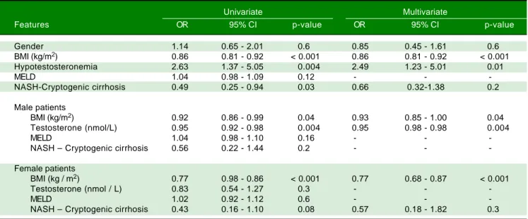
By multivariable regression analysis including sex, BMI, hypotestosteronemia, MELD score, and etiology of the cirrhosis, only hypotestosteronemia (RR = 2.76, p = 0.005) and BMI (RR = 0.88, P < 0.001 were independent-ly associated with sarcopenia. These results were replicat-ed in a model that includreplicat-ed only male patients, whereas in female patients only BMI was associated with sarcopenia (Table 3).
Analysis of myosteatosis
The attenuation index for determination of myosteato-sis was available in 87 patients; of these, 55 (63%) had myo-steatosis. It was more commonly identified in sarcopenic than in non-sarcopenic patients (79% vs. 53%, p = 0.01). Although myosteatosis was more common in male pa-Figure 3.
Figure 3.Figure 3.
Figure 3.Figure 3. Scatter plot of L3 skeletal muscle index against testosterone levels in male (AAAAA) and female (BBBBB) patients.
A AA AA
L3 SMI cm
2 / m
2
80
70
60
50
40
30
0 20 40 60 80
Testosterone level (nmol/L)
No sarcopenia Sarcopenia
r = 0.18, p = 0.04 B BB B B
L3 SMI cm
2 / m
2
70
60
50
40
30
20
0 1 2 3 4 5
Testosterone level (nmol/L)
r = 0.17, p = 0.1 No sarcopenia Sarcopenia
tients with sarcopenia (78 vs. 42%, p = 0.008), this was not the case in female patients with sarcopenia (82% vs. 71%, p = 0.7). By multiple regression analysis, myosteatosis was associated with sarcopenia (RR 2.85, p = 0.046), but not with hypotestosteronemia (RR 2.18, p = 0.1).
DISCUSSION
In this study, we demonstrate an association between testosterone levels and sarcopenia. This association was independent of well-known factors that contribute to tes-tosterone levels, such as age, the stage of liver disease, and the etiology of cirrhosis.
Sarcopenia in cirrhosis is an independent predictor of mortality before transplantation2 and morbidity after liver
transplantation.14 Currently, cross-sectional imaging
stud-ies, including CT scan or MRI, are the gold standard tools to quantify skeletal muscle mass and hence constitute a good resource for objective, detailed and reproducible nu-tritional assessment of patients.12 A meta-analysis
evaluat-ing the impact of CT-assessed sarcopenia includevaluat-ing 19 studies, with more than 3,000 patients, showed an inde-pendent association between sarcopenia and waiting list and post-liver transplant mortality.15 More recently, a
mul-ticenter study across five academic transplant centers in North America including almost 400 adult patients with cirrhosis listed for liver transplant validated the associa-tion of sarcopenia and mortality and established similar cutoff values in cirrhotic patients awaiting liver transplan-tation using L3 SMI.16
Hypotestosteronemia is one of several mechanisms im-plicated in sarcopenia in cirrhosis.1 We found testosterone
levels were significantly lower in male patients with sarco-penia, but the correlation between L3-SMI and testoster-one levels was weak, suggesting that there are many factors implicated in sarcopenia and that the degree of hypotesto-steronemia is not intimately associated with the degree of muscle wasting. These findings are consistent with the re-sults of recent studies by Sinclair et al. that demonstrated also a weak, but significant correlation between testoster-one levels and muscle mass, and that low testostertestoster-one levels were associated with higher mortality in male patients with cirrhosis.11,17 We did not find this association in females.
The absolute difference between testosterone levels in sar-copenic and non-sarsar-copenic females was small (0.2 vs. 7.3 nmol/L in males). Moreover, as only 39% of our sample was comprised of women and only 16% of them had hypotesto-steronemia, it is possible that our study was underpowered to evaluate this association in female patients. Studies in postmenopausal women have shown that low testosterone levels are associated with a lower muscle mass.18 In
addi-tion to this, we used an immunoassay to determine testo-sterone levels, which is known to have reduced sensitivity in case of female patients, who usually have much lower concentrations when compared to males, further limiting the interpretation of the results found in women. The asso-ciation between sarcopenia and hypotestosteronemia in women requires evaluation in future studies.
Sarcopenia in liver cirrhosis is multifactorial; hence, it is improbable that a single treatment will be able to reverse it.1
Treatment approaches so far have focused on nutritional supplementation, replacing calories and proteins by differ-ent routes of administration, with limited benefit, suggest-ing that cirrhosis is a state of anabolic resistance. Exercise Table 3. Features associated with sarcopenia by multivariable regression analysis.
Univariate Multivariate
Features OR 95% CI p-value OR 95% CI p-value
Gender 1.14 0.65 - 2.01 0.6 0.85 0.45 - 1.61 0.6
BMI (kg/m2) 0.86 0.81 - 0.92 < 0.001 0.86 0.81 - 0.92 < 0.001
Hypotestosteronemia 2.63 1.37 - 5.05 0.004 2.49 1.23 - 5.01 0.01
MELD 1.04 0.98 - 1.09 0.12 - -
-NASH-Cryptogenic cirrhosis 0.49 0.25 - 0.94 0.03 0.66 0.32-1.38 0.2
Male patients
BMI (kg/m2) 0.92 0.86 - 0.99 0.04 0.93 0.85 - 1.00 0.04
Testosterone (nmol/L) 0.95 0.92 - 0.98 0.004 0.95 0.98 - 0.98 0.004
MELD 1.04 0.98 - 1.10 0.16 - -
-NASH – Cryptogenic cirrhosis 0.56 0.22 - 1.44 0.2 - -
-Female patients
BMI (kg / m2) 0.77 0.98 - 0.86 < 0.001 0.77 0.68 - 0.87 < 0.001
Testosterone (nmol / L) 0.83 0.54 - 1.27 0.3 - -
-MELD 1.02 0.92 - 1.12 0.6 - -
-NASH – Cryptogenic cirrhosis 0.43 0.16 - 1.10 0.08 0.57 0.18 - 1.82 0.3
and physical activity may be of benefit but are limited by fa-tigue in this population.19,20 The significant association
be-tween sarcopenia and testosterone and the data from recent supplementation trials in cirrhosis21, 22 indicate that
replace-ment therapy may be a potential therapeutic strategy to im-prove muscle mass. Additionally, testosterone has the advantage of improving bone mineral density, which could be of much benefit in patients with cirrhosis, who have an increased risk of fractures.23 Future testosterone
replace-ment studies in cirrhosis will be needed to confirm the ef-fect of supplementation not only on muscle mass but also on muscle quality, to assess whether there may also be a role for testosterone supplementation in female patients, and to study the safety of such intervention. There are sev-eral potential adverse effects that have been associated with testosterone supplementation. Observational studies have linked testosterone levels with hepatocellular carcinoma, but results have been inconsistent.24-26 There were no
hepa-tocellular carcinoma cases in the clinical trials mentioned earlier, but these were small studies probably insufficiently powered to study that outcome.21,22 When using
testoster-one replacement therapy to increase btestoster-one mineral density, clinical guidelines on the management of osteoporosis in cirrhosis recommend discussing risks/benefits with the pa-tient before starting therapy.27 Testosterone replacement is
not recommended in patients with prostate cancer, erythro-cytosis, hyperviscosity, sleep apnea, or heart failure.18,28,29 In
addition to this, as suggested by the European Working Group on Sarcopenia in Older People (EWGSOP),30 it may
be inappropriate to define sarcopenia only considering mass, as there is a nonlinear correlation between muscle mass and function, so therapies exclusively focused on mus-cle mass may turn to be ineffective unless some strategies are considered to improve function as well.1,4
Myosteatosis has been recognized as an independent predictor of long-term mortality in patients with liver cir-rhosis.3 There is fewer data about the association between
fatty infiltration of the muscle and testosterone. In patients with prostate cancer, androgen deprivation therapy has been associated with myosteatosis.7 Despite
hypotesto-steronemia being more frequent in patients with myostea-tosis, the difference was not statistically significant. Of note, we could assess muscle quality in a limited number of patients, so the interpretation of this result must be done with caution, as a type 2 error cannot be ruled out. The association between testosterone and myosteatosis and the role of testosterone replacement in improving muscle quality in this population is largely unknown and will need to be assessed in future trials.
Our study has both strengths and limitations. To our knowledge, this is the first study that has evaluated the as-sociation between testosterone levels and sarcopenia in both male and female patients with cirrhosis, and also the
first to assess the relation between myosteatosis and testo-sterone. Moreover, muscle mass was determined by cross-sectional imaging, which is currently considered by some as the gold standard in this population.12 Although
we have shown that there is a significant association be-tween testosterone levels and sarcopenia in male patients, due to the lack of a randomized interventional study de-sign we are unable to draw conclusions about causality. We also did not determine other hormone levels which may play a role in this association such as estrone, estradi-ol, prolactin and, free testosterone. In fact, results from other studies have been inconsistent about whether free testosterone or total testosterone should be measured in these patients and some studies suggest that estrone and the testosterone/estradiol might also have clinical and prognostic implications. Patients with compensated cir-rhosis have higher levels of the sex hormone binding glob-ulin when compared with healthy controls, while patients that are decompensated have low levels, suggesting free testosterone may be a more reliable marker. However, the equations that are normally used to indirectly calculate free testosterone, have not been validated in patients with cirrhosis.21,24,31 In the case of women patients, because of
their lower testosterone levels, the sensitivity of immu-noassays is compromised and some authors consider it completely unreliable. However, the assay that we used has been shown to have good agreement with liquid chro-matography – mass spectrometry.32 Another limitation is
that we could only retrieve the information about myost-eatosis in 41% of the patients, which may limit the inter-pretation of this result.
In conclusion, low testosterone levels are associated with sarcopenia in male patients with cirrhosis. Testoster-one replacement therapy might be a potential therapeutic strategy to improve muscle mass in male cirrhotic pa-tients, and its role in improving muscle quality remains to be defined. The safety and efficacy of this treatment re-quire further prospective evaluation.
ABBREVIATIONS
• BMI: body mass index. • CT: computed tomography. • HBV: hepatitis B virus. • HCV: hepatitis C virus. • HU: hounsfield unit. • L3: third lumbar vertebra.
• L3 SMI: L3 skeletal muscle index. • NAFLD: non-alcoholic fatty liver disease.
CONFLICT OF INTEREST
GRANTS
Carlos Moctezuma-Velazquez received a grant from Gilead Canada to complete his hepatology fellowship.
ACKNOWLEDGMENTS
Presented in part at the International Liver Congress of the European Association for the Study of Liver Diseases, April 24, 2015; Vienna, Austria.
REFERENCES
1. Dasarathy S, Merli M. Sarcopenia from mechanism to diagno-sis and treatment in liver disease. J Hepatol 2016; 65: 1232-44.
2. Montano-Loza AJ, Meza-Junco J, Prado CM, Lieffers JR, Baracos VE, Bain VG, Sawyer MB. Muscle wasting is asso-ciated with mortality in patients with cirrhosis. Clin
Gastro-enterol Hepatol 2012; 10: 166-73.
3. Montano-Loza AJ, Angulo P, Meza-Junco J, Prado CM, Saw-yer MB, Beaumont C, Esfandiari N, et al. Sarcopenic obesity and myosteatosis are associated with higher mortality in pa-tients with cirrhosis. J Cachexia Sarcopenia Muscle 2016; 7: 126-35.
4. Montano-Loza AJ. Clinical relevance of sarcopenia in patients with cirrhosis. World J Gastroenterol 2014; 20: 8061-71.
5. Basualto-Alarcon C, Varela D, Duran J, Maass R, Estrada M. Sarcopenia and androgens: a link between pathology and treatment. Front Endocrinol 2014; 5: 217.
6. von Haehling S. The wasting continuum in heart failure: from sarcopenia to cachexia. Proc Nutr Soc 2015; 74: 367-77. 7. Chang D, Joseph DJ, Ebert MA, Galvao DA, Taaffe DR,
Den-ham JW, Newton RU, et al. Effect of androgen deprivation therapy on muscle attenuation in men with prostate cancer. J
Med Imaging Radiat Oncol 2014; 58: 223-8.
8. Gluud C. Serum testosterone concentrations in men with al-coholic cirrhosis: background for variation. Metabolism
1987; 36: 373-8.
9. Van Thiel DH, Kumar S, Gavaler JS, Tarter RE. Effect of liver transplantation on the hypothalamic-pituitary-gonadal axis of chronic alcoholic men with advanced liver disease. Alcohol
Clin Exp Res 1990; 14: 478-81.
10. Sinclair M, Grossmann M, Gow PJ, Angus PW. Testosterone in men with advanced liver disease: abnormalities and impli-cations. J Gastroenterol Hepatol 2015; 30: 244-51.
11. Sinclair M, Gow PJ, Grossmann M, Shannon A, Hoermann R, Angus PW. Low serum testosterone is associated with ad-verse outcome in men with cirrhosis independent of the model for end-stage liver disease score. Liver Transpl
2016; 22: 1482-90.
12. Giusto M, Lattanzi B, Albanese C, Galtieri A, Farcomeni A, Giannelli V, Lucidi C, et al. Sarcopenia in liver cirrhosis: the role of computed tomography scan for the assessment of muscle mass compared with dual-energy X-ray absorptiom-etry and anthropomabsorptiom-etry. Eur J Gastroenterol Hepatol 2015; 27: 328-34.
13. Martin L, Birdsell L, Macdonald N, Reiman T, Clandinin MT, McCargar LJ, Murphy R, et al. Cancer cachexia in the age of obesity: skeletal muscle depletion is a powerful prognostic factor, independent of body mass index. J Clin Oncol 2013; 31: 1539-47.
14. Montano-Loza AJ, Meza-Junco J, Baracos VE, Prado CM, Ma M, Meeberg G, Beaumont C, et al. Severe muscle deple-tion predicts postoperative length of stay but is not associ-ated with survival after liver transplantation. Liver Transpl
2014; 20: 640-8.
15. van Vugt JL, Levolger S, de Bruin RW, van Rosmalen J, Metselaar HJ, JN IJ. Systematic Review and Meta-Analysis of the Impact of Computed Tomography-Assessed Skeletal Muscle Mass on Outcome in Patients Awaiting or Under-going Liver Transplantation. Am J Transplant 2016; 16: 2277-92.
16. Carey EJ, Lai JC, Wang CW, Dasarathy S, Lobach I, Mon-tano-Loza AJ, Dunn MA. A multi-center study to define sar-copenia in patients with end-stage liver disease: From the Fitness, Life Enhancement, and Exercise in Liver Transplan-tation (FLEXIT) Consortium. Liver Transpl 2017; 23: 625-33. 17. Sinclair M, Grossmann M, Angus PW, Hoermann R, Hey P,
Scodellaro T, Gow PJ. Low testosterone as a better predic-tor of mortality than sarcopenia in men with advanced liver disease. J Gastroenterol Hepatol 2016; 31: 661-7.
18. Zanandrea V, Giua R, Costanzo L, Vellas B, Zamboni M, Ce-sari M. Interventions against sarcopenia in older persons.
Curr Pharm Des 2014; 20: 5983-6006.
19. Dasarathy S. Cause and management of muscle wasting in chronic liver disease. Curr Opin Gastroenterol 2016; 32: 159-65.
20. Zenith L, Meena N, Ramadi A, Yavari M, Harvey A, Carbon-neau M, Ma M, et al. Eight weeks of exercise training in-creases aerobic capacity and muscle mass and reduces fatigue in patients with cirrhosis. Clin Gastroenterol Hepatol
2014; 12: 1920-6 e1922.
21. Sinclair M, Grossmann M, Hoermann R, Angus PW, Gow PJ. Testosterone therapy increases muscle mass in men with cirrhosis and low testosterone: A randomised controlled
tri-al. J Hepatol 2016; 65: 906-13.
22. Yurci A, Yucesoy M, Unluhizarci K, Torun E, Gursoy S, Baskol M, Guven K, et al. Effects of testosterone gel treat-ment in hypogonadal men with liver cirrhosis. Clin Res
Hepatol Gastroenterol 2011; 35: 845-54.
23. Santos LA, Romeiro FG. Diagnosis and Management of Cir-rhosis-Related Osteoporosis. Biomed Res Int 2016; 2016: 1423462.
24. Tanaka K, Sakai H, Hashizume M, Hirohata T. Serum testosterone:estradiol ratio and the development of hepato-cellular carcinoma among male cirrhotic patients. Cancer Res 2000; 60: 5106-10.
25. Yu MW, Chen CJ. Elevated serum testosterone levels and risk of hepatocellular carcinoma. Cancer Res 1993; 53: 790-4.
26. Ganne-Carrie N, Chastang C, Uzzan B, Pateron D, Trinchet JC, Perret G, Beaugrand M. Predictive value of serum sex hormone binding globulin for the occurrence of hepatocellu-lar carcinoma in male patients with cirrhosis. J Hepatol
1997; 26: 96-102.
27. Collier JD, Ninkovic M, Compston JE. Guidelines on the man-agement of osteoporosis associated with chronic liver dis-ease. Gut 2002; 50 (Suppl. 1): i1-9.
28. Snyder PJ, Bhasin S, Cunningham GR, Matsumoto AM, Stephens-Shields AJ, Cauley JA, Gill TM, et al. Effects of Testosterone Treatment in Older Men. N Engl J Med 2016; 374: 611-24.
30. Cruz-Jentoft AJ, Baeyens JP, Bauer JM, Boirie Y, Cederholm T, Landi F, Martin FC, et al. Sarcopenia: European consensus on definition and diagnosis: Report of the European Working Group on Sarcopenia in Older People. Age Ageing 2010; 39: 412-23.
31. Goldman AL, Bhasin S, Wu FCW, Krishna M, Matsumoto AM, Jasuja R. A Reappraisal of Testosterone’s Binding in Circula-tion: Physiological and Clinical Implications. Endocr Rev
2017; Doi: 10.1210/er.2017-00025 [Epub ahead of print]. 32. Brandhorst G, Streit F, Kratzsch J, Schiettecatte J, Roth HJ,
Luppa PB, Körner A, et al. Multicenter evaluation of a new automated electrochemiluminescence immunoassay for the
quantification of testosterone compared to liquid chromatog-raphy tandem mass spectrometry. Clin Biochem 2011; 44: 264-7.
Correspondence and reprint request:
Aldo J. Montano-Loza, M.D, Ph.D. Department of Medicine. University of Alberta