Long-Term, Low-Dose Exposure to
Microcystin-LR Does not Cause or
Increase the Severity of Liver Disease in Rodents
Meaghan Labine,* Yuewen Gong,** Gerald Y. Minuk* * Department of Pharmacology and Therapeutics, Faculties of Medicine. University of Manitoba, Winnipeg, Manitoba, Canada. ** Pharmacy. University of Manitoba, Winnipeg, Manitoba, Canada.
November-December, Vol. 16 No. 6, 2017: 959-965
INTRODUCTION
Cyanobacteria (blue-green algae) contamination of drinking water is an increasingly common public health
concern throughout the world.1,2 Previous case reports
and animal studies have documented that acute expo-sure to various cyanobacterial toxins (cyanotoxins) and microcystins (MC) in particular, can cause severe
hepa-tocyte injury and liver failure.3-5 Based on these
find-ings, the World Health Organization (WHO)
designated 1.0 μg/L of MC-LR as the maximum safe
concentration of cyanotoxin permissible in human
drinking water.6
The mechanism whereby cyanotoxins induce hepa-tocyte injury is unclear but appears to involve increased oxidative stress, inhibition of serine/threonine phos-phatase activity and/or activation of Akt and p38/ERK/
JNK signaling.7-11 There are also data to suggest that
The Official Journal of the Mexican Association of Hepatology, the Latin-American Association for Study of the Liver and
the Canadian Association for the Study of the Liver
Manuscript received: Manuscript received:Manuscript received:
Manuscript received:Manuscript received: December 15, 2016. Manuscript accepted:Manuscript accepted:Manuscript accepted:Manuscript accepted:Manuscript accepted: April 16, 2017.
DOI:10.5604/01.3001.0010.5288
A B S T R A C T A B S T R A C T A B S T R A C T A B S T R A C T A B S T R A C T
Background. Background.Background. Background.
Background. Acute exposure to high concentrations of microcystin-LR (MC-LR) can cause significant hepatocyte injury. Aim. To document the effects of long-term, low-dose MC-LR exposure on hepatic inflammation and fibrosis in mice with healthy and dis-eased livers. Material and methods.Material and methods.Material and methods.Material and methods. Male CD1 mice (N = 20/group) were exposed to 1.0 Material and methods. μg/L of MC-LR in drinking water; 1.0
μg/L MC-LR plus 300 mg/L of the hepatotoxin thioacetamide (MC-LR/TAA); or 300 mg/L TAA alone for 28 weeks. Liver biochemis-try and histology were documented at the end of the study period. In addition, hepatic stellate cells (HSCs), were exposed in vitro to MC-LR (0.1-10,000 μg/L) and monitored for changes in cell metabolism, proliferation and activation. Results.Results.Results.Results.Results. Liver biochemistry and histology were essentially normal in MC-LR alone exposed mice. MC-LR/TAA and TAA alone exposed mice had significant hepatic inflammation and fibrosis but the extent of the changes were similar in the two groups. In vitro, MC-LR had no effect on HSC me-tabolism, proliferation or activation. Conclusion.Conclusion.Conclusion.Conclusion. Long-term, low-dose exposure to MC-LR is unlikely to lead to chronic liver diseaseConclusion. in the setting of a normal liver or exacerbate existing liver disease in the setting of ongoing hepatitis.
Key words. Key words.Key words. Key words.
Key words. Cyanobacteria. Blue-green algae. Cyanotoxins. Microcystins. Hepatic fibrosis. Cirrhosis. Chronic liver disease. Hepat-ic stellate cells.
sub-lethal exposures to cyanotoxins enhance hepatic lipotoxicity and are fibrogenic in the liver and possibly
the heart by activating tissue myofibroblasts.11-15
In the present study we documented the effects of long-term, low-dose exposure to MC-LR in mice with healthy livers and those with thioacetamide
(TAA)-in-duced liver injury. We also documented the in vitro
ef-fects of a range of MC-LR concentrations on the metabolic activity, proliferation and activation of hepat-ic stellate cells (HSC), the myofibroblasts of the liver principally responsible for hepatic fibrogenesis.
MATERIAL AND METHODS
were maintained on a 12 h light:dark cycle throughout the study.
Four study groups (N = 20/group) were analyzed: those allowed free access to water alone, water
contain-ing 1.0 μg/L of MC-LR (Sigma Aldrich, Oakville, ON,
Canada), 1.0 μg/L of MC-LR and 300 mg/L of TAA
(Sig-ma Aldrich) or 300 mg/L of TAA alone. Following 28 weeks of exposure, mice were euthanized, body/liver weights recorded, blood tested by standard biochemical techniques for evidence of hepatic inflammation (se-rum alanine aminotransferase - ALT) and dysfunction (serum total bilirubin - TB) and hepatic histology was
examined. The latter consisted of staining 5 μm slices of
paraffin embedded tissue with Hemotoxylin and Eosin (H&E) and Picric Acid Sirius Red for evidence of in-flammatory activity and fibrosis respectively. H&E slides were graded for inflammation according to the metavir inflammation scoring scale using the following units; 0 = no inflammation, 1 = minimal inflammation/ occasional spotty necrosis, 2 = mild inflammation/little hepatocellular damage, 3 = moderate inflammation with noticeable hepatocellular damage and 4 = severe inflammation with prominent diffuse hepatocellular damage. Picric Acid and Sirius Red stained slides were graded for fibrosis according to the metavir fibrosis grade scale using the following units; 0 = no scarring, 1 = fibrosis confined to the portal tracts, 2 = fibrosis ex-tending beyond the portal tracts, 3 = bridging fibrosis; fibrosis spreading and connecting to central veins or other portal tracts and 4 = cirrhosis or advanced scar-ring of the liver.
In vitro studies were performed with a rodent hepatic
stellate cell (HSC) line (CFSC-2G), a gift from Dr. Y. Gong, Faculty of Pharmacy, University of Manitoba. Cells were cultured in Dulbecco’s Modified Eagle’s medium (DMEM) (Gibco Laboratories, Grand Island, NY, USA) supplemented with 10% fetal bovine serum
(FBS), 100 units/mL of penicillin and 100 μg/mL
strep-tomycin and incubated at 37 ºC (in a humidifier in a 5%
C02 atmosphere) until tested for oxidative stress,
ser-ine/threonine phosphatase activity, proliferation and ac-tivation (transition to a myofibroblast phenotype).
Oxidative Stress
CFSC-2G cells were plated in 96 well plates at a
density of 10,000 cells per well in 100 μ/L of
supple-mented DMEM medium and allowed to adhere over-night. Medium was replaced with medium containing
various concentrations of MC-LR (0.1-10,000 μg/L), 0.1
μg/L of okadaic acid (OA) (positive control) or
medi-um alone. After 24 h of incubation at 37 ºC, CellRox Deep Red reagent oxidative stress dye (Life
Technolo-gies, Grand Island, NY, USA) was added to each well at
a concentration of 5 μM and incubated at 37 ºC for 30
min according to manufacturer’s instructions. Cells were then washed x3 using sterile PBS. Readings were taken using a Ziess Axio observer Z1 fluorescent mi-croscope at a 40x objective against untreated cells
con-taining PBS and 5 μM of CellRox dye and analyzed
using Ziess AxioVision 4 software.
Serine/Threonine Phosphatase Inhibition
CFSC-2G cells were seeded at 200,000 cells/well in DMEM supplemented medium in 6 well plates and al-lowed to attach overnight. Medium containing various
concentrations of MC-LR (0.1- 10,000 μg/L), okadaic
acid (positive control) or medium alone was then add-ed. Cells were allowed to incubate for an additional 24 hrs, washed with sterile PBS, and lysed using radioim-munoprecipitation (RIPA) lysis buffer plus protease in-hibitor. Cell lysates were then added to a RediPlate 96 EnzChek Serine/threonine Phosphatase Assay Kit Plate (Molecular Probes, Eugene, OR, United States) as per the manufacturer’s instructions. Fluorescence readings were measured using a BioTek Synergy 4 microplate reader (BioTek Instruments, Winoosky, VT, USA) at an excitation/emission maxima of 358/452.
Cellular Proliferation
WST-1[2-(4-Iodophenyl)-3-(4-nitrophenyl)-5-(2,4-disulfophenyl)-2H-tetrazolium] (WST-1) (Roche Ap-plied Science, Laval, QC, Canada) was employed to document CFSC-2G proliferative activity. Briefly, 96 well plates were seeded with CFSC-2G cells at a densi-ty of 2,000 cells/well in DMEM medium and allowed to attach for 4 h. Medium was then replaced with medi-um containing various concentrations of MC-LR
(0.1-1,000 μg/L), Epidermal Growth Factor (100 μg/L) or
Platelet Derived Growth Factor (100 μg/L) (positive
controls). Following 24, 48, 72 and 96 h incubation at 37 ºC, cells were washed with PBS and fresh medium
containing 10 μ/L of the WST-1 reagent was added to
each well. Cells were then incubated for 2 h at 37 ºC, and absorbance read at 450 nm (reference wavelength 630 nm) using a BioTek plate reader (BioTek Instru-ments). Readings were compared against cell free blanks containing medium alone and WST-1 reagent.
Activation
smooth muscle actin alpha (SMAA) protein. Following exposure to MC-LR or positive controls as described above for cell proliferation, CFSC-2G cells were lysed using RIPA lysis buffer plus protease inhibitor. Protein levels were quantified using a Bradford Protein Assay Kit as per the manufacturer’s instructions (Bio-Rad Laboratories, Mississauga, ON, Canada). Readings were taken at an absorbance of 560 nm (reference wavelength optical density 630 nm) using a BioTek plate reader (BioTek Instruments). Aliquots were electrophoresed through 10% polyacrylamide-SDS gels and resolved proteins transferred to Nitro-pins 2,000 membranes (Micron Separations, Westborough, MA, USA). Mem-branes were blocked with 5.0% skim milk in tris-buff-ered saline for 1 h at room temperature, incubated overnight at 4 ºC in the presence of 1:1000 anti-SMAA (Roche Molecular Biochemicals, Laval, QC, Canada), diluted in PBS + 1% Tween-20. Membranes were then washed in PBS + 1% Tween-20 5x, exposed to peroxi-dase-conjugated sheep anti-mouse secondary antibody (1:1000 diluted in PBS + 1% Tween-20) (Amersham-Pharmacia Biotech, Piscataway, NJ, USA) for 1 h at
room temperature and washed in PBS +1% Tween 20 5x. Bands were detected using an enhanced chemilumi-nescence system (Amersham-Pharmacia Biotech, Pis-cataway, NJ, USA).
STATISTICS
Body/liver weights and plasma chemistry compari-sons were performed with Sigma Plot software apply-ing one-way anova analysis. Liver histology results for inflammation and fibrosis were analyzed for signifi-cance using the Kruskal-Wallis one-way anova on ranks method, comparing the treatment groups to negative controls. A two-sided P < 0.05 was considered signifi-cant.
RESULTS
Table 1 provides the results of body and liver weight determinations at the time of sacrifice. Compared to water alone exposed mice, MC-LR exposed mice had similar body and liver weights. However, MC-LR/TAA
Table 1. Body Weight, Liver Weight and Percent Liver to Body Wight in Mice Exposed to Water Alone (control), MC-LR Alone, MC-LR plus TAA or TAA Alone for 28 Weeks.
Treament group BWT LWT LWT % BWT
Control 53.8 ± 5.3 g 2.8 ± 0.3 g 5.0 ± 0.1%
MC-LR 55.8 ± 5.2 g 2.7 ± 0.6 g 4.8 ± 0.1%
MC-LR + TAA 45.7 ± 4.1 g* 3.2 ± 0.6 g 6.0 ± 0.1%**
TAA 47.4 ± 3.5 g* 3.1 ± 0.6 g 6.5 ± 0.2%**
* P < 0.05, ** P < 0.01, BWT: Body weight. LWT: Liver weight. MC-LR: Microcystin-LR. TAA: Thioa cetamide.
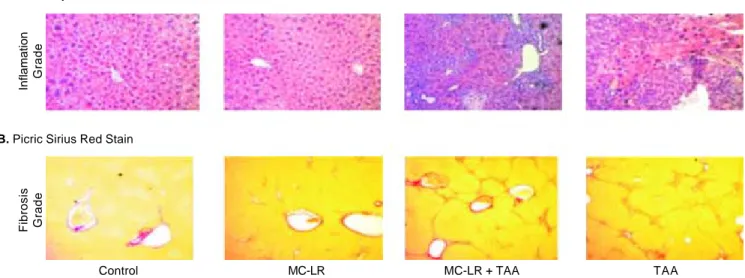
Figure 1. Figure 1.Figure 1.
Figure 1.Figure 1. Hematoxylin & Eosin and Picric Cirius stains of livers of male CD1 mice exposed to water alone (control) microcystin-LR (MC-LR) alone, MC-LR plus thioacetamide (TAA) or TAA alone for 28 weeks.
A. Hemotoxylin & Eosin Stain
Inflamation
Grade
Fibrosis Grade
Control MC-LR MC-LR + TAA TAA
and TAA alone exposed groups had significantly lower body weights and higher liver/body weight ratios when compared to water alone exposed controls (p < 0.01 re-spectively).
Serum ALT and TB determinations were also meas-ured at the time of sacrifice. Levels were similar in wa-ter and MC-LR alone exposed mice but significantly elevated in the MC-LR/TAA and TAA alone exposed cohorts (p < 0.05). The extent of the elevations in ALT and TB values were similar in the latter two cohorts.
The results of H&E staining for hepatic inflamma-tion are provided in figure 1 and semi-quantitatively in table 2. There was either no or minimal evidence of he-patic inflammatory activity in the livers of water and MC-LR alone exposed mice while inflammation was significantly increased (but to a similar extent) in the MC-LR/TAA and TAA alone cohorts (METAVIR scores: 2.6 ± 1.0 and 3.3 ± 0.7 respectively) when com-pared to water alone controls (p < 0.05 respectively).
Picric Sirius Red staining for fibrosis (Figure 1 and Table 2) indicated no differences in the extent of fibro-sis in livers of water and MC-LR alone exposed mice but significant increases (again, to a similar extent) in
Table 2. Serum Alanine Aminotransferase, Total Bilirubin, Histologic Grade and Stage of Liver Disease in Mice Exposed to Water Alone (control), MC-LR Alone, MC-LR + TAA or TAA Alopne for 28 weeks.
Treatment group ALT Total bilirubin Inflammation grade Fibrosis grade
(< 50) (0.4) (0.4) (0.4)
Control 43.61 ± 17.48 0.11 ± 0.03 mg/dL 0.88 ± 0.89 0.06 ± 0.25
MC-LR 48.35 ± 25.32 0.12 ± 0.05 mg/dL 1.16 ± 0.5 0.26 ± 0.45
MC-LR + TAA 69.12 ± 23.16* 0.22 ± 0.04 mg/dL** 2.57 ± 0.99** 2 ± 1.03**
TAA 68.71 ± 25.71* 0.21 ± 0.03 mg/dL** 3.26 ± 0.65** 1.79 ± 0.71**
* P < 0.05, ** P < 0.01, ALT: Alanine aminotransferase. MC-LR: Microcystin-LR. TAA: Thiocentamide.
Figure 2. Figure 2. Figure 2. Figure 2.
Figure 2. Oxidative stress of CFSC-2G hepatic stellate cells following 24 hours exposure to varying concentrations of microcystin-LR.
Mean signal intensity
250
200
150
100
50
0
0 0 1 10 100 1,000 5,000 10,000 0.1 0A
Microcystin-LR Concentration (μg/L) P < 0.001***
PP2A Activity (%)
120
100
80
60
40
20
0
0 0 1 10 100 1,000 5,000 10,000 0.1 0A
Microcystin-LR Concentration (ug/L)
Figure 3. Figure 3. Figure 3. Figure 3.
Figure 3. Serine/threonine phosphatase 2A (PP2A) activity in CFSC-2G hepatic stellate cells following 24 hours exposure to varying concentrations of microcystin-LR.
P < 0.001***
the MC-LR/TAA and TAA alone cohorts (METAVIR scores: 2.0 ± 1.0 and 1.8 ± 0.7 respectively) when com-pared to water alone controls (p < 0.01 respectively).
The results of HSC exposure to a range of MC-LR
concentrations in vitro are provided in figures 2-5. As
shown in figures 2 and 3, only at the highest
concentra-tions of MC-LR tested (5,000 and 10,000 μg/L) were
sig-nificant increases in oxidative stress and inhibition of serine/threonine phosphatase activity observed. Re-garding proliferative activity, there were no increases in HSC proliferation after 24, 48 and 96 h of exposure to
MC-LR at concentrations as high as 1,000 μg/L
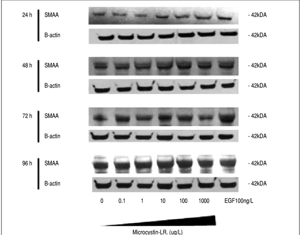
com-pared to buffer alone controls (Figure 4). Similarly, SMAA protein expression remained unaltered follow-ing exposure to the same concentrations of MC-LR for identical durations of time (Figure 5).
DISCUSSION
The results of this study indicate that healthy mice exposed to a low concentration of MC-LR in their drinking water for a total of 28 wks (approximately 30% of their anticipated life span) do not develop enlarged
*** ***
***
***
***
Figure 4. Figure 4.Figure 4.
Figure 4.Figure 4. Proliferative activity of CFSC-2G hepatic stellate cells following 96 h exposure to varying concentrations of microcystin-LR. 2.0
1.5
1.0
0.5
0.0
P < 0.001*** P < 0.01** P < 0.05*
Absorbance (450 nm)
0 h 24 h 48 h 72 h 96 h
Time (h) 0 ug MCLR
0.1 ug MCLR 1 ug MCLR
10 ug MCLR 100 ug MCLR 1,000 ug MCLR
Figure 5. Figure 5.Figure 5.
Figure 5.Figure 5. Smooth muscle alpha actin pro-tein (SMAA) expression in CFSC-2G hepatic stellate cells following 96 h exposure to vary-ing concentrations of microcystin-LR.
livers, biochemical evidence of active hepatic inflam-mation or dysfunction and have essentially normal liver histology. The results also suggest that long-term, low-dose exposure does not potentiate hepatic inflammation or fibrosis in the setting of active (TAA induced) liver disease. These findings were supported by the results of
in vitro experiments which demonstrated that low
con-centrations of MC-LR do not induce oxidative stress,
inhibit serine/threonine phosphatase activity, enhance the proliferative activity or activate HSCs to undergo transformation to a myofibroblast phenotype.
Although the above results are reassuring, it must be noted they are not consistent with previous findings
from other investigators. Specifically, Elleman, et al.
de-scribed hepatocyte degeneration, scattered lobular necrosis, mononuclear cell infiltration and progressive
24 h SMAA - 42kDA
B-actin - 42kDA
48 h SMAA - 42kDA
B-actin - 42kDA
72 h SMAA - 42kDA
B-actin - 42kDA
96 h SMAA - 42kDA
B-actin - 42kDA
0 0.1 1 10 100 1000 EGF100ng/L
Microcystin-LR. (ug/L)
100 ng TGf-b
** * ***
*** *** ***
*
fibrosis in mice treated daily for six weeks with “sub-lethal” intraperitoneal injections of purified MC-LR at
25%, 50% and 75% of the LD100 dose.13 In another
study, Frangez, et al. reported increased peri-portal
in-flammation and fibrosis in female New Zealand rabbits treated every other day for three weeks with intraperi-toneal injections of 7.5 mg/kg cyanobacterial lysates
containing 1 mg/g of MC-RR.14 Finally, He, et al.
de-scribed changes in keeping with nonalcoholic
steato-hepatitis in BALB/c mice exposed to low-dose (40 μg/
kg) MD-LR for 90 days.11
The reason(s) for the discrepancy between the above reports and our own findings remain to be determined. Whether differences in species (mice versus rabbits), strains of mice (CD1 versus BALB/c), nutrition (in the above studies MC-exposed mice lost significant amounts of weight) and routes of administration (oral
versus intraperitoneal) explain the different outcomes
requires further research. Also to be considered are dif-ferences in the concentrations of MC-LR employed. Unfortunately, the previous studies provided insuffi-cient data to calculate the molarity of MC-LR required for comparative analyses. Finally, it is possible that ex-posure to more than one MC congener is required to cause inflammation and/or fibrosis, as suggested by Frangez, et al.15
The need to determine whether cyanotoxins enhance hepatic injury in the setting of existing liver disease was driven by the growing epidemic of obesity and high prevalence of viral hepatitis which together, have result-ed in large segments of the general population being di-agnosed with chronic liver disease. Thus, whether long-term, low-dose cyanotoxin exposure enhances he-patic inflammation and fibrosis in the setting of existing liver disease is an important clinical question that had yet to be addressed. Here again, the results of the present study are reassuring. Biochemical and histolog-ic evidence of hepathistolog-ic inflammation and fibrosis were similar in MC-LR/TAA compared to TAA alone ex-posed mice. Whether the same findings would be ob-tained in other models of chronic liver disease remains to be determined.
There are a number of limitations to this study that warrant emphasis. First, only the most common and well-studied cyanotoxin, MC-LR, was employed. Sec-ond, the amount of water spillage in the animal holding cages was not measured and therefore, precise determi-nations of MC-LR exposure could not be made. Third, the in vitro studies involved HSCs alone and perhaps co-cultures with hepatocytes or other non-parenchymal cells would have provided different results. Finally, properly designed studies in humans where cyanotoxin contamination of the drinking water has been
docu-mented (and quantitated) are required to address the question whether these encouraging findings in rodents can be extrapolated to humans.
In conclusion, the results of the present study do not support concerns that long-term, low-dose exposure to cyanotoxins cause hepatic inflammation or fibrosis in healthy livers or exacerbate either feature in the setting of existing liver disease.
ACKNOWLEDGEMENTS
This research was supported by a grant from the Public Health Agency of Canada. Ms M. Labine is a re-cipient of a Manitoba Health Research Council fellow-ship award. The authors would also like to thank Ms R. Vizniak for her prompt and accurate typing of the man-uscript.
COMPLIANCE WITH ETHICAL STANDARDS
There was no conflict of interest. Approval for the study was obtained from the University of Manitoba an-imal ethics committee. Funding was received from a graduate studentship from Manitoba Water Stewardship for Meaghan Labine.
REFERENCES
1. Duy TN, Lam PK, Shaw GR, Connell DW. Toxicology and risk assessment of freshwater cyanobacterial (blue-green algal) toxins in water. Rev Environ Contam Toxicol
2000; 163: 113-85.
2. Carmichael WW. Health Effects of Toxin-Producing Cy-anobacteria: The CyanoHABs. Human and Ecological Risk Assessment. An International Journal 2001; 7: 1393-407. 3. Pouria S, de Andrade A, Barbosa J, Cavalcanti RL, Barre-to VT, Ward CJ, Preiser W, et al. Fatal microcystin inBarre-toxi- intoxi-cation in haemodialysis unit in Caruaru, Brazil. Lancet
1998; 352: 21-6.
4. Yuan M, Carmichael WW, Hilborn ED. Microcystin analysis in human sera and liver from human fatalities in Caruaru, Brazil 1996. Toxicon 2006; 48: 627-40.
5. Falconer IR, Burch MD, Steffensen DA, Choice M, Coverd-ale OR. Toxicity of the blue green alga (cyanobacterium) Microcystis aeruginosa in drinking water to growing pigs, as an animal model for human injury and risk assessment.
Environ Toxicol Water Qual 1994; 9: 131-9.
6. WHO. Algae and cyanobacteria in fresh water. In: Guide-lines for safe recreational water environments. Vol 1. Coastal and fresh waters. Geneva, Switzerland: World Health Organization; 2003, p. 136-58.
7. Guzman RE, Solter PF. Hepatic oxidative stress following prolonged sublethal microcystin LR exposure. Toxicol Pathol 1999; 27: 582-8.
9. Malbrouck C, Trausch G, Devos P, Kestemont P. Effect of microcystin-LR on protein phosphatase activity and glyco-gen content in isolated hepatocytes of fed and fasted ju-venile goldfish Carassius auratus L. Toxicon 2004; 44: 927-32.
10. Buratti FM, Scardala S, Funari E, Testai E. Human glutath-ione transferases catalyzing the conjugation of the hepa-toxin microcystin-LR. Chem Res Toxicol 2011; 24: 926-33.
11. Carvalho GM, Oliveira VR, Casquilho NV, Araujo AC, Soares RM, Azevedo SM, Pires KM, et al. Pulmonary and hepatic injury after sub-chronic exposure to sublethal doses of microcystin-LR. Toxicon 2016; 112: 51-8. 12. He J, Li G, Chen J, Lin J, Zeng C, Chen J, Deng J, et al.
Prolonged exposure to low-dose microcystin induces non-alcoholic steatohepatitis in mice: a systems toxicology study. Arch Toxicol 2017; 91: 465-80.
13. Elleman TC, Falconer IR, Jackson AR, Runnegar MT. Isola-tion, characterization and pathology of the toxin from a
Microcystis aeruginosa (= Anacystis cyanea) bloom. Aust J Biol Sci 1978; 31: 209-18.
14. Frangez R, Kosec M, Sedmak B, Beravs K, Demsar F, Juntes P, Pogacnik M, et al. Subchronic liver injuries caused by microcystins. Pflugers Arch 2000; 440: R103-104. 15. Milutinovic A, Zorc-Pleskovic R, Petrovic D, Zorc M, Suput
D. Microcystin-LR induces alterations in heart muscle. Fo-lia Biol (Praha) 2006; 52: 116-8.
Correspondence and reprint request: G.Y. Minuk, M.D.
Morberg Family Chair in Hepatology University of Manitoba.
John Buhler Research Centre 715 McDermot Ave. Winnipeg, MB R3E 3P4