P R O B L E M–B A S E D
P H Y S I O L O G Y
Robert G. Carroll, PhD
Professor of Physiology
Brody School of Medicine
1600 John F. Kennedy Blvd. Ste 1800
Philadelphia, PA 19103-2899
PROBLEM-BASED PHYSIOLOGY ISBN 978-1-4160-4217-4
Copyright © 2010 by Saunders, an imprint of Elsevier Inc.
All rights reserved. No part of this publication may be reproduced or transmitted in any form or by any means, electronic or mechanical, including photocopying, recording, or any information storage and retrieval system, without permission in writing from the publisher. Permissions may be sought directly from Elsevier’s Rights Department: phone: (+1) 215 239 3804 (US) or (+44) 1865 843830 (UK); fax: (+44) 1865 853333; e-mail: healthpermissions@elsevier.com. You may also complete your request on-line via the Elsevier website at www.elsevier.com/permissions.
Library of Congress Cataloging in Publication Data Carroll, Robert G.
Problem-based physiology / Robert G. Carroll. — 1st ed. p. cm.
Includes bibliographical references and index. ISBN 978-1-4160-4217-4
1. Physiology, Pathological—Case studies. I. Title.
[DNLM: 1. Pathology—Case Reports. 2. Physiology—Case Reports. QT 104 C319p 2010] RB113.C25 2010
616.07—dc22 2008040746
Acquisitions Editor: William R. Schmitt
Developmental Editor: Barbara Cicalese
Publishing Services Manager:Hemamalini Rajendrababu
Project Manager: Srikumar Narayanan
Design Direction:Gene Harris
Printed in China
Last digit is the print number: 9 8 7 6 5 4 3 2 1 Notice
Medical knowledge is constantly changing. Standard safety precautions must be followed, but as new research and clinical experience broaden our knowledge, changes in treatment and drug therapy may become necessary or appropriate. Readers are advised to check the most current product information provided by the manufacturer of each drug to be administered to verify the recommended dose, the method and duration of administration, and contraindications. It is the responsibility of the practitioner, relying on experience and knowledge of the patient, to determine dosages and the best treatment for each individual patient. Neither the Publisher nor the Editors assume any liability for any injury and/or damage to persons or property arising from this publication.
To Bettie Ann and to my children Anne Corinne, Elise, and Graham,
for 24 years of love, joy, and friendship
Richard H. Ray Ph.D. Professor of Physiology Brody School of Medicine
ix
A medical student’s ability to retain and apply physi-ological knowledge is improved when information is presented in a contextual format. For medical students, clinical scenarios provide an appropriate springboard for exploring the pathophysiology that leads to the development of specific symptoms, the progression of the disease process, and the appropriate clinical and therapeutic interventions that can be used to treat a patient.
The 88 clinical cases presented in this book are grouped into 10 sections, each loosely tied to an organ system. Each section has an introduction and concept map to help emphasize the interrelatedness of the cases related to that organ system. The integrative nature of physiology and medicine, however, results in complex clinical cases that often extend across multiple organ systems. The advantage of this complexity is that stu-dents interpret information across multiple organ sys-tems, as is appropriate for each clinical case.
The individual cases follow a typical clinical sce-nario. Each case begins with the presenting complaint, followed by a medical history that is limited to events pertinent to the complaint. The physical examination reports vital signs and key physical findings, which is followed by the results from subsequent diagnostic tests. The pathophysiology section expands on the patient presentation and laboratory results, outlin-ing the physiological mechanisms of both the symp-tom development and the progression of the disease. When appropriate, the pathophysiology section ends with a discussion of the logic that underlies the treat-ment options and the therapeutic outcomes.
The following aspects of this book make it a unique learning aid:
● Each case study is presented as an unknown to pro-mote active learning. The gradual unfolding of the clinical scenario allows students to test their predic-tive abilities and to provoke appropriate questions for sequential learning issues and further investigation.
● The integration of basic and clinical sciences is de-signed to help develop a “big picture” of disease processes.
● Over 135 full-color illustrations reinforce basic physiological mechanisms.
● The 10 section introductions provide a concept map to help organize the physiology underlying multiple clinical presentations.
● Two clinical vignette-based practice questions are derived from each of the 88 cases, illustrating how patient scenarios can be used as a basis for integra-tive physiology examination questions.
Although medical students are anticipated to be the main benefactors of this book, the format of patient presentation allows the book to be useful to a variety of advanced undergraduate, graduate, and professional students in the health-related professions. The health profession team deals with the same patient, and the clinical scenarios provide useful starting points for dis-cussion by all members of the team.
It is my hope that all students will develop a deeper understanding of concepts of medical physiology but also develop an enthusiasm for the lifelong learning process initiated by complex patient-based problem presentations. Clinical practice is continually improv-ing based on outcomes of medical and physiological research. Health practitioners need to become com-fortable with their role as perpetual students.
I invite the readers’ suggestions for improvement of this work. The preferred way to submit suggestions or corrections is electronic mail. Enthusiastic readers are advised to include “Problem-Based Physiology” as the subject line and send submissions by email to: carrollr@ecu.edu
xi
As with most major undertakings, Problem-Based Physiology could not be completed without the help of many individuals. First and foremost, I would like to thank my colleagues within the teaching community of the American Physiological Society, the Interna-tional Association of Medical Science Educators, and the International Union of Physiological Sciences for educating me about the effectiveness of a “student-centered” teaching style. Teaching and curriculum modifications need to be informed by the results of educational research, and I have benefited from both the knowledge and the enthusiasm of these groups.
For this project, I am particularly indebted to Swapan Nath and Sanjay Revanker for the innovative structure of this series as executed in the “Problem-Based Microbiology” text. I followed their format as closely as possible, but incorporated the unique as-pects of physiology.
I am also extremely grateful to my colleagues at the Brody School of Medicine at East Carolina Univer-sity for the information and expertise they have shared over the years, particularly Dick Ray, Mike Van Scott, Bob Lust, Chris Wingard, Greg Iams, Jitka Virag, Steve Wood, and Ed Seidel. Each of these educators has helped simultaneously reveal the complexity and simplicity of the organ systems and helped me under-stand the clinical implications of the field of physiol-ogy. Dick Ray, in particular, was a huge help in the
neurophysiology section, as well as in composing the Practice Questions and Answers tied to each of the clinical cases.
I am particularly grateful to the graduate students in the Physician Assistant Program Pathophysiology courses of 2006 and 2007 for both the clinical scenar-ios and clinical fact checking for the cases used in this book. Teaching is always a learning experience, and it has been fun learning with this group of talented students.
Many at Elsevier deserve recognition for their role in the production of this book. My heartfelt thanks go to William Schmitt, Acquisitions Editor of Medical Textbooks, for over a decade of support to the teach-ing section of the American Physiological Society. Barbara Cicalese expertly balanced the demands as Developmental Editor in encouraging timely comple-tion of tasks but also allowed time to develop a quality product.
Finally, I wish to acknowledge the medical and graduate students, past and present, who provided support for the framework of the text in the inception of ideas for this project and provided encouragement and stimulation for completion of Problem-Based Physiology.
xiii Problem-Based Physiology was written for students with
various study goals and learning styles and can be used both in a physiology course and as a review resource for the USMLE Step 1 examination. It might also be used as a reference during the clinical years. There are three parts to each case: patient presentation, physical examination and laboratory results, and pathophysiol-ogy. Practice questions at the end of the book allow students to assess their knowledge base. Each of these parts is described next. At the end is a special discus-sion with suggestions for three ways or tracks that this book might be used, depending on the study goals, preferred learning style, and background of the indi-vidual student.
FEATURES
Section Overviews
At the beginning of each section is an overview that in-cludes a concept map of the specific organ system and illustrates where the individual cases fit in the func-tional aspect of the organ system. This background is built on using the problem-solving exercises in the cases that follow.
Cases
Cases are presented in an “unknown format” intended to immediately engage the reader in thinking about physiology in clinical terms, emphasizing the applica-tion facts. Each case includes important pieces of in-formation about the patient’s problem that is under discussion.
CASE DESCRIPTION. To obtain the greatest ben-efit from the problem-based nature of this book, it is advisable that the reader focus separately on each part of the first page of the case, making sure that the infor-mation is synthesized and digested before going on to the next. The entirety of the case description has been
kept to the first page of the case as much as possible. In those cases where it has by necessity flowed onto the next page, it is suggested that the reader purposely not read the pathophysiology until a proper synthesis of the case description has been attained.
Clinical Scenario. Each case begins with a present-ing symptom in an appropriate clinical settpresent-ing and includes pertinent history of the patient’s problem.
Physical Examination and Laboratory Studies. These features give further information about the spe-cific case and offer a framework for evaluating the situation.
PATHOPHYSIOLOGY OF KEY SYMPTOMS. This
section illustrates the mechanisms underlying the pre-sentation of the symptoms and the progression of the disease. Whenever possible, either clinical outcome or treatment options are provided.
FURTHER READING. A short bibliography of current resources is provided to allow the student to more fully explore the case.
Practice Questions and Answers
xiv ● HOW TO USE THIS BOOK
TRACKS OF STUDY
Before plunging into the first case, readers are encouraged to evaluate their fund of knowledge and think about how they should best approach this book. Three “Tracks” of study are described here, designed to give readers an ap-propriate experience, based on the level of understanding.
Self-motivated learners with a strong basic science background who enjoy the freedom of a non-traditional curriculum and who learn best through self-directed reading and problem solving may wish to approach the book using the Track 1 philosophy. These individuals might read the Section Over-views for each organ system and then delve into the investigative study of a patient problem. The goal would be to focus on the clinical scenario and related information available on the first page of the case, using them to define the diagnosis before reading on to verify the diagnosis. Once the mechanism of the disease has been determined, students can turn to the pathophysiology section, which expands on the underlying physiology and mechanisms of the thera-peutic options available for treating this particular patient’s problem.
Track 2 can be used by small groups of students
in a PBL setting, where student-centered, self-di-rected learning is emphasized. Here, a faculty facili-tator might present the first page of a case over the
course of three discussion sessions. For instance, the presenting symptoms and medical/social history could be presented and progressively disclosed in Session 1 and physical examination and laboratory results in Session 2. This would leave the follow-up materials for student self-study and group discus-sion in Sesdiscus-sion 3. During the first sesdiscus-sion, students might also discuss open-ended learning issues that are outside the scope of this book. Students might further refine their learning in the context of the problem and the open-ended issues by focusing on biology, diagnostics, pathophysiology, and manage-ment in the second, and possibly a third, session. This PBL track is appropriate for students who are flexible in their learning goals and who want a strong clinical context for their learning.
Track 3 is appropriate for students who learn
well from a more traditional combination of pre-sentations (e.g., lectures) and readings and are more comfortable in a teacher-directed environment. Students using Track 3 may save the first-page patient scenario and other information for the end of their study to be used for assessment of recall and application of processed information.
xv SECTION I Nerves and Muscles
Introduction . . . 2
Case 1: Amyotrophic Lateral Sclerosis . . . 4
Case 2: Taxol-Induced Peripheral Neuropathy . . . 7
Case 3: Multiple Sclerosis . . . 10
Case 4: Botulism . . . 12
Case 5: Myasthenia Gravis . . . 15
Case 6: Duchenne Muscular Dystrophy . . . 17
SECTION II Cardiology Introduction . . . 20
Case 7: Hemorrhagic Shock . . . 22
Case 8: Congestive Heart Failure . . . 27
Case 9: Pericardial Tamponade . . . 30
Case 10: Mitral Valve Regurgitation . . . 33
Case 11: Patent Ductus Arteriosus . . . 35
Case 12: Aortic Valve Stenosis . . . 38
Case 13: Paroxysmal Supraventricular Tachycardia. . . 40
Case 14: Essential Hypertension . . . 42
Case 15: Coronary Artery Disease . . . 45
Case 16: Intermittent Claudication . . . 48
SECTION III Nephrology and Electrolyte Balance Introduction . . . 52
Case 17: Renal Artery Stenosis . . . 54
Case 18: Malignant Hypertension . . . 56
Case 19: Kidney Stones. . . 59
Case 20: Poststreptococcal Glomerulonephritis . . . 62
Case 21: Diabetes Neuropathy . . . 65
Case 22: Acute Tubular Necrosis . . . 67
Case 23: Neurogenic Bladder . . . 70
Case 24: Nephrogenic Diabetes Insipidus. . . 73
SECTION IV Hematology Introduction . . . 76
Case 25: Coumadin-type Herbal Supplement. . . 78
Case 26: Hemophilia A . . . 80
Case 27: Thrombocytopenia. . . 82
Case 28: Iron Deficiency Anemia . . . 84
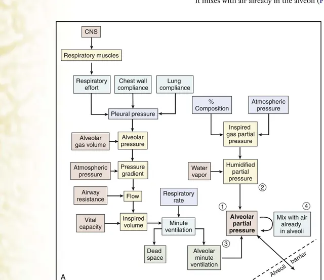
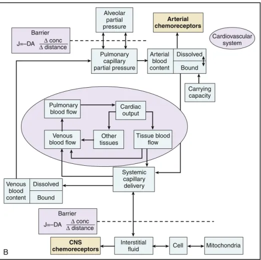
SECTION V Respiration
Introduction . . . 90
Case 30: Chronic Obstructive Pulmonary Disease . . . 93
Case 31: Pneumothorax . . . 95
Case 32: Asthma . . . 97
Case 33: Barbiturate Overdose . . . 99
Case 34: Acute Mountain Sickness . . . 101
Case 35: Pulmonary Edema . . . 103
Case 36: Carbon Monoxide Poisoning . . . 105
Case 37: Exercise Intolerance . . . 107
Case 38: Cyanide Poisoning . . . 109
Case 39: Pulmonary Embolism. . . 111
Case 40: Pneumonia . . . 113
SECTION VI Neurology Introduction . . . 116
Case 41: Traumatic Injury (Coma). . . 117
Case 42: Bitemporal Hemianopsia . . . 119
Case 43: Astrocytoma of the Vermis of the Cerebellum . . . 121
Case 44: Parkinson’s Disease . . . 123
Case 45: Symptomatic Epilepsy . . . 126
Case 46: Alzheimer’s Disease . . . 128
Case 47: Expressive Aphasia after a Stroke. . . 131
Case 48: Vertigo from Acoustic Neuroma . . . 133
Case 49: Syringomyelia . . . 135
Case 50: Bell’s Palsy . . . 137
Case 51: Carpal Tunnel Syndrome. . . 139
SECTION VII Gastroenterology Introduction . . . 142
Case 52: Achalasia . . . 144
Case 53: Peptic Ulcer . . . 146
Case 54: Gastroesophageal Reflux Disease (GERD) . . . 150
Case 55: Pernicious Anemia . . . 152
Case 56: Pancreatitis . . . 153
Case 57: Celiac Disease. . . 155
Case 58: Impaired Fat Absorption . . . 157
Case 59: Secretory Diarrhea . . . 159
Case 60: Fecal Incontinence . . . 161
SECTION VIII Metabolism Introduction . . . 164
Case 61: Diabetes Mellitus Type 1 . . . 165
Case 62: Hypoglycemia . . . 168
Case 63: Graves’ Disease . . . 170
Case 64: Hypothyroidism (Hashimoto’s Thyroiditis) . . . 172
Case 65: Hyperparathyroidism . . . 174
Case 66: Starvation (Anorexia Nervosa) . . . 176
Case 67: Obesity . . . 178
Case 68: Influenza. . . 181
Case 69: Hypothermia . . . 183
SECTION IX Endocrinology
Introduction . . . 186
Case 70: Acute Adrenal Insufficiency (Addisonian Crisis) . . . 189
Case 71: Cushing’s Syndrome (Primary Hypercortisolism from Adrenal Tumor) . . . 191
Case 72: Conn’s syndrome (Hyperaldosteronism) . . . 193
Case 73: Pheochromocytoma . . . 195
Case 74: Acromegaly. . . 198
Case 75: Hyperprolactinemia . . . 201
Case 76: Panhypopituitarism . . . 203
SECTION X Reproduction Introduction . . . 206
Case 77: Female Menopause. . . 208
Case 78: Hypothalamic Amenorrhea . . . 210
Case 79: Polycystic Ovary Syndrome . . . 212
Case 80: Endometriosis . . . 214
Case 81: Ectopic Pregnancy . . . 216
Case 82: Placental Abruption . . . 218
Case 83: Eclampsia . . . 220
Case 84: Gestational Diabetes . . . 222
Case 85: Secondary Hypogonadotropic Hypogonadism . . . 224
Case 86: Cryptorchidism . . . 226
Case 87: Benign Prostatic Hyperplasia. . . 229
Case 88: Erectile Dysfunction . . . 231
Appendix A Practice Questions and Answers . . . 233
Appendix B Credits. . . 269
Nerves and Muscles
S
ECTION
I
ATP required
Ca Ca
Ca++ Ca++
Action potential
Calcium pump Sarcolemma
Voluntary movement requires a successful interplay of central nervous system components, peripheral nervous system components, muscles, and joints of the skeleton. Abnormalities in movement include disorders of coordination as well as of muscle weakness. Move-ment disorders provide a complex diagnostic challenge because defects in the chain of events can occur in any one region or multiple locations. Consequently, this introduction is organized around the physiology of normal voluntary muscle movement involved in picking up a pencil from a desk.
Voluntary movement originates in the planning regions of the accessory motor cortex. The first region of the brain to become activated is the sensory cortex, specifically the area receiving proprioception input from the periphery. Before movement can be initiated, it is first necessary to know where in three-dimensional space both the hand and the object are located. Proprioception plays a continuing and impor-tant role throughout voluntary muscle movement.
Voluntary muscle activity also integrates activity from the cerebellum and the basal ganglia. Coordina-tion of central nervous system activities and symptoms arising from specific defects are addressed in neurosci-ence textbooks. Here, clinical problems arising from defects in the upper motor neuron, spinal cord, alpha (α)-motor neuron, and muscle are addressed.
Cell electrical activity underlies neuromuscular function. Both nerves and muscle cells have negative resting membrane potentials, resulting from both the differential distribution of ions across the cell mem-brane and the relative conductance for these ions based on ion channels. At rest, the cell is most permeable to K+ and, consequently, resting membrane potential is
close to the K+Nernst (equilibrium) value of −90 mV.
Any changes in plasma K+values can impact both nerve
and muscle function.
Depolarization of the cell can result in a local potential or an action potential. Local potentials are more common and are found in the neuron dendrites, cell body, and regions of the axon covered by the myelin sheath. Action potentials are a marked depolarization of the axon regions not covered by myelin (or the entire axon in unmyelinated axons), due to the opening of voltage-gated Na+channels. Local potentials decrease
over time and distance, but action potentials regenerate without any decrease in amplitude. Consequently, action potentials allow neuronal electrical activity to be conducted over long distances. Myelinated nerves have the fastest conduction velocity (see Case 3).
The axons of upper motor neurons extend from the motor cortex to the spinal cord, where they synapse with both an α-motor neuron and spinal cord inter-neurons (see Case 1). The axon of the upper motor
CNS motor cortex Other inter-neurons Spinal cord -motor neuron Action potential Depolarization Presynaptic axon terminal Central nervous system Synaptic cleft Skeletal muscle cell Ca++ entry ACh vesicles Vesicle exocytosis ACh ACh Receptor End plate potential T tubules Sarcoplasmic reticulum Ca++ Reuptake Troponin C Actin Action potential Phosphocreatine ATP Myosin Sarcomere shortening Muscle shortening Length-tension relationship Temporal summation tetany Tension Skeleton Movement Vesicles Vesicles Choline Acetate ACh esterase Mito-chondria Glucose, O2, fatty acids Heat
Section I ●NERVES AND MUSCLES 3
neuron is myelinated, and the action potential is trans-mitted by saltatory conduction.
The α-motor neuron, sometimes called the lower motor neuron, receives descending input from the upper motor neurons of the corticospinal tract, as well as multiple inputs from spinal cord interneurons. Spinal cord interneurons coordinate and integrate a variety of muscle activities, from the simple mono-synaptic patellar tendon stretch reflex to the more complex polysynaptic flexor withdrawal reflex.
The axon of the α-motor neuron extends from the spinal cord to the muscle fibers (see Case 2). The
α-motor neuron is myelinated, and action potentials are transmitted by saltatory conduction.
The axon and all of the muscle fibers that it inner-vates represent the “motor unit.” Some axons, such as the motor units of the hand that are used in writ-ing, innervate only a few muscle fibers and provide fine control. Other axons innervate a large number of muscle fibers, such as the motor units of the leg that are used for lifting.
Theα-motor neuron synapses with a skeletal muscle cell at the neuromuscular junction. The neuromuscu-lar junction consists of the presynaptic axon terminal, the synaptic cleft, and the postsynaptic “end plate” region of the skeletal muscle cell.
Neuromuscular transmission begins with the action potential arriving at the axon terminal of the
α-motor neuron (Fig. I-1). Depolarization of the axon terminal opens voltage-sensitive calcium channels, leading to the influx of calcium into the axon termi-nal. Calcium entry causes the docking and fusion of
acetylcholine-containing vesicles, resulting in the exocytosis of acetylcholine (see Case 4). Acetylcholine diffuses across the synaptic cleft, where it binds with a nicotinic receptor on the skeletal muscle cell (see Case 5). Receptor binding opens a nonspecific cation channel, leading to a depolarization of the end plate region and the development of an action potential within the skeletal muscle cell.
The action potential in a skeletal muscle cell is transmitted to the interior of the muscle cell along the T tubules. Depolarization of the T tubules stimulates the release of calcium from the sarcoplasmic reticulum. Calcium binds with troponin C, exposing the active sites on the actin protein. The myosin binds to actin and, in the presence of adenosine triphosphate (ATP), contracts according to the sliding filament theory, causing the muscle to develop tension and to shorten (see Case 6).
Skeletal muscle contraction occurs by a tetany or sustained contraction (Fig. I-2). For an individual fiber, the strength of contraction is determined in part by the length-tension relationship (Fig. I-3). Increases in contractile force for an entire muscle can also be achieved by activating a larger number of motor units and, consequently, contracting a larger number of fibers within the muscle.
Rate of stimulation (times per second)
5 10 15 20 25 30 35 40 45 50 55
Strength of muscle contraction
Tetanization
FIGURE I–2 Frequency summation and tetanization.
3 4 2 1 0 100 50 0
Length of sarcomere (micrometers) A B C D C B A D T ension de v eloped (per cent)
4
PHYSICAL EXAMINATION
VS: T 37°C, P 68/min, R 14/min, BP 110/60 mm Hg, BMI 25
PE: Neuromuscular examination reveals a positive Babinski sign on the right foot but not the left foot. Patient has some slurring of speech (dysarthria) and awkwardness when asked to run down a hallway. Patellar tendon reflex on the right leg is exagger-ated (hyperreflexia). There were no other signifi-cant findings. Patient is referred for muscle biopsy and neuromuscular study.
LABORATORY STUDIES
Muscle biopsy: Normal
Electromyography: As disease progresses, muscle fibrillation, increased amplitude and duration of action potentials may be found.
Nerve conduction studies: Normal early, then defec-tive as disease progresses to a lower motor neuron disease
DIAGNOSIS
Amyotrophic lateral sclerosis (ALS)
PATHOPHYSIOLOGY OF KEY
SYMPTOMS
The diagnosis of ALS is achieved by eliminating the variety of other causes of muscle weakness. This diagnosis is complicated by the fact that the appearance of clinical symptoms and the time course for the progression of the disease are quite variable. One diag-nostic approach involves beginning with skeletal muscle function and assessing function from the periphery back to the central nervous system.
Skeletal muscles elsewhere in the body are func-tioning normally. This indicates that the muscles are capable of maintaining a normal resting membrane
potential and that cellular and extracellular electrolyte concentrations are within the normal range. The muscle biopsy rules out any protein-related defects in the muscle contractile and structural proteins. Elec-tromyography confirms that the muscle is still able to contract when directly electrically stimulated. The defect in nerve traffic, however, eventually leads to atro-phy of the skeletal muscle and exacerbates the weakness associated with the progression of the disease.
An appropriate patellar tendon reflex requires affer-ent input from the muscle spindles, transmission of the afferent action potential to the spinal cord, a mono-synaptic reflex within the spinal cord, efferent action potential transmission by the alpha (α)-motor neuron, transmission across the neuromuscular synapse, activation of the skeletal muscle, and, finally, contrac-tion (Fig. 1-1). This patient shows a hyperreflexive response in the right leg. Normally, the α-motor neu-rons receive descending inhibitory input from upper motor neurons and both inhibitory and stimulatory input from interneurons (Figs. 1-2 and 1-3). Hyper-reflexia can be an indication that the normal descending input is diminished, which is characteristic of an upper motor neuron disease.
The finding of a positive Babinski sign is also characteristic of a defect in the upper motor neurons.
1
1
The patient indicates that he noticed trouble with cramping and weakness in his right leg 4 months ago but attributed that to a change in his exercise habits. The weakness is progressing despite continued exercise, and now the left leg is beginning to show signs of weakness. He was last seen for a routine physical examination 3 years ago, and no abnormalities were noted at that time.Motor nerve
Stretch reflex Muscle spindle Sensory nerve
CASE 1 5
The Babinski sign is elicited by stroking the sole of the foot. When only lower motor neurons are functional, the big toe extends as part of a spinal cord reflex. The upper motor neurons usually inhibit the flaring response. When the upper motor neurons are func-tional, stroking the sole of the foot causes the big toe to
contract. The Babinski sign is present in infants until about 2 years of age, when the upper motor neuron development becomes complete. Prior to the complete development of the upper motor neurons, stroking the sole of the foot causes the big toe to contract.
Nerve conduction velocity assessment is normal, indicating that the transmission of the action potential along the α-motor neuron by saltatory conduction is normal.
ALS is a motor neuron disease and does not involve other types of neurons. Consequently, ALS is characterized by progressive muscle weakness but no defects in intelligence, memory, or sensory discrimi-nation. ALS begins as an upper motor neuron disease and then progresses to a complete loss of upper motor neuron input, resulting in a spastic or rigid paralysis. As the disease progresses, lower motor neurons may become involved, leading first to muscle weakness, then to fasciculations, and ultimately to a complete loss of muscle control and spinal cord reflexes.
The progressive loss of motor neurons ultimately involves the phrenic nerve, which innervates the diaphragm. Consequently, death results from respira-tory muscle paralysis unless the patient is placed on an artificial ventilator.
Three to 10 percent of ALS cases are due to a domi-nant genetic defect, some of which involve a mutation of the superoxide dismutase 1 gene. The etiology of the remaining 90% to 97% of the cases is unknown.
Patients with ALS sometimes show elevated levels of glutamate in blood and spinal fluid. Excessive activation of the excitatory glutaminergic nerves is associated with neuronal damage, and, consequently, glutamate is suspected to be a contributing or causative agent.
OUTCOME
Riluzole (Rilutek), a glutamate antagonist, slows the progression of ALS. Although the exact mechanism of benefit is unknown, riluzole inhibits glutamine release and also blocks the activity of excitatory amino acids. Riluzole also inactivates voltage-gated sodium channels and interferes with intracellular events that follow trans-mitter binding and excitatory amino acid receptors.
Riluzole is the only approved treatment for ALS. Patient management is directed at relieving the symp-toms and maintaining the quality of life. Physical therapy can help maintain some muscle and joint motility. Ultimately, death results from the loss of phrenic nerve function and respiratory paralysis.
FLEXOR REFLEX
CROSSED EXTENSOR REFLEX Painful
stimulus from hand
Polysynaptic circuit Excited
RECIPROCAL INHIBITION
Inhibited
Excited Inhibited
FIGURE 1–2 Flexor reflex, crossed extensor reflex, and reciprocal inhibition.
Sensory neurons
Propriospinal tract Interneurons Corticospinal tract from pyramidal cells of cortex
Rubrospinal tract Reticulospinal tract
Anterior motor neuron
Motor nerve Tectospinal and reticulospinal tracts Vestibulospinal and reticulospinal tracts
F U R T H E R R E A D I N G
Web Sources
http://www.ninds.nih.gov/disorders/amyotrophiclateralsclerosis/ detail_amyotrophiclateralsclerosis.htm
Rilutek information. Available at http://products.sanofi-aventis.us/ rilutek/rilutek.html
MDConsult: ALS: Possible role of environmental influences. Neurol Clin 23:2, 2005.
MDConsult: Protein interaction may be key to apoptosis in ALS. Lancet Neurol 3:9, 2004.
Text Sources
Carroll RG: Elsevier’s Integrated Physiology. Philadelphia, Elsevier, 2007.
Copstead L, Banasik J: Pathophysiology, 3rd ed. Philadelphia, Saunders, 2005.
Dudek RW, Louis TM: High-Yield Gross Anatomy, 3rd ed. Philadelphia, Lippincott Williams & Wilkins, 2008, p 21. Guyton AC, Hall JE: Textbook of Medical Physiology, 11th ed.
Philadelphia, Saunders, 2006.
7
C A S E
PHYSICAL EXAMINATION
VS: T 37°C, P 75/min, R 18/min, BP 128/84 mm Hg, BMI 34
PE: Sensory neuropathy (tingling) present in the hands. Deep tendon reflexes were diminished bilaterally.
LABORATORY STUDIES
None ordered
DIAGNOSIS
Taxol-induced peripheral neuropathy
COURSE
The sensory and motor neuropathies resolved over the next 5 days.
PATHOPHYSIOLOGY OF KEY
SYMPTOMS
Normal neuromuscular transmission requires the release of acetylcholine from the synaptic vesicles in the alpha (α)-motor neuron presynaptic nerve terminal (Fig. 2-1), diffusion across the synaptic cleft, and bind-ing to the receptors at the motor end plate region of the skeletal muscle cell. The acetylcholine is degraded by the enzyme acetylcholinesterase into acetate and choline. The choline is transported back into the pre-synaptic terminal and returned to the recycled portion of the vesicle. Acetylcholine is resynthesized, and the vesicle is ready for reuse.
The vesicles and associated vesicle proteins are synthesized in the cell body of the α-motor neuron.
The vesicles are transported to the axon terminal by microtubules. Once reaching the axon terminal, the vesicles are acidified and loaded with acetylcholine for synaptic release.
Cisplatin and paclitaxel both disrupt microtubule function by polymerizing the tubules and, therefore, block cellular mitosis. Consequently, rapidly dividing cells are targeted and killed. Neuromuscular symp-toms develop because of the depletion of the vesicles that release acetylcholine. Disruption of the microtu-bules reduces the rate of vesicle arrival at the presyn-aptic axon and can lead to a reduction in the number of vesicles released during depolarization of the axon terminal.
Muscle electrical activity consists of miniature end plate potentials and end plate potentials. The minia-ture end plate potentials result from the release of a single vesicle of acetylcholine, usually causing a 0.4-mV depolarization in the muscle cell. The magnitude of the miniature end plate potentials will not be altered, because each vesicle contains a normal amount of ace-tylcholine, but their frequency may be diminished.
A normal α-motor neuron action potential normally causes the release of about 125 vesicles. That quantity of acetylcholine released into the synaptic cleft causes a 40-mV depolarization at the muscle end plate, which is sufficient to elicit an action potential and a con-traction of the muscle. In this patient, the end plate potentials will be diminished because the total number of vesicles released during any one action potential will be diminished. Consequently, every α-motor neuron action potential does not result in a muscle cell con-traction. This results in the weakness in the muscles.
Paclitaxel can also damage Schwann cells, lead-ing to diminished nerve conduction velocity of both motor and sensory nerves, particularly the large myelinated nerves.
Sensory nerve conduction is more acutely altered, because the cell body of the sensory nerves is not behind the blood-brain barrier and, consequently, is more completely exposed to the paclitaxel.
2
2
A 54-year-old woman returns to her oncologist for continuing treatment of recurrentovarian cancer. It is 2 days since treatment, and she is now complaining of paresthesia of the hands and feet and difficulty in fastening buttons due to muscle weakness in the hands.
The patient began treatment for recurrent ovarian cancer 2 months ago. The treatment regimen includes paclitaxel, 150 mg/m2; gemcitabine, 800 mg/m2;
and cisplatin, 75 mg/m2, three times a week for 6 cycles. She was diagnosed with
Symptoms develop gradually, as the majority of the neurotransmitter vesicles in the axon are recycled and recovered after exocytosis. Only a small portion of the vesicles are lost and have to be replaced by vesicles synthesized in the cell body. Consequently, the onset of muscle weakness occurs 1 to 3 or more days after paclitaxel treatment.
The muscle groups involved also illustrate the pathophysiology. Axons innervating distal muscles have the longest distance for vesicle transport and,
consequently, are more sensitive to microtubule disruption.
OUTCOME
Paclitaxel neuropathies normally (but not always) resolve over 1 week. Excessive alcohol use and diabe-tes are risk factors for the development of permanent neuropathies.
EXOCYTOSIS
Active zone
H+
H+
Transporter
A nonpeptide neurotransmitter is synthesized in the nerve terminal and transported into a vesicle.
ATP
B
Nonpeptide neurotransmitters are synthesized and transported into vesicles in the nerve terminal.
1 2 3
AXON Nerve
terminal Endoplasmic
reticulum
cis
Golgi transGolgi
Myelin sheath
Endosomes (vesicles)
Synaptic cleft Microtubules
CELL BODY BIOGENESIS
Nucleus
A
Vesicles travel through the axon on microtubule tracks via fast axonal transport. Peptide neurotransmitters are already in some vesicles. Vesicle and peptide
neurotransmitter precursors and enzymes are synthesized in the cell and are released from Golgi.
9 CASE 2 9
F U R T H E R R E A D I N G
Web Sources
Argyriou A, Polychronopoulos P, Koutras A, et al: Clinical and electrophysiological features of peripheral neuropathy induced by administration of cisplatin plus paclitaxel-based chemotherapy. Eur J Cancer Care 16:231-237, 2007. Available at http:// gateway.tx.ovid.com.jproxy.lib.ecu.edu/gw1/ovidweb.cgi
Postma TJ, Vermorken JB, Liefting AJ, et al: Paclitaxel-induced neuropathy. Ann Oncol 6:489-494, 1995. Available at http:// gateway.tx.ovid.com.jproxy.lib.ecu.edu/gw1/ovidweb.cgi
Text Sources
Brunton L, Lazo J, Parker K: Goodman & Gilman’s The Pharmacological Basis of Therapeutics, 11th ed. New York, McGraw-Hill Professional, 2005.
Carroll RG: Elsevier’s Integrated Physiology. Philadelphia, Elsevier, 2007.
Copstead L, Banasik J: Pathophysiology, 3rd ed. Philadelphia, Saunders, 2005.
Guyton AC, Hall JE: Textbook of Medical Physiology, 11th ed. Philadelphia, Saunders, 2006.
10
PHYSICAL EXAMINATION
VS: T 36.5°C, P 70/min, R 15/min, BP 124/80 mm Hg, BMI 27
PE: Muscle strength testing: Normal. Deep tendon reflexes: Normal. When asked to walk, the patient had difficulty in balancing and maintaining a nor-mal gait. Patellar tendon reflexes were nornor-mal. The muscles, however, were weak.
LABORATORY STUDIES
MRI: Gadolinium-enhancing brain lesions, as well as one lesion in the brain infratentorium and three periventricular lesions
Cerebrospinal fluid analysis: Oligoclonal IgG band Edrophonium (Tensilon) test: Normal
Nerve conduction velocity test: Normal Electromyography: Normal
DIAGNOSIS
Multiple sclerosis
PATHOPHYSIOLOGY OF KEY
SYMPTOMS
Outflow from the upper motor neurons of the cerebral motor cortex travels by the corticospinal tracts to syn-apse with an alpha (α)-motor neuron. Each muscle fiber is innervated by a single α-motor neuron; however, a single α-motor neuron can innervate one or many muscle fibers. The motor unit consists of the α-motor neuron and all of the muscle fibers that it innervates. The strength of muscle contraction depends in part on the number of motor units activated. Smaller motor units are activated first, followed by larger ones.
Complex activities such as walking require the coor-dinated, sequential activation of multiple motor units. The sequence of activation is determined by neurons in the cerebellum, basal ganglia, and premotor cortex
that synapse with the upper motor neurons. This results in the appropriate sequential activation of the muscles involved in walking and in balance, producing a smooth gait.
Action potential transmission depends on the diameter of the axon and on the presence of myelina-tion. Myelination restricts the regions of the axon capable of producing an action potential to the nodes of Ranvier, where the myelin sheath is absent (Fig. 3-1). The wave of depolarization that initiates in the axon hillock travels by local conduction in the region of the axon beneath the myelin and is regenerated as an action potential at the nodes. This form of transmis-sion is called “saltatory” conduction, because the action potential appears to jump from node to node along the length of the axon. Myelinated axons have much faster conduction velocities than do axons that lack myelin.
Multiple sclerosis is thought to be an autoimmune disease that leads to loss of myelin from central ner-vous system axons. The lost myelin is replaced by scar tissue called sclerosis. The central nervous system damage results in multiple lesions that are visible on magnetic resonance imaging.
Demyelinating diseases diminish the speed of axonal transmission. Consequently, this negatively impacts the coordinated and sequential activation of motor neurons necessary for walking. The difficulty
3
3
walking.On questioning, the patient indicates she had trouble walking for 5 days last month and also that she has experienced pain and prickly sensations that come and go, as well as occasional muscle weakness.
Myelin sheath
1 2 3
Node of Ranvier Axoplasm
FIGURE 3–1 Saltatory conduction along a myelinated axon. Flow of electrical current from node to node is illustrated by the
CASE 3 11
in walking is likely due to damage to the myelinated axons of the basal ganglia or the cerebellum.
Deep tendon reflexes are normal because the neuro-muscular transmission is working normally in the early stage of the disease. Similarly, muscle weakness may not be apparent. However, over time, the degen-eration of the nerves will lead to muscle atrophy and weakness.
OUTCOME
Currently there is no cure for multiple sclerosis. The frequency and intensity of symptoms, however, can be diminished with interferon drugs that diminish the activity of the immune system. In addition, relapses may be shortened by treatment with corticosteroids. Symptoms can be helped by physical therapy.
Web Sources
Campellone J: U.S. National Library of Medicine. Nerve con-ductivity test. Accessed April 30, 2007. Available at http:// www.nlm.nih.gov/medlineplus/ency/article/003927.htm
Goetz C: Textbook of Clinical Neurology, 3rd ed., Philadelphia, Saunders, 2007. Available at http://www.mdconsult.com/das/ book/body/79885428-3/0/1488/402.html?#4-u1.0-B978-1-4160-3618-0.10048-7–s0030_3250
Multiple Sclerosis Foundation. Accessed October 16, 2007. Available at http://www.msfocus.org/info_diagnosed.php
Wrongdiagnosis.com. Deep tendon reflexes. Accessed October 2007. Available at http://www.wrongdiagnosis.com/t/tendinitis/ book-diseases-14b.htm
Text Sources
Carroll RG: Elsevier’s Integrated Physiology. Philadelphia, Elsevier, 2007.
Copstead L, Banasik J: Pathophysiology, 3rd ed. Philadelphia, Saunders, 2005.
Guyton AC, Hall JE: Textbook of Medical Physiology, 11th ed. Philadelphia, Saunders, 2006.
McPhee SJ, Papadakis MA, Tierney LM Jr: Current Medical Diag-nosis and Treatment, 46th ed. New York, McGraw-Hill, 2007.
12
PHYSICAL EXAMINATION
VS: T 37°C, P 90/min, R 19/min, BP 145/100 mm Hg
PE: Physical examination reveals double vision and drooping of the eyelids. The patient is slurring his speech and has difficulty swallowing. He shows signs of muscle weakness. Deep tendon reflexes are diminished in the legs and arms.
LABORATORY STUDIES
CBC, electrolytes: Normal
Brain CT and MRI: Normal (rule out cerebrovascular accident)
Cerebrospinal fluid examination: Normal
Electromyography: Nerve conduction velocity and amplitude normal; neuromuscular junction impaired Edrophonium (Tensilon) test: Normal (rule out
myas-thenia gravis, myasthenic syndrome)
Stool sample: Positive for Clostridium botulinum
DIAGNOSIS
Botulism toxicity
COURSE
Botulism is a paralytic illness caused by a neurotoxin released from Clostridium botulinum. There are seven types of C. botulinum bacteria, identified by the letters A through G. The bacteria grow best in low oxygen conditions, and food-borne botulism is usually caused by eating contaminated foods canned at home.
PATHOPHYSIOLOGY OF KEY
SYMPTOMS
Acetylcholine is stored in vesicles in the presynap-tic terminal of alpha (α)-motor neurons. After an action potential arrives, calcium enters the presynaptic
4
4
and has difficulty swallowing.The patient is not from the area but is visiting friends who live on a commune. On questioning, the patient indicates that approximately 24 hours earlier he ate some green beans that were canned at the commune. Symptoms began 3 hours ago, and the decision to come to the emergency department was made because of his difficulty in swallowing. The patient is now anxious and easily excited.
Calcium channels Neural
membrane
Muscle membrane Release
sites Vesicles
Dense bar
Basal lamina and
acetylcholine-sterase Acetylcholine receptors
Subneural cleft
FIGURE 4–1 Release of acetylcholine from synaptic vesicles at the neural membrane of the neuromuscular junction. Note the proxim-ity of the release sites in the neural membrane to the acetylcholine re-ceptors in the muscle membrane, at the mouths of the subneural clefts.
Milliseconds
Milliv
olts
0 15 30 45 60 75
C B
A +60 +40 +20 0 –20 –40 –60 –80 –100
CASE 4 13
4 5
6
FUSION AND EXOCYTOSIS +
Ca2+
RECYCLING OF SNAREs
DISASSEMBLY OF TERNARY SNARE COMPLEX
-SNAP
-SNAP
NSF
NSF
ADP ATP
FORMATION OF TERNARY COMPLEX OF SNARES
INITIAL STATE TIGHTENING OF TERNARY
SNARE COMPLEX Synaptic
vesicle
n-Sec–1
n-Sec–1 Synaptotagmin
Synaptobrevin
SNAP-25 Syntaxin Nerve terminal membrane
1 2 3
With the endocytosis of the vesicle, the synap-tobrevin is effectively recycled. The syntaxin and SNAP-25 are now free for an additional cycle of vesicle fusion.
-SNAP and the ATPase NSF bind
to the ternary SNARE complex and use the energy of ATP hydrolysis to disassemble the SNAREs.
The entry of Ca2+and its binding
to synaptotagmin triggers fusion. The three SNAREs–synaptobrevin, syntaxin and SNAP-25–continue to
form a tight bundle of helices,
drawing the vesicle and presynaptic membranes into close apposition. n-sec-1 dissociates from syntaxin, allowing
the syntaxin and SNAP-25 to form a complex. The distal end of synaptobrevin begins to wind around the syntaxin/SNAP-25 complex, forming a ternary complex. Vesicles with synaptotagmin and
synaptobrevin (a v-SNARE) move to the nerve terminal membrane, which contains syntaxin and SNAP-25 (both t-SNAREs).
terminal, causing the vesicles to migrate toward the presynaptic membrane. There are numerous proteins involved in vesicle exocytosis. The SNAP-SNARE mechanism includes proteins that mediate vesicle docking and fusion, including synapsin 1, synapto-brevin, SNAP-25, syntaxin, synaptotagmin, and syn-aptophysin. The series of SNAP and SNARE proteins allow docking of the vesicles with the cell membrane, fusion, and, finally, exocytosis of the acetylcholine into the synaptic cleft (Fig. 4-1). The acetylcholine diffuses across the synaptic cleft and binds to receptors on the end plate region of the skeletal muscle cell.
Botulinum toxin destroys one or more of the family of “SNAP” proteins that are essential for docking and fusion of the acetylcholine vesicles with the presynap-tic membrane of the α-motor neuron. In the absence of these proteins, acetylcholine is not released by action potentials arriving at the neuromuscular junction and flaccid paralysis occurs. The paralysis is first evident in the cranial muscles and exhibits a symmetrical weak-ening, leading to paralysis. The impairment spreads to other proximal muscles and finally will include the distal muscles.
Impaired acetylcholine release by botulinum toxin depresses the end plate potential amplitude, an event tied to the exocytotic release of acetylcholine-containing vesicles (Fig. 4-2). Miniature end plate potential amplitude, which is a function of the quan-tity of acetylcholine in each vesicle, is not affected. The toxin is extremely potent, with a lethal dosage of 2 to 3 mg. There are many forms of botulinum toxin, but all cleave one of the proteins involved in vesicle priming (synaptobrevin, syntaxin, and SNAP-25) (Fig. 4-3) and subsequent inhibition of Ca++-induced
vesicle release from presynaptic terminals and degen-eration of the terminals.
Very dilute preparations of botulinum toxin (Botox) can be used to treat disorders involving hyperactivity of neuromuscular junctions. In addition, botulinum toxin is used cosmetically to paralyze muscles, which reduces the wrinkles in the face resulting from con-traction of the underlying muscles. Recovery of func-tion after exposure to botulinum toxin requires weeks to months and depends on the synthesis of new dock-ing and fusion proteins in the nucleus of the neuron as well as transport along the axon for incorporation at the presynaptic nerve terminal.
The loss of function at the neuromuscular junction is the basis for all of the patient’s symptoms. Droopy eyelids result from paralysis of the levator palpebrae superioris (eyelid) muscle. Difficulty in swallowing results from paralysis of the skeletal muscle of the upper esophagus. Impaired α-motor neuron function also accounts for the diminished deep tendon reflexes. The double vision and the blurred vision are the result of partial paralysis of the ocular muscles.
OUTCOME
Early diagnosis allows treatment with an antitoxin directed against the botulinum toxin. Untreated severe botulism leads to respiratory paralysis and death. However, the patient can be placed on a venti-lator for several weeks while the affected proteins are resynthesized and normal neuromuscular transmission is reestablished.
Administration of the antitoxin resulted in a stabi-lization of the patient but did not reverse any damage already present. The patient was admitted to the hos-pital for observation for 4 days and was released when he began to show improvement of the symptoms.
F U R T H E R R E A D I N G
Web Source
http://www.bt.cdc.gov/agent/botulism/
Text Sources
Carroll RG: Elsevier’s Integrated Physiology. Philadelphia, Elsevier, 2007.
Copstead L, Banasik J: Pathophysiology, 3rd ed. Philadelphia, Saunders, 2005.
Goldman L, Ausiello D: Clostridial infections. Cecil Medicine, 23rd ed. Philadelphia, Saunders, 2007.
Guyton AC, Hall JE: Textbook of Medical Physiology, 11th ed. Philadelphia, Saunders, 2006.
Horowitz B: Botulinum toxin. Crit Care Clin 21:825–839, 2005. McPhee SJ, Papadakis MA, Tierney LM Jr: Current Medical
Diag-nosis and Treatment, 46th ed. New York, McGraw-Hill, 2007. Salzman M, Madsen JM, Greenberg MI: Toxins: Bacterial and
15
C A S E
PHYSICAL EXAMINATION
VS: T 37°C, P 75/min, R 15/min, BP 130/90 mm Hg, BMI 28
PE: Neurologic examination indicated normal sen-sory responses. Deep tendon reflexes were normal, although a small amount of muscle weakness was present.
LABORATORY STUDIES
Edrophonium (Tensilon) test: Positive
Repetitive nerve stimulation test on the biceps: Response to the fifth stimulus in the train of stimuli was decreased by 20%.
Plasma testing: Presence of antibodies directed against the acetylcholine receptor (normal < 0.03 mmol/L)
DIAGNOSIS
Myasthenia gravis
COURSE
Myasthenia gravis is an autoimmune disease in which antibodies attack the acetylcholine receptors on the motor end plate region of the muscle cell. The symp-toms are due to both the inactivation of the acetylcho-line receptors and to the disruption of the histology of the motor end plate region.
PATHOPHYSIOLOGY OF KEY
SYMPTOMS
Neuromuscular transmission requires the release of an appropriate amount of acetylcholine into the synaptic cleft, the diffusion of the acetylcholine across the cleft, and the binding of the acetylcholine to the receptors on the motor end plate region of the skeletal muscle cell. Binding of acetylcholine to the receptors opens
a cation channel that is equally selective for Na+ and
K+, and there is a subsequent depolarization of the end
plate region to −15 mV. The depolarization gener-ates an action potential that spreads along the skeletal muscle cell, causing Ca++ release from the
sarcoplas-mic reticulum and inducing a contraction.
Myasthenia gravis is a chronic autoimmune disease leading to destruction of the acetylcholine receptors on the motor end plate region of muscle cells. Ace-tylcholine release from the α-motor neuron synapse, however, remains normal.
Normally, the amount of acetylcholine released by an α-motor neuron action potential is in excess of the amount needed to generate a skeletal muscle action potential. The threshold for an action potential in skeletal muscle is about −40 mV, so there is a large safety factor in neuromuscular transmission. Conse-quently, a noticeable impairment of neuromuscular transmission does not occur until approximately 70% of the acetylcholine receptors have been damaged.
Although acetylcholine release is normal, the absence of functional receptors on the motor end plate region of the muscle cell means that the bio-logic response is diminished. Normally, acetylcholine is degraded in the synaptic cleft by the activity of the enzyme acetylcholinesterase. Drugs such as edro-phonium inhibit acetylcholinesterase. Therefore, an improvement in function after edrophonium confirms a defect in acetylcholine/receptor interaction.
Sequential nerve stimulation results in a reduced amount of acetylcholine released from the nerve ter-minal. This is not normally evident because of the large safety factor for neuromuscular transmission. However, when the number of receptors on the muscle cell is diminished there is a reduction in the strength of contraction that is detected by the repetitive nerve stimulation test.
The diagnosis of myasthenia gravis is based on the presence of antibodies against the acetylcholine recep-tor. The disruption of the motor end plate region of the skeletal muscle cell can also be detected histologi-cally from a biopsy.
Myasthenia gravis is often associated with a tumor of the thymus gland. For these patients, thymectomy
5
5
A 65-year-old man comes to his primary care physician complaining of a decreasingability to read that is more pronounced in the evening. The difficulty is due to a combination of diplopia and blurred vision.
can provide significant relief or complete remission of symptoms.
OUTCOME
Acutely, symptoms can be diminished by increasing the amount of acetylcholine in the synaptic cleft. This is done by administering pyridostigmine, an acetylcho-linesterase inhibitor. Blocking the degradation of ace-tylcholine acts to increase the effective concentration of acetylcholine in the synapse and therefore activates
a greater percentage of the remaining functional acetylcholine receptors.
Plasma exchange has caused an acute diminishment of symptoms as the acetylcholine receptor antibodies are removed from the circulation.
Chronically, treatments that diminish the activity of the immune system will diminish the destruction of the acetylcholine receptors, allowing the muscle cell to replace the receptors by synthesis of new receptor proteins. These treatments include corticosteroids, immunosuppressive drugs, or thymectomy.
F U R T H E R R E A D I N G
Web Sources
http://www.emedicine.com http://www.medscape.com
Ocular aspects of myasthenia gravis. Available at http:// www.medscape.com/viewarticle/410859
Thymectomy and myasthenia gravis. Available at http:// www.medscape.com/viewarticle/524436
Thymus and lesions. Available at http://www.emedicine.com/radio/ topic693.HTM
Text Sources
Carroll RG: Elsevier’s Integrated Physiology. Philadelphia, Elsevier, 2007.
Copstead L, Banasik J: Pathophysiology, 3rd ed. Philadelphia, Saunders, 2005.
Guyton AC, Hall JE: Textbook of Medical Physiology, 11th ed. Philadelphia, Saunders, 2006.
LeBlond R, DeGowin R, Brown D: DeGowin’s Diagnostic Exami-nation, 8th ed. New York, McGraw-Hill, 2004.
McPhee SJ, Papadakis MA, Tierney LM Jr: Current Medical Diag-nosis and Treatment, 46th ed. New York, McGraw-Hill, 2007. Pagana K, Pagana T: Mosby’s Diagnostic and Laboratory Test
17
C A S E
PHYSICAL EXAMINATION
VS: T 37°C, P 80/min, R 15/min, BP 90/70 mm Hg, BMI 22, height and weight 40th percentile, normal growth curve
PE: Muscle coordination is low for age. Muscles are weak, and grip strength is weak. Muscles of the calf appear hypertrophied.
LABORATORY STUDIES
Plasma analysis: Elevated creatine kinase level
Muscle biopsy: Confirmed the diagnosis of Duchenne muscular dystrophy
DIAGNOSIS
Duchenne muscular dystrophy
PATHOPHYSIOLOGY OF KEY
SYMPTOMS
Muscle tension development depends on the con-traction of the muscle filaments as described by the sliding filament model. Depolarization of the muscle cell causes Ca++ to be released from the sarcoplasmic
reticulum (Fig. 6-1). The Ca++ binds to troponin C,
pulling the tropomyosin away from the G-actin site. Myosin binds to the exposed actin site, and the myosin head pivots using adenosine triphosphate (ATP) as an energy source. The pivoting of the myosin head causes the actin filaments to slide past the myosin filaments, shortening the muscle (Fig. 6-2). This shortening is transmitted to the muscle cell cytoskeleton by a number of proteins, including the elastic protein titin.
Dystrophin is a protein that anchors the cyto-skeleton of the muscle cell to the extracellular matrix. An abnormal dystrophin molecule alters the trans-mission of tension from the contracting muscle
6
6
A 3-year-old boy is brought to the clinic for physical examination. His mother reportsthat the child has difficulty in walking and he appears clumsy compared with other boys his age.
The patient learned to stand and walk about 8 months after the three boys in his play group who are the same age. Both parents appear normal, and the father has no known family history of musculoskeletal problems. The mother was adopted and does not know the medical history of her family of origin.
ATP required
Ca Ca
Ca++ Ca++
Action potential
Calcium pump Sarcolemma
Actin filaments Myosin filaments
filaments through the cell membrane to the extracel-lular matrix. The contractile actin and myosin proteins still function and shorten normally. The abnor-mal dystrophin results in both muscle weakness and damage to the cell membrane. Creatine kinase leaks from the damaged muscle cells and consequently
is found in abnormally high levels in the plasma. Diagnosis is confirmed by detecting the abnormal dys-trophin protein from a muscle biopsy.
The pseudohypertrophy of the calf muscles is due to an inflammatory response generated by the dam-aged muscle cells and the replacement of damdam-aged muscle cells with scar tissue. Consequently, although the muscles appear hypertrophied, there is actually a deficit in functioning contractile filaments in the mus-cle and the musmus-cles are weak.
Duchenne muscular dystrophy is transmitted from mother to son as an X-linked recessive trait. As a recessive trait, women with one copy of the defective gene are carriers who exhibit no signs of the disease. There is a 50% chance of the transmission of the disease to sons and a 50% chance that daughters will be asymptomatic carriers.
OUTCOME
There is currently no cure for any form of muscular dystrophy. Current treatment is designed to relieve the symptoms, including diminishing deformities in the joints and spine. Severe cardiac and respiratory prob-lems develop during the late teen years and early 20s.
F U R T H E R R E A D I N G
Text Sources
Behrman RE, Kliegman RM, Jenson HB: Nelson Textbook of Pediatrics, 16th ed. Philadelphia, Saunders, 2000.
Brambrink AM, Kirsch JR: Perioperative care of patients with neuro-muscular disease and dysfunction. Anesthesiol Clin 25:483-509, 2007.
Carroll RG: Elsevier’s Integrated Physiology. Philadelphia, Elsevier, 2007.
Copstead L, Banasik J: Pathophysiology, 3rd ed. Philadelphia, Saunders, 2005.
Deconinck N, Dan B: Pathophysiology of Duchenne muscular dystrophy: current hypotheses. Pediatr Neurol 36:1-7, 2007. Guyton AC, Hall JE: Textbook of Medical Physiology, 11th ed.
Philadelphia, Saunders, 2006.
McPhee SJ, Papadakis MA, Tierney LM Jr: Current Medical Diag-nosis and Treatment, 46th ed. New York, McGraw-Hill, 2007.
I A I
Z Z
I A
Relaxed
Contracted I
Z Z
S
ECTION
II
Cardiovascular
Physiology
Right lung
Head and upper extremities
Trunk and lower extremities
Left lung
Pulmonary artery
Aorta Ductus arteriosus
Left pulmonary
The functional role of the cardiovascular system is to provide an adequate tissue blood flow to support cellular metabolism. The cardiovascular system trans-ports nutrients to the tissues of the body and removes metabolic waste products. Compounds are exchanged between the blood and the cells mostly at the capillary level by the processes of diffusion and filtration. Diffu-sional movement of a compound, described by Fick’s law of diffusion,
J DA( Concentration/ Distance)$ $
is increased by increasing the concentration gradi-ent, by increasing the surface area participating in exchange, and by decreasing the distance. Filtration is a net movement of water based on the balance of hydrostatic and oncotic pressures, as described by the Starling hypothesis (Fig. II-1).
Tissue blood flow depends on the pressure gradi-ent and the vascular resistance, as described by the equation
Flow = pressuregradient/resistance Q = P/R.$
Resistance to blood flow is locally determined in the microcirculation by the contraction or relaxation of vascular smooth muscle, which controls arteriolar diameter (see Case 11). Depletion of nutrients, or accumulation of metabolic wastes, leads to a dilation of the vascular smooth muscle and an increase in tissue blood flow.
Adequate blood flow depends on a relatively con-stant arterial pressure to provide the pressure gradi-ent. Arterial pressure is a regulated variable in the cardiovascular system (see Case 7). The arterial
B
STARLING HYPOTHESIS
Filtration
Reabsorption
A
c iP
cPcis the major filtration force, 32 mm Hg at arteriole and 15 mm Hg at venule.
c is the major reabsorption force, 25 mm Hg at both arteriole and venule.
P
iNet filtration = 5 L/day 7200
L/day
Pc > c Pc = c c> Pc
25 mm Hg = 25 mm Hg 25 mm Hg > 15 mm Hg
32 mm Hg > 25 mm Hg
Venule Arteriole
Filter Filter = reabsorb Reabsorb 7195
L/day
Section II ●CARDIOVASCULAR PHYSIOLOGY 21
baroreceptor reflex provides acute (within seconds) control of blood pressure by appropriately adjusting the balance of sympathetic and parasympathetic ner-vous system output (see Case 13). Endocrine control systems, particularly angiotensin II, provide blood pressure control in the intermediate time frame, and the renal regulation of circulating blood volume provides chronic regulation of blood pressure.
Arterial blood pressure is determined by the vol-ume of blood entering the arteries (cardiac output), the volume of blood exiting the arteries (determined by total peripheral resistance), and arterial compliance (decreases with age and atherosclerosis). Contraction
of cardiac muscle generates the pressure in the arterial system, and, consequently, impaired pumping ability in the heart causes a drop in arterial blood pressure (see Cases 10 and 12). The major determinants of cardiac output are preload in the ventricle (see Case 9), after-load created by arterial blood pressure (see Case 14), and contractility (Case 8) (Fig. II-2).
Ischemia occurs when tissue blood flow is insuffi-cient to match tissue metabolic needs (see Case 16). Ischemia results in impaired organ function and possibly cellular death. The coronary and the cerebral circulations are particularly susceptible to interrup-tions in blood flow (see Case 15).
Arterial blood pressure
Cardiac output
Stroke volume
Systolic volume
Ventric. ejection
Coronary blood flow Diastolic
volume
Heart rate
Tissue vascular resistance Tissue
blood flow
Vasomotor tone
Ventricular filling
Ventricular distensibility
Pacemaker frequency
Local control
2
6
5 4 3
2 1
Vasodilator Vasoconstrictor
Contractility
Venous capacity Blood volume
Total peripheral resistance
FIGURE II–2 Map of the cardiovascular system. This diagram illustrates the causal relationships of various cardiovascular parameters.
22
PHYSICAL EXAMINATION
VS: T 35.5°C, P 120/min and weak, R 22/min and shallow, BP 80/60 mm Hg
PE: Height 68 in, weight 155 lb (70 kg), BMI 25 The patient was pale, diaphoretic, and anxious. He
was transferred to the trauma room, where arterial, Swan-Ganz, and bladder catheters were inserted and a pulse oximeter was placed on the fourth finger of the left hand.
LABORATORY STUDIES
Pulse oxymetry: 92% saturated
Arterial blood gases: Po2 90 mm Hg, Pco2 32 mm Hg, pH 7.45
Venous blood gases: Po2 25 mm Hg, Pco2 47 mm Hg, pH 7.32
Cardiac output: 3 L/min Hematocrit: 35%
Plasma protein concentration: 5 mg/dL Urine production: Minimal
DIAGNOSIS
Hemorrhagic shock
COURSE
The patient was infused with 2 L of typed and cross-matched blood. This restored the blood pressure toward normal, and pulse and respiratory rates declined. Mixed venous blood gas values returned toward normal. The patient was transferred to surgery for repair of the damaged artery.
PATHOPHYSIOLOGY OF KEY
SYMPTOMS
Arterial blood pressure is the regulated variable in the cardiovascular system. Arterial pressure results from the accumulation of blood in the aorta and large arter-ies. Consequently, arterial blood pressure represents a balance between the volume entering the aorta (cardiac output of the left ventricle) and the volume leaving the artery and flowing into the capillaries (determined by total peripheral resistance).
Cardiac output is determined by the pumping abil-ity of the heart and is limited by the venous return. Pumping ability of the heart is a function of heart rate and stroke volume, and stroke volume is a function of the ventricular preload and the cardiac contractility. Venous return ultimately limits cardiac output because as cardiac output exceeds venous return the preload on the ventricle falls, resulting in a reduced cardiac output. The heart cannot pump more blood than the volume that flows into it from the vena cava.
Arterial blood pressure is sensed by the stretch recep-tors of the aortic arch and the carotid sinus, collectively called arterial baroreceptors. A drop in arterial blood pres-sure unloads the baroreceptors and causes a sympathetic activation and a parasympathetic inhibition (Fig. 7-1). Sympathetic activation causes increases in heart rate, ventricular contractility, and total peripheral resis-tance and a decrease in venous capaciresis-tance (Table 7-1).
7
7
a laceration to the left thigh in an industrial accident that cut the femoral artery. The patient was working in a metal fabrication plant. A falling piece of steel lacerated the artery in his left thigh, causing the loss of 1.5 L of blood, an estimated 30% of blood volume. Bleeding was controlled by direct pressure, and the patient received 2 L of 0.9% saline during transport.Number of impulses from carotid
sinus ner
v
es per second
80
0 160 244
ΔI
ΔP = maximum
Arterial blood pressure (mm Hg)