Canine leishmaniasis in Mexico: the detection of a new focus of canine
leishmaniasis in the state of guerrero correlates with an increase of
human cases
a
rticleDulce Rosete-Ortíz,1 Miriam del Socorro Berzunza-Cruz,1 Norma Lilia Salaiza-Suazo,1
Camila González,2 Nancy Treviño-Garza,3 Adriana Ruiz-Remigio,1 Marco Elías Gudiño-Zayas,1
Sandra Beltrán-Silva,4 José Luis Romero-Zamora,5 Alfonso Ugarte-Soto,6 Beatriz Rivas-Sánchez,1
and Ingeborg Becker1
ABSTRACT
Background. In Mexico, a steady increase of patients with visceral leishmaniasis has been reported, especially in the states of Chiapas and Guerrero, yet only limited information exists on canine leishmaniasis in areas of visceral leishmaniasis in Mexico. A veterinary report of dogs with nonhealing cutaneous lesions in Pungarabato, Guerrero led us to investigate the possible presence of Leishmania infection in an area where Lutzomyia longipalpis and Lutzomyia evansi, both vectors of Leishmania infantum, have been described.
Methods. We analyzed skin lesions of 25 dogs by immunohistochemistry and PCR.
Results. We found a 60% prevalence of Leishmania-infected dogs, the infection rate being higher in males than females. Thus, we es-tablished a new focus of canine leishmaniasis, and although to date no patients have been reported in this municipality, it is close to and shares the same ecological characteristics of dry tropical forests as regions where visceral leishmaniasis has been reported in Mexico. We also include updated information of localities of visceral leishmaniasis in Mexico as well as the distribution of possible sand fly vectors.
Conclusions. Our data show the need to ascertain the magnitude of this new focus in view of the current data on human visceral leish-maniasis, a disease that is surging in Mexico.
Key words: canine leishmaniasis, patients, visceral leishmaniasis, Guerrero, Mexico.
1 Departamento de Medicina Experimental, Facultad de Medicina,
Universidad Nacional Autónoma de México (UNAM), México, D.F., México
2 Departamento de Zoología, Instituto de Biología, UNAM,
Méxi-co, D.F., México
3 Dirección de Enfermedades Transmitidas por Vector,
CENAVE-CE, Secretaría de Salud, México, D.F., México
4 Hospital General Regional Dr. Rafael Pascacio Gamboa, Tuxtla
Gutiérrez, Chiapas
5 Departamento de Infectología, Hospital Infantil de México
Federico Gómez, Servicio de Infectología, Hospital General de México, Departamento de Microbiología y Parasitología, Facultad de Medicina, UNAM, México D.F., México
6 Facultad de Medicina Veterinaria y Zootecnia, Universidad
Autónoma de Guerrero, Guerrero, México
Received for publication: 2-3-11
Accepted for publication: 2-8-11
InTRODuCTIOn
Visceral leishmaniasis (VL) is transmitted by sandflies and caused by various species of Leishmania parasites. These parasites cause a wide spectrum of diseases and it is estimated that the annual occurrence of human VL
world-wide is 500,000 with more than 50,000 related deaths. In several parts of the world there is a clear increase of VL patients, including urbanization of VL due to changes in demographic and ecological factors. In the New World, VL is caused by Leishmania infantum (or L. chagasi) and transmitted to humans by Lutzomyia longipalpis and
Lut-zomyia evansi.1Lu. longipalpis is considered the principal
vector of L. infantum and ranges from Mexico to Argen-tina, whereas Lu. evansi is distributed from Mexico to Colombia and Venezuela.2-12 VL cases have been reported
in Mexico in the states of Chiapas, Guerrero, Puebla, Oaxaca, Morelos, and Veracruz since 1952, yet the highest prevalence of the disease has been reported in the state of Chiapas and Guerrero.13-19 Domestic dogs have been
shown to be the reservoirs and have been associated with VL outbreaks in Brazil, Paraguay, and Argentina where disease spread has been found to be the consequence of socioenvironmental factors such as deforestation as well as human and domestic dog migration.20-22 Dogs have
prevalence of 58.3% was reported in a pilot study carried out in the municipalities of Tuxtla Gutierrez and Chiapa de Corzo, a region where the majority of VL patients of Chiapas have been reported.23 Despite the growing number
of patients with VL in Guerrero, canine leishmaniasis has not been explored in this state due partly to the lack of specific diagnostic tools.
In this study we analyzed a novel focus of canine leishmaniasis in the state of Guerrero using immuno-histochemistry and PCR and found a high prevalence of
Leishmania-infected dogs in a geographic area where
patients with VL as well as Leishmania infantum vectors
(Lu. longipalpis and Lu. evansi) have been described.
MATERIALS AnD METHODS
Study Location
The analysis of infected dogs was carried out in the mu-nicipality of Pungarabato, Guerrero (18°N, 100°W), a state located in southwest Mexico characterized by tropical dry forest, with an annual rainfall between 800 and 1200 mm and an average annual temperature of 27.8°C. The study was carried out between January and April.
Data of patients with VL were taken from the litera-ture13-19 as well as from data provided from the pediatric
ward of the Hospital General Regional Dr. Rafael Pascacio Gamboa of Tuxtla Gutierrez, Chiapas by one of the authors (S.B.S.), from the Hospital Infantil de México Federico Gómez (J.L.R.Z.), and from the Boletín Epidemiológico de la Secretaría de Salud.
The geographic coordinates were obtained for each locality where human VL cases, Lu. longipalpis and Lu.
evansi species3 and infected dogs had been reported,
based on the Instituto Nacional de Estadística y Geografía (INEGI) 2000 locality database.24 Distributional maps
were built by including the geographic information into a geographic information system using the ArcGis 3.2 software (Esri, Redlands, CA).
Animal Samples
We analyzed 28 dogs (15 males and 13 females), 25 of which presented cutaneous lesions and three were appar-ently healthy. The age of the dogs ranged from 6 months to 15 years, the mean age being 4 years. These dogs were kept outdoors at all times. All the dogs had owners who gave their consent to have their dogs included in the study. All
dogs underwent a clinical examination by a veterinarian searching for signs related to canine leishmaniasis such as skin lesions, onychogryphosis, alopecia, ulcers and conjunctivitis. Punch skin biopsies or skin scrapings were taken from the lesions with a 4-mm biopsy punch after the dogs were tranquilized and locally anesthetized. Part of the biopsy was fixed in 10% neutral formaldehyde for immunohistochemistry and part was placed in Tris-EDTA (10 mM−1 mM) for PCR analysis.
Immunohistochemistry
Tissues fixed in 10% neutral formaldehyde were embedded in paraffin and cut at 4 μm. For immunostaining, the slides were blocked with a solution containing 5% skim milk powder and 0.1% Tween 20 in PBS (pH 7.4) for 30 min at room temperature. Five-min washes with 10 mM Tris-HCl, pH 7.4, 150 mM NaCl were followed by 1 h incubation with a polyclonal rabbit anti-Leishmania antibody diluted 1:5000 in blocking buffer. As secondary antibody, a mouse anti-rab-bit antibody was used at a 1:1000 dilution for 30 min. Slides were washed with 100 mM Tris-HCl, pH 7.4, 150 mM NaCl and were incubated with avidin-biotin complex coupled to peroxidase and revealed with diaminobenzidine. Addition-ally, smears were done with the punch biopsies in order to visualize intracellular parasites. The smears were stained using the previously mentioned method.
Molecular Diagnosis
Leishmania molecular typing was carried out by PCR
with primers using sequences of gp63 conserved in all
Leishmania species that we previously developed for the
identification of parasites belonging to the Leishmania
genus.25 The forward sequence was LM9 (5′-GGA CGA
GCT CAT GGC GCC-3′), and the reverse sequence was LM12 (5′-CTG GCA CAC CTC CAC GTA C-3′). PCR
was performed using 50 μl of the reaction mixture: 20 mM Tris–HCl, pH 8.4; 50 mM KCl; 1.5 mM MgCl2; 125 μM
RESuLTS
All 28 dogs were malnourished and 25 of the dogs presented one or more clinical signs related to canine leishmaniasis such as skin lesions (four dogs had cutaneous ulcers of 1 cm and one of the dogs presented alopecia). In three of the dogs we found onychogryphosis and in one dog we found conjunctivitis (Figure 1).
Immunohistochemical stains were carried out in 25 dogs (15 males and 10 females) with signs of possible leishmaniasis. Additionally, a biopsy was taken from three apparently healthy female dogs. In 12/15 male and in 5/10 female biopsies we found Leishmania parasites (Figure 1). PCR analysis revealed that 14 males and seven female dogs were positive. The sensitivity of the PCR analysis was higher, showing that 84% of the dogs tested positive, whereas immunohistochemistry analysis showed that 68% were positive. A concordance of positive results by both methods was found in 15/25 biospsies (60%). The number of positive dogs found in the state of Guerrero is slightly higher than that reported in Chiapas (Table 1).
In order to associate this new focus of canine leish-maniasis with the current information of VL in Mexico,
we reviewed and updated the information of VL patients in Mexico and recorded their geographic location. We found that VL has been reported in six Mexican States: Chiapas, Guerrero, Puebla, Oaxaca, Morelos and Vera-cruz (Table 2). We found that after the state of Chiapas, the Balsas River basin of Guerrero State has the second highest report of human VL, where the proven vector Lu.
longipalpis has also been collected (Table 3). The
geo-graphical analysis of reported cases of patients with VL,
Leishmania-infected dogs and distribution of Lu.
longi-palpis and Lu. evansi colocalized in areas of dry tropical
forests of Guerrero (Figure 2).
DISCuSSIOn
The first Mexican case of VL was reported in a 5-year-old child described in 1952 in Huitzuco, Guerrero. By 1990, further cases of VL were reported in Chiapas and new foci were beginning to surge in Guerrero. These data reflect the progressive expansion of VL in Mexico. Yet few data exist on canine leishmaniasis in Mexico,23 partly due to
lack of specific diagnostic tools. In this work we analyzed canine tissue biopsies by immunohistochemistry and by PCR, finding a prevalence of canine leishmaniasis of 60% (using both techniques) in the endemic zone of dry forests of Guerrero where the vector Lu. longipalpis has been de-scribed. Our data are in accordance with the description of canine leishmaniasis in a focus of VL of Chiapas,23 albeit
in the VL focus of Chiapas both Lu. longipalpis and Lu.
evansi have been found (Table 4).3 This pilot study
pro-vides evidence of a new VL focus in the state of Guerrero, yet further work is needed to ascertain its magnitude and to prevent further human VL cases, especially in view of the fact that human epidemics of VL are usually preceded by, or concomitant with, high infection rates in the canine
Figure 1. Dogs with cutaneous lesions and onychogryphosis. Punch biopsy of skin lesions shows abundant Leishmania parasites.
Table 1. Dogs with leishmaniasis
State Locality No. of dogs
Chiapas Tuxtla Gutiérrez 14 Guerrero Pungarabato 17
Total 31
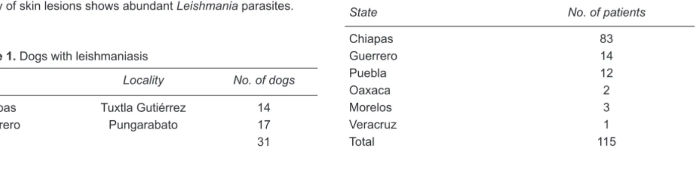
Table 2. Total number of patients with visceral leishmaniasis reported in different Mexican states (1952-2010)
State No. of patients
Chiapas 83
Guerrero 14
Puebla 12
Oaxaca 2
Morelos 3
Veracruz 1
Figure 2. Known occurrence points of patients with visceral leish-maniasis (VL) (red squares), dogs infected with Leishmania (green stars), Lutzomyia longipalpis (black dots) and Lutzomyia evansi
(yellow triangle shown by arrow).
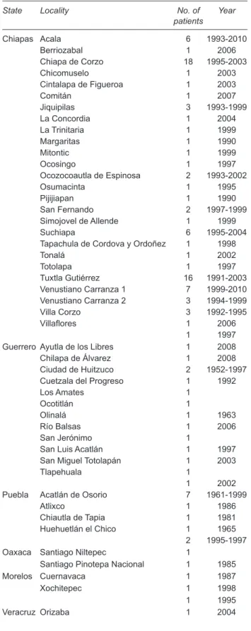
Table 3. Patients with visceral leishmaniasis reported in Mexico
State Locality No. of
patients Year
Chiapas Acala 6 1993-2010 Berriozabal 1 2006 Chiapa de Corzo 18 1995-2003 Chicomuselo 1 2003 Cintalapa de Figueroa 1 2003
Comitán 1 2007
Jiquipilas 3 1993-1999 La Concordia 1 2004 La Trinitaria 1 1999
Margaritas 1 1990
Mitontic 1 1999
Ocosingo 1 1997
Ocozocoautla de Espinosa 2 1993-2002
Osumacinta 1 1995
Pijijiapan 1 1990
San Fernando 2 1997-1999 Simojovel de Allende 1 1999 Suchiapa 6 1995-2004 Tapachula de Cordova y Ordoñez 1 1998
Tonalá 1 2002
Totolapa 1 1997
Tuxtla Gutiérrez 16 1991-2003 Venustiano Carranza 1 7 1999-2010 Venustiano Carranza 2 3 1994-1999 Villa Corzo 3 1992-1995 Villaflores 1 2006
1 1997 Guerrero Ayutla de los Libres 1 2008 Chilapa de Álvarez 1 2008 Ciudad de Huitzuco 2 1952-1997 Cuetzala del Progreso 1 1992
Los Amates 1
Ocotitlán 1
Olinalá 1 1963
Río Balsas 1 2006
San Jerónimo 1
San Luis Acatlán 1 1997 San Miguel Totolapán 1 2003
Tlapehuala 1
1 2002 Puebla Acatlán de Osorio 7 1961-1999
Atlixco 1 1986
Chiautla de Tapia 1 1981 Huehuetlán el Chico 1 1965
2 1995-1997 Oaxaca Santiago Niltepec 1
Santiago Pinotepa Nacional 1 1985 Morelos Cuernavaca 1 1987
Xochitepec 1 1998
1 1995 Veracruz Orizaba 1 2004
Table 4. Patients with visceral leishmaniasis in the state of Chia-pas (1990-2010)
Mean age (months) 33
Male 38
Female 33
Not reported 12
population.20-22 We found that the distribution of patients
with VL and dogs infected with Leishmania overlaps with the distribution area of Lu. longipalpis and Lu. evansi, two sand fly species that are vectors of Leishmania infantum.
Thus, our study provides useful information of a potential risk area of VL in Mexico.
It is noteworthy that VL caused by L. mexicana has also been reported in the state of Tabasco where cutane-ous leishmaniasis is prevalent and dogs infected with L.
mexicana were found to present cutaneous lesions.26,27
These lesions contrast with those of dogs infected with L.
infantum because the latter can present a variety of signs
It is noteworthy that Lu. longipalpis has also been reported in the Yucatan Peninsula from different sam-pling sites; however, parasite occurrence in this area still remains unknown due to the lack of clinical reports and parasite detection.
Although our present work has limitations of not being a population-based random sampling study and therefore does not provide an accurate view of the prevalence of
Leishmania infection in the canine population of this zone
of Guerrero, it does indicate that a well-established focus exists. Our preliminary data on the prevalence of canine leishmaniasis equals the results of some investigators from other parts of the world. This converts the dog as a possible natural reservoir of leishmaniasis that could play an impor-tant role in the ecoepidemiology of the disease causing a potential public health problem that needs to be addressed.
Acknowledgments
This work was supported by grants CONACyT 102155 and PAPIIT, DGAPA-IN 220109 to I.B.
Correspondence: Ingeborg Becker, MD, PhD
Departamento de Medicina Experimental, Facultad de Medicina Universidad Nacional Autónoma de México Mexico, D.F., Mexico E-mail: [email protected]
REFEREnCES
1. Mauricio IL, Howard MK, Stothard JR, Miles MA. Genomic diversity in the Leishmania donovani complex. Parasitology 1999;119:237-246.
2. Maingon RD, Ward RD, Hamilton JG, Bauzer LG, Peixoto AA. The Lutzomyia longipalpis species complex: does population sub-structure matter to Leishmania transmission? Trends Parasitol2008;24:12-17.
3. Ibáñez-Bernal S, Rodríguez-Domínguez G, Gómez-Hernández CH, Ricárdez-Esquinca JR. First record of Lutzomyia evansi
(Nuñez-Tovar 1924) in Mexico (Diptera: Psychodidae, Phle-botominae).Mem Inst Oswaldo Cruz2004;99:127-129. 4. Travi BL, Vélez ID, Brutus L, Segura I, Jaramillo C, Montoya J.
Lutzomyia evansi, an alternate vector of Leishmania chagasi
in a Colombian focus of visceral leishmaniasis. Trans R Soc Trop Med Hyg1990;84:676-677.
5. Zeledón R, Murillo J, Gutiérrez H. Observaciones sobre la ecología de Lutzomyia longipalpis (Lutz & Neiva, 1912) y posibilidades de existencia de leishmaniasis visceral en Costa Rica. Mem Inst Oswaldo Cruz 1984;79:455-459.
6. Montoya-Lerma J, Cadena H, Oviedo M, Ready PD, Barazarte R, Travi BL, et al. Comparative vectorial efficiency of Lutzomyia evansi and Lu. longipalpis for transmitting Leishmania chagasi. Acta Trop 2003;85:19-29.
7. Salomón OD, Quintana MG, Bruno MR, Quiriconi RV, Cabral V. Visceral leishmaniasis in border areas: clustered distribution of phlebotomine sand flies in Clorinda, Argentina. Mem Inst Oswaldo Cruz2009;104:801-804.
8. Andrade Barata R, França-Silva JC, Mayrink W, Costa da Silva J, Prata A, Seixas Lorosa E, et al. Aspectos da ecologia e do comportamento de flebotomíneos em área endêmica de leishmaniose visceral, Minas Gerais. Rev Soc Brasil Med Trop 2005;38:421-425.
9. Pech-May A, Escobedo-Ortegón FJ, Berzunza-Cruz M, Rebo-llar-Téllez EA. Incrimination of four sandfly species previously unrecognized as vectors of Leishmania parasites in Mexico. Med Vet Entomol 2010;24:150-161.
10. Young DG, Duncan MA. Guide to the Identification and Geogra-phic Distribution of Lutzomyia Sand Flies in Mexico, the West Indies, Central and South America (Diptera: Psychodidae). Memoirs of the Entomological Institute No. 54. Gainesville: Associated Publishers; 1994.
11. Travi BL, Montoya J, Gallego J, Jaramillo C, Llano R, Velez ID. Bionomics of Lutzomyia evansi (Diptera: Psychodidae) vector of visceral leishmaniasis in northern Colombia. J Med Entomol 1996;33:278-285.
12. Lainson R, Ward RD, Shaw JJ. Experimental transmission of
Leishmania chagasi, causative agent of neotropical visceral leishmaniasis, by the sandfly Lutzomyia longipalpis. Nature 1977;266:628-630.
13. Cruz-López O, Tamariz-Cruz O, Gándara-Ramírez JL, Rojas-Domínguez R, Cárdenas-Perea ME. A case of visceral leish-maniasis. Rev Invest Clin 1997;49:231-235.
14. Trejo-Pérez JA, Miranda-Novales MG, Solórzano-Santos F, Cabrera-Muñoz L, Díaz-Ponce H. Kala-azar in Mexico: report of 2 cases. Bol Med Hosp Infant Mex 1993;50:662-665. 15. Dorantes-Mesa S. Five cases of kala-azar collected in Mexico.
Bol Med Hosp Infant Mex 1988;45:546-551.
16. Aguirre A, Biagi F, Hernández Nieto A. Second autochtonous case of kala-azar in Mexico. Visceral leishmaniasis. Bol Med Hosp Infant Mex 1963;20:317-333.
17. Baez Villaseñor J, Ruiloba J, Rojas E, Treviño A, Campillo C.A case of kala-azar in Mexico. Bol Oficina Sanit Panam 1953;34:23-30.
18. Velasco-Castrejón, O, Guzmán-Bracho C, Rivas-Sánchez B, Aguilar-Torrentera F. Las Leishmaniasis, con Especial Refe-rencia a México. Publicación Técnica del INDRE n° 4, 1994. México: INDRE/SSA.
19. Montalvo Vázquez AM. Leishmaniasis en pediatría. Reporte de cinco casos en el Hospital Infantil de México Federico Gó-mez. Tesis para la Especialidad en Pediatría Médica, UNAM, Hospital Infantil de México Federico Gómez. México; 1995. 20. Margonari C, Freitas CR, Ribeiro RC, Moura ACM, Timbó M,
Gripp AH, et al. Epidemiology of visceral leishmaniasis through spatial analysis, in Belo Horizonte municipality, state of Minas Gerais, Brazil. Mem Inst Oswaldo Cruz 2006;101:31-38. 21. Werneck GL, Costa CHN, Walker AM, David JR, Wand
22. Queiroz PVS, Monteiro GRG, Macedo VPS, Rocha MAC, Batista LMM, Quieroz JW, et al. Canine visceral leishmania-sis in urban and rural areas of Northeast Brazil. Res Vet Sci 2009;86:267-273.
23. Ricárdez-Esquinca R, Gómez-Hernández C, Guevara A. En-cuesta rápida de Leishmaniasis visceral en caninos en un área endémica en Chiapas. REDVET 2005;6. Available at: http:// www.veterinaria.org/revistas/redvet/n080805B/080503B.pdf 24. Instituto Nacional de Estadística y Geografía (INEGI). Available
at: http://www.inegi.gob.mx
25. Berzunza-Cruz M, Bricaire G, Salaiza-Suazo N, Pérez-Montfort R, Becker I. PCR for identificaction of species causing
Ame-rican cutaneous leishmaniasis. Parasitol Res 2009;104:691-699.
26. Velasco-Castrejón O, Rivas-Sánchez B, Munguía-Saldaña A, Hobart O. Leishmaniasis cutánea de perros en México. Enf Inf Microbiol 2009;29:135-140.
27. Monroy-Ostria A, Hernández-Montes O, Barker DC. Aetiology of visceral leishmaniasis in Mexico.Acta Trop 2000;75: 155-161.