Vaccine–Induced Immunity by Blocking Binding and
Functions of Cross-Reactive Antibodies
Xuqing Zhang1, Maria Eugenia Rodrı´guez2, Eric T. Harvill1*
1Department of Veterinary and Biomedical Sciences, Pennsylvania State University, University Park, Pennsylvania, United States of America,2Department of Chemistry, Centre of Applied Biotechnology (CINDEFI, CCyT La Plata), School of Science, La Plata University, La Plata, Argentina
Abstract
Although the prevalence ofBordetella parapertussisvaries dramatically among studies in different populations with different vaccination regimens, there is broad agreement that whooping cough vaccines, composed only ofB. pertussisantigens, provide little if any protection againstB. parapertussis. In C57BL/6 mice, aB. pertussiswhole-cell vaccine (wP) provided modest protection againstB. parapertussis, which was dependent on IFN-c. The wP was much more protective against an isogenicB. parapertussisstrain lacking O-antigen than its wild-type counterpart. O-antigen inhibited binding of wP–induced antibodies toB. parapertussis, as well as antibody-mediated opsonophagocytosisin vitroand clearancein vivo. aP–induced antibodies also bound betterin vitroto the O-antigen mutant than to wild-typeB. parapertussis, but aP failed to confer protection against wild-type or O antigen–deficient B. parapertussis in mice. Interestingly, B. parapertussis–specific antibodies provided in addition to either wP or aP were sufficient to very rapidly reduceB. parapertussisnumbers in mouse lungs. This study identifies a mechanism by which one pathogen escapes immunity induced by vaccination against a closely related pathogen and may explain whyB. parapertussisprevalence varies substantially between populations with different vaccination strategies.
Citation:Zhang X, Rodrı´guez ME, Harvill ET (2009) O Antigen AllowsB. parapertussisto EvadeB. pertussisVaccine–Induced Immunity by Blocking Binding and Functions of Cross-Reactive Antibodies. PLoS ONE 4(9): e6989. doi:10.1371/journal.pone.0006989
Editor:Ulrich A. Maus, Hannover School of Medicine, Germany
ReceivedJune 11, 2009;AcceptedAugust 19, 2009;PublishedSeptember 14, 2009
Copyright:ß2009 Zhang et al. This is an open-access article distributed under the terms of the Creative Commons Attribution License, which permits unrestricted use, distribution, and reproduction in any medium, provided the original author and source are credited.
Funding:This work was supported by NIH grant GM083113 (ETH) and ANPCyT PICT559 (MER). The funders had no role in study design, data collection and analysis, decision to publish, or preparation of the manuscript.
Competing Interests:The authors have declared that no competing interests exist.
* E-mail: [email protected]
Introduction
Whooping cough is an acute, highly contagious, paroxysmal coughing illness [1]. The first whooping cough vaccines consisting of whole inactivatedB. pertussiswere licensed in the mid-1940s and led to a dramatic decrease of disease incidence [1,2]. However, the potential health risk associated with whole cell vaccines led to the development of acellular vaccines, consisting of some combination of B. pertussis antigens including pertussis toxin (PT), pertactin, filamentous hemaglutinin (FHA) and 2 fimbriae serotypes. Despite maintenance of high vaccine coverage, the reported whooping cough incidence has been increasing over the past 20 years in some developed countries [3,4], although a large portion of whooping cough infections are thought to remain unreported [5]. Both B. pertussis and B. parapertussis are causative agents of whooping cough [1,6] that appear to have evolved independently from distinct lineages of B. bronchisepticathrough rearrangements and large scale gene loss, with B. parapertussis emerging more recently thanB. pertussis[7,8]. Although they are closely related, a few striking differences exist between the human-adapted bordetellae. For example,B. parapertussislipopolysaccharide (LPS) includes a repetitive membrane-distal O-antigenic structure, while B. pertussis only expresses lipid A and a branched-chain core oligosaccharide with a complex trisaccharide modification, but lacks O-antigen [9,10].B. pertussisexpresses PT, butB. parapertussis
does not due to mutations in the promoter region [11,12]. Since differential diagnosis of B. pertussis and B. parapertussis does not affect the course of treatment, it is rarely performed in clinical settings [13,14]. The CDC does not list B. parapertussis as reportable [3], but a few epidemiological studies have reported the percentage of whooping cough cases caused byB. parapertussis to be from 1% to 98%, most commonly 4–40% [13]. AlthoughB. parapertussisappears to contribute substantially to disease, whoop-ing cough vaccines are solely derived fromB. pertussis[1].
Clinical and experimental data indicate that whooping cough vaccines are very efficacious againstB. pertussisbut not againstB. parapertussis[14–18], however, a mechanistic understanding of this phenomenon has not been described. While whooping cough vaccines may fail to generate efficient cross-immunity againstB. parapertussis, it is also possible that cross-reacting adaptive immunity is generated but is evaded byB. parapertussis. Recently, our lab showed that the O-antigen ofB. parapertussisshields it from B. pertussis-infection-induced antibodies [19], relevant to the natural immune-mediated competition between B. pertussis and B. parapertussis in unvaccinated population. However, nearly all people in industrialized countries are vaccinated, changing the immune landscape of the host population and the immune-mediated competition between these two human pathogens.
cell vaccine (wP) had some effect, but a commercial acellular vaccine (aP) had no effect againstB. parapertussisgrowth in mouse lungs. IFN-c
contributes to the protection againstB. parapertussisby wP. O-antigen shieldedB. parapertussisfrom the binding of vaccine-induced antibodies, interfered with opsonophagocytosis ofB. parapertussismediated by aP and wP-induced antibodies and blocked antibody-mediated clearance in vivo. wP conferred more protection against an isogenicB. parapertussis strain lacking O-antigen, indicating that O-antigen contributed to the evasion of wP-induced immunity. aP, however, failed to induce cross-protection againstB. parapertussiswith or without the hindrance of O-antigen. InB. pertussisvaccinated hosts, supplement ofB. parapertussis-specific, but not B. pertussis-specific, antibodies conferred protection against B. parapertussis, indicating that the lack of proper antibody responses causes the failure of these vaccines against B. parapertussis. Together these results explain the clinical finding thatB. parapertussis avoids clearance by the current vaccines, and provides a mechanistic understanding that will guide new approaches to overcoming this problem.
Materials and Methods
Bacterial strains and growth
B. pertussis strain 536, B. parapertussis strain CN2591 and its isogenic mutant lacking O-antigen, CN2591Dwbm, have been described previously [10,20]. For opsonization, attachment and phagocytosis experiments, these strains were transformed with plasmid pCW505 (kindly supplied by Dr. Alison Weiss, Cincinnati, Ohio) which induces cytoplasmic expression of GFP without affecting growth, or antigen expression [21]. Bacteria were maintained on Bordet-Gengou agar (Difco) supplemented with 10% sheep’s blood (Hema Resources) and 20mg/mL
streptomy-cin (Sigma-Aldrich). Liquid cultures were grown overnight in Stainer-Scholte broth at 37uC to mid-log phase [22,23].
Cells
Peripheral blood polymorphonuclear leukocytes (PMNs) were isolated from heparinized venous blood using Ficoll-Histopaque (Sigma, St Louis, MO) gradient centrifugation. PMNs were harvested and the remaining erythrocytes were removed by hypotonic lysis. Cell viability was.99% as determined by Trypan Blue exclusion. Prior to functional assays, PMNs were washed twice with Dulbecco’s modified Eagle medium (DMEM) (Hyclone) supplemented with 10% of fetal calf serum (FCS) (Hyclone), resuspended, and used immediately. All experiments were carried out with freshly isolated PMNs lacking FccRI (CD64) expression, as monitored by FACS analysis using a fluorescence-activated cell sorter FACScan (Becton Dickinson, San Jose, CA) with anti-FccRI mAb 22 [24].
Opsonization
GFP-expressing strains were opsonized by incubation at 37uC with 5% heat-inactivated wP-induced/naive or aP/adjuvant-induced serum samples for 30 min in a final volume of 50mL. Serum opsonized bacteria were incubated with R-phycoerythrin (RPE)–labeled goat F(ab9)2fragments of anti-mouse IgG (Southern
Biotechnology, Birmingham, AL) for 30 min at 4uC. Opsonization of eachBordetellastrain was determined by FACS analysis [25].
Attachment and phagocytosis
Attachment and phagocytosis of the Bordetella strains were evaluated as previously described with a few modifications [26]. Briefly, serum opsonized GFP-expressing bacteria were incubated with PMNs at multiplicity of infection (MOI) of 30 for 20 min at 37uC to allow binding. After extensive washing to remove non-attached bacteria, an aliquot was maintained on ice to be used for
bacterial attachment control. Another aliquot was further incubated for 1 h at 37uC to allow internalization. Phagocytosis was stopped by placing PMNs on ice. Cell surface bound bacteria in both aliquots (before and after 1 hour incubation at 37uC) were detected by incubation with RPE–labeled goat F(ab9)2fragments of anti-mouse
IgG at 4uC for 30 min. To avoid eventual nonspecific binding of antibodies, all incubations were done in the presence of 25% heat-inactivated human serum. After washing, samples were analyzed by flow cytometry. Ten thousand cells were analyzed per sample. Green fluorescence intensity associated with PMNs maintained at 37uC for 20 min has previously been shown to represent bacterial attachment [25]. Phagocytosis was calculated from the drop in mean red fluorescence intensity of green-positive cells after incubation for additional1h at 37uC as described [25].
Animal experiments
C57BL/6 mice were obtained from Jackson Laboratories (Bar Harbor) and bred in our Bordetella-free, specific pathogen-free breeding rooms at The Pennsylvania State University. 4–6 week old mice were sedated with 5% isoflurane (Abbott Laboratory) in oxygen and vaccinated by intraperitoneally (i.p.) injection of 16108 CFU of heat-inactivated bacteria in 1 mL of phosphate balanced saline (PBS, Omnipur) (wP), 1/5 human dose of Adacel (Sanofi Pastuer) (0.5mg PT, 1mg FHA, 0.6mg pertactin, 5mg
fimbriae 2 and 3 per mouse) with Imject Alum (Thermo Scientific) (aP) or only Imject Alum in 200mL PBS on day 14 and 28 prior to challenge [27]. For challenge, mice were sedated and inoculated by pipetting 50mL PBS containing 56105CFU of the indicated bacteria onto the external nares [28]. This method reliably distributes the bacteria throughout the respiratory tract [29]. For adoptive transfer of immune serum, mice were vaccinated with the indicated bacteria on day 0 and 14 and sera were collected on day 28 or sera were collected from naı¨ve animals. 200mL of sera were
i.p. injected at the time of inoculation [30,31]. For quantification of bacteria numbers, mice were sacrificed via CO2inhalation and
the lung, trachea, and nasal cavity were excised. Tissues were homogenized in PBS, serial diluted and plated onto Bordet-Gengou agar plates with 20mg/mL streptomycin, and colonies were counted after incubation at 37uC for 3–5 days [30]. Gamma interferon (IFN-c) was depleted by i.p. injections of 5 mg of the antibody from hybridoma XMG1.2 one day prior to challenge [32]. The lower limit of detection was 10 CFU. For all experiments, protocols were reviewed and approved by the Pennsylvania State University IACUC and all animal were handled in accordance with institutional guidelines.
Splenocyte re-stimulations
Spleens were excised from groups of 3–4 C57BL/6 mice after vaccination. Splenocytes were isolated as previously described [27,33]. In brief, spleens were homogenized and red blood cells were lysed with 0.84% ammonium chloride. 26106cells were
re-suspended in DMEM supplemented with 10% FCS, 1 mM sodium pyruvate (HyClone), and 100mg/mL penicillin and streptocycin (HyClone) and placed into each well of a 96-well tissue culture plate. Splenocytes were stimulated with either media alone or media containing 107CFU (MOI of 5) of the indicated bacteria that had been heat-killed [27,33]. After three days, the supernatants were collected and analyzed for IFN-c and interleukin-10 (IL-10) production via Enzyme-linked immunosorbent assays (ELISA) as per the manufacturers’ instructions (R&D Systems).
Titer ELISAs
bacteria were diluted to 56107CFU/mL in a 1:1 mix of 0.2 M
sodium carbonate and 0.2 M sodium bicarbonate buffers. These antigens were coated onto 96-well plates, incubated for 2 h at 37uC in a humidified chamber, washed and blocked. A1:50 or 1:10 dilution of wP-induced/naive or aP/adjuvant-induced serum samples from an individual mouse was added to the first well of each row and serially diluted 1:2 across the plates. Plates were incubated for 2 h at 37uC, washed and probed with 1:4000 dilution of goat anti-mouse Ig horseradish peroxidase (HRP)-conjugated antibodies (Southern Biotech) for 1 h and visualized with 2,29-Azino-bis (3-ethylbenzothiazoline-6-sulfonic acid) dia-mmonium salt in phosphate-citrate buffer with hydrogen peroxide at an absorbance of 405 nm. Titers were determined via the endpoint method based on optimal density of identically treated wells probed with naı¨ve or adjuvant-induced sera.
Western blot analysis
Lysates containing 56105CFU of indicated heat-killed bacteria
were run on 7% sodium dodecyl sulfate-polyacrylamide gel electrophoresis (SDS-PAGE) gels in denaturing conditions. Poly-vinylidene Fluoride (PVDF) membranes (Millipore)were probed overnight with either naı¨ve serum or serum from vaccinated mice
at a 1:10 or 1:100 dilution for aP and wP-induced serum respectively. 1:10,000 dilution of goat anti-mouse Ig HRP-conjugated antibodies (Southern Biotech) was used as the detector antibody [19,36]. The membrane was visualized with ECL Western Blotting Detection Reagent (Pierce Biotechnology).
Statistical analysis
The means+/2standard error (error bars) were determined for all appropriate data. Two-tailed, unpaired Student’s T-tests were used to determine statistical significance between groups. Results were also analyzed by ANOVA and Tukey simultaneous test in Minitab with similar significance. All experiments were performed at least twice with similar results.
Results
B. parapertussisO-antigen contributes to the evasion of wP–induced immunity
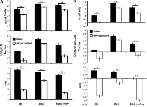
[image:3.612.61.553.314.671.2]To examine whether wP is cross-protective against B. parapertussis and whether O-antigen interferes with its cross-protection, naı¨ve or wP vaccinated C57BL/6 mice were challenged with 56105CFU of B. pertussis, B. parapertussis or an
Figure 1.B. parapertussisis more susceptible to wP–induced immunity in the absence of O-antigen.Groups of four naı¨ve (black) or wP vaccinated (white) C57BL/6 mice were challenged with the indicated bacteria. (A) The number of CFUs recovered from the respiratory tract on day 3 post-challenge is expressed as the Log10mean6the standard error. Decreases in Log10CFU in vaccinated mice compared to naı¨ve mice on day 3
post-challenge are indicated underneath thexaxes. (B) The change in CFU number over the first 3 days after challenge is expressed as change in Log10mean6the standard error. * indicates P#0.05. ** indicates P#0.01. The limit of detection is indicated as the lower limit of theyaxes.
isogenic B. parapertussis strain lacking O-antigen (BppDwbm). In comparison to naı¨ve mice, wP treatment reduced B. pertussis numbers by 91.9%, 97.8% and .99.9% in the nasal cavity, trachea and lungs by day 3 post challenge; naı¨ve mice having about 7000 fold more bacteria in the lungs than vaccinated mice (Figure 1A). wP vaccination reduced B. parapertussis loads by 76.6%, 83.0% and 97.6% in the nasal cavity, trachea and lung; naı¨ve mice having about 40 fold more bacteria in the lungs than vaccinated mice (Figure 1A). These results are consistent with multiple clinical studies showing whole cell vaccines confer good protection againstB. pertussisbut relatively little protection against B. parapertussis [16,17,37]. Interestingly, compared to naı¨ve mice, wP reduced numbers of an O-antigen deficient B. parapertussis strain by 89.4%, 99% and .99.9% in the nasal cavity, trachea and lung; naı¨ve mice having around 2000 fold more bacteria in the lungs than vaccinated mice (Figure 1A). The fold protection (reduction of bacterial number in each individual vaccinated mouse compared to mean number of bacteria in naive mice on day 3 post-challenge) of wP against O-antigen deficient B. parapertussis was significantly higher than against wild-type B. parapertussis in both trachea and lung (lung: P = 0.0006, trachea: P = 0.018), indicating that wP is more efficacious against O-antigen deficientB. parapertussis.
To understand how vaccination affects the infection, it is important to examine these effects in the context of the dynamic infectious process. B. pertussis and B. parapertussis increased in numbers throughout the respiratory tract of naı¨ve mice over 3 days, reflecting effective colonization and bacterial growth (Figure 1B). O-antigen deficient B. parapertussisgrew in the nasal cavity and trachea but not in the lungs of naı¨ve mice due to its increased susceptibility to complement [38] (Figure 1B). wP decreased the numbers ofB. pertussisin all respiratory organs over 3 days, resulting in a net decline in numbers, particularly in the lower respiratory tract (LRT). Vaccination did not decrease B. parapertussis numbers as efficiently in the lung andB. parapertussis actually grew in numbers in the trachea and nasal cavity, reflecting successful colonization and expansion despite vaccination (Figure 1B). Interestingly, wP vaccination decreased O-antigen deficientB. parapertussisnumbers as efficiently as it didB. pertussis numbers in the trachea and lung (Figure 1B). Together these results are consistent with clinical studies showing that wP vaccination confers relatively little protection againstB. parapertussis and show that O-antigen is required for B. parapertussisto avoid efficient wP vaccine-induced immunity.
Many developed countries have switched to acellular vaccines, although these provide even less protection againstB. parapertussis [16,18,37]. We therefore determined if the O-antigen of B. parapertussis contributes to the evasion of aP-induced immunity. While aP reducedB. pertussiscolonization in both lung and trachea of vaccinated mice by 99.7% and 96.8% compared to the mice given just adjuvant, aP had no effects on eitherB. parapertussisor O-antigen deficientB. parapertussis colonization (Figure 2). These data indicate that aP does not induce protective immunity against B. parapertussis.
wP induces T cells that cross react withB. parapertussis
To investigate why aP is less effective than wP against B. parapertussis and why wP confers different levels of protection against B. pertussis, O-antigen deficient and wild-type B. paraper-tussis, we compared their induction of T cell responses known to be important for control and clearance of B. parapertussis [34]. Splenocytes from mice that were naı¨ve, vaccinated with wP, aP or adjuvant were stimulated with heat-killed B. pertussis or wild-type or O-antigen deficientB. parapertussis. After 3 days, IFN-cand
IL-10 production, representative of TH1 and TH2 responses, was
measured. aP vaccination did not affect IFN-c or IL-10 levels, which were similar to those induced by naı¨ve and adjuvant-treated controls (Figure 3). In contrast, splenocytes from wP-vaccinated mice responded similarly to heat-killedB. parapertussisorB. pertussis, producing significantly higher IFN-cand IL-10 than splenocytes from naı¨ve or adjuvant-treated mice (Figure 3). IFN-cand IL-10 production was abolished in TCRb/d2/2mice (data not shown), indicating that vaccine-induced IFN-c and IL-10 production is dependent on T cells. These data indicate that the T-cell response to wP is cross-reactive toB. parapertussisand that O-antigen did not affect the T cell response.
IFN-ccontributes to wP–induced protection againstB. parapertussis
[image:4.612.319.552.60.380.2]In the vaccination studies above (Figure 1, Figure 2), protection againstB. parapertussiscorrelates with the high IFN-cresponses of wP but not aP vaccinated animals (Figure 3). IFN-c has been shown to contribute to leukocyte recruitment and the reduction of bacterial numbers during B. parapertussis infection (D.N. Wolfe, A.T. Karanikas, S.E. Hester, M.J. Kennett, E.T. Harvill, submitted for publication). To determine how the cross-reactive IFN-c response after wP vaccination might contribute to its protection against B. parapertussis, naı¨ve or wP vaccinated mice Figure 2. aP does not reduceB. parapertussisnumbers.Groups of four C57BL/6 mice were vaccinated with adjuvant only (black) or aP (white), challenged with the indicated bacteria and sacrificed on day 3 post-challenge. The numbers of CFUs recovered from the respiratory tract are expressed as the Log10mean6the standard error. ** indicates
P#0.01. The limit of detection is indicated as the lower limit of they
axes.
were left untreated or depleted of IFN-c, challenged with B. pertussis orB. parapertussis and sacrificed 3 days later for bacterial enumeration. Vaccination or IFN-cdepletion had no effects on colonization of the nasal cavity (Figure 4). However, B. pertussis numbers were reduced by.99.9% and.99.5% in the lung and trachea of vaccinated mice compared to naı¨ve mice regardless of the presence or absence of IFN-c(Figure 4). Although wP reduced B. parapertussisnumbers in the lung and trachea by about 98.6% and 99.6%, this effect was abolished in mice given IFN-c
neutralizing antibodies (Figure 4), indicating that IFN-c contrib-utes to the protection conferred by wP againstB. parapertussis.
Serum antibody responses to aP and wP are cross-reactive to denatured but not liveB. parapertussis
We have previously shown that antibodies are required for anamnestic immunity toB. parapertussis[34]. To determine whether B. pertussisvaccines induce antibodies that recognize and/or protect againstB. parapertussis, we first tested whether antibody responses are cross-reactive between strains and if O-antigen affects the responsiveness of antibodies. In a western blot, whole cell extracts ofB. pertussis, wild-type and O-antigen deficientB. parapertussiswere probed with serum antibodies from aP, adjuvant only, wP vaccinated or naı¨ve mice. Compared to the control, aP vaccination induced serum antibodies that recognized distinct antigens present in all three bacteria, although the size and intensity of bands
appeared to differ betweenB. pertussisandB. parapertussis. While wP-induced serum antibodies recognized four major bands in denatured B. parapertussis, they recognize more antigens in B. pertussis, especially those of high molecular weights (Figure 5A).
To quantify the cross-reactivity of antibodies, we determined the titers of aP or wP-induced serum antibodies by ELISA using heat-inactivated bacteria as antigens. wP-induced sera had much higher titers than aP-induced sera (Figure 5B). For both aP and wP-induced serum antibodies,B. pertussis-specific antibody titers were much higher than B. parapertussis-specific antibody titers regardless of the presence or absence of O-antigen (Figure 5B). Since heat killing releases many antigens that are not exposed on the surface of live bacteria, similar experiments were performed with live bacteria as antigens. Interestingly, both wP and aP-induced serum antibodies bound to live B. parapertussis more efficiently in the absence of O-antigen (Figure 5C). These data suggest that O-antigen interferes with the binding ofB. pertussis vaccine-induced antibodies to liveB. parapertussis.
O-antigen inhibits aP– and wP–induced, antibody-mediated opsonization and subsequent attachment to, and phagocytosis by, PMNs
[image:5.612.59.383.62.401.2]To determine whether O-antigen interferes with key functions of antibodies, we assessed its effects on opsonization and subsequent attachment to and phagocytosis by PMNs mediated Figure 3. Splenic production of IFN-cand IL-10 is cross-reactive.Splenocytes from groups of four naı¨ve C57BL/6 mice or mice vaccinated
with the indicated vaccine were stimulated with media only (vertically hatched), heat-killedB. pertussis(black),B. parapertussis(white) or O-antigen deficientB. parapertussis(horizontally hatched) for 3 days. The concentration of IFN-cand IL-10 in the supernatant is expressed as the mean6the standard error. ND indicates none detected.
by aP or wP induced antibodies. Consistent with the ELISA results (Figure 5C),B. pertussis was efficiently opsonized by both aP and wP-induced serum antibodies (Figure 6A, 6B, and 6D). Although B. parapertussiswas efficiently opsonized by whole cellB. parapertussis (wPP)-induced serum antibodies (Figure 6C and 6D), aP or wP-induced antibodies failed to opsonize this bacterium (Figure 6A, 6B, and 6D). In contrast, O-antigen deficientB. parapertussis was efficiently opsonized by aP or wP-induced antibodies (Figure 6A, 6B, and 6D), indicating that O-antigen hinders the opsonization of B. parapertussisby both aP and wP-induced serum antibodies.
To examine if O-antigen blocking of opsonization results in inhibitory effects on attachment of B. parapertussisto phagocytes, bacteria opsonized with vaccine-induced antibodies were further incubated with polymorphonuclear leukocytes (PMN) for 20 mins and cell surface bound bacteria numbers were identified by flow cytometry. aP or wP-induced-antibody-opsonized B. pertussis
attached to PMNs efficiently (Figure 7A, 7B, and 7D). B. parapertussisattached to PMN after opsonization with wPP-induced serum antibodies (Figure 7C and 7D), but neither aP nor wP-induced antibodies mediated attachment of this bacterium to PMNs (Figure 7A, 7B, and 7D).B. parapertussislacking O-antigen, opsonized with aP or wP-induced antibodies, efficiently attached to phagocytes (Figure 7A, 7B, and 7D), indicating that O-antigen also inhibits this process.
To determine if O-antigen interferes with aP or wP-induced antibodies ability to mediate phagocytosis of B. parapertussis by PMNs, an aliquot of cells from the attachment experiment was incubated for one extra hour and phagocytosis was measured. No significant internalization of naı¨ve serum-opsonized bacteria was observed.B. pertussisopsonized with aP or wP-induced antibodies was efficiently phagocytosed, but similarly treated B. parapertussis was not (Figure 8). In contrast, wPP-induced antibody-opsonized B. parapertussiswas efficiently internalized by PMNs (Figure 8).B. parapertussis lacking O-antigen opsonized by aP or wP-induced antibodies was efficiently phagocytosed by PMNs. Together these results indicate that O-antigen protectsB. parapertussisfrom aP or wP-induced serum antibody mediated opsonization, attachment to phagocytes and subsequent internalization by these cells.
O-antigen blocks antibody-mediated clearance ofB. parapertussisfrom mouse lungs
Since O-antigen interferes with the binding of B. pertussis vaccine-induced antibodies to liveB. parapertussisand phagocytosis dependent on these antibodies in vitro, we tested if O-antigen inhibits antibody-mediated clearance in vivo. Serum from naı¨ve (NS), wP- or wPP-vaccinated mice were transferred to naı¨ve animals. Bacterial loads in the lungs were determined on day 14 postB. parapertussischallenge since antibodies have no effect until B. parapertussisspecific T cell responses are generated around day 14 [30,39] (D.N. Wolfe unpublished data). Serum from wPP vaccinated animals significantly lowered numbers of both wild-type and O-antigen deficient B. parapertussis compared to naı¨ve serum treated animals and modestly reducedB. pertussisnumbers (Figure 9). This indicates that although lower level of phagocytosis of O-antigen deficientB. parapertussis mediated by wPP-induced serum antibodies were observed compared to wild-type strain (Figure 8), this low level of phagocytosis and/or other biological activities of passively transferred antibodies might be sufficient to reduce O-antigen deficient strain in vivo (Figure 9). wP-induced serum completely clearedB. pertussisfrom the lung by day 14 post-challenge but did not reduceB. parapertussisnumbers. These serum antibodies did, however, reduce the numbers of O-antigen deficientB. parapertussisby more than 90% (Figure 9), indicating that O-antigen prevents the wP-induced serum antibody mediated reduction ofB. parapertussisnumbersin vivo.
B. parapertussis–specific antibodies augmentB. pertussis vaccine-induced protective immunity againstB.
parapertussis
[image:6.612.62.297.61.451.2]We have previously shown that both antibodies and T cells are required for anamnestic protective immunity againstB. parapertussis [34]. Although wP vaccination induced T cell responses that were cross-reactive (Figure 4) and antibodies that bound antigens from heat-killed B. parapertussis (Figure 5B), O-antigen decreased the binding of these antibodies to liveB. parapertussis (Figure 5), the opsonophagocytosis mediated by these antibodiesin vitro(Figure 6, Figure 7, Figure 8) and their antibody-mediated clearancein vivo (Figure 9). Based on these observations, we hypothesized that wP induces sufficient T cell response but the antibody response is not Figure 4. IFN-c contributes to the protection against B.
parapertussis by wP.Groups of four naı¨ve (black and horizontally hatched) or wP vaccinated (white and vertically hatched) C57BL/6 mice were untreated (2) (black and white) or i.p. injected with (+) (horizontally and vertically hatched) anti-IFN-c antibody, challenged withB. parapertussisand sacrificed 3 days post-challenge. The number of CFUs throughout the respiratory tract is expressed as the Log10mean
6the standard error. * indicates P#0.05. ** indicates P#0.01. The limit of detection is indicated as the lower limit of theyaxes.
Figure 5. O-antigen inhibits the binding ofB. pertussisvaccine-induced antibodies to live, but not denatured,B. parapertussiscells.
(A) Western blots were performed on lysates of indicated bacteria probed with serum antibodies collected from mice that were vaccinated with aP, adjuvant only (Adj) or wP. (B) Heat-inactivated or (C) liveB. pertussis(black),B. parapertussis(white) or O-antigen deficientB. parapertussis(hatched) were coated on ELISA plate. Serum antibody titer of mice vaccinated with aP or wP is expressed as mean of the end point titers of four independent samples6the standard error. ** indicates P#0.01. The dashed line indicates the limit of detection.
sufficient to clear B. parapertussis because O-antigen protects B. parapertussisfrom wP-induced antibodies. If this were the case, then adding antibodies that bind liveB. parapertussisshould render wP induced immunity sufficient to rapidly clearB. parapertussis. To test this, mice were vaccinated with aP (Figure 10A) or wP (Figure 10B), these vaccinated mice were left untreated or given naı¨ve, wP or wPP-induced serum antibodies at the time of B. parapertussischallenge and sacrificed three days after challenge. To compare the protective immunity to that generated by B. parapertussis, a group of mice were vaccinated with wPP and adoptively transferred with wPP-induced antibodies. Less than 100 CFUs ofB. parapertussiswere recovered in the LRT of this group of mice by day 3 post-challenge (Figure 10B). The very high titers of this wPP-induced serum antibodies alone had modest effect on reducing B. parapertussis numbers (Figure 10B), consistent with prior results with convalescent serum [30,39]. In both aP and wP vaccinated mice, naı¨ve sera had no effect, indicating that components in the serum other than those induced by vaccination do not affect bacteria numbers. Transfer of wP-induced sera had
no effect onB. parapertussiscolonization throughout the respiratory tract. However,B. parapertussisnumbers were significantly lower in the LRT of both aP and wP vaccinated animals given wPP-induced sera than in those given naı¨ve sera (Figure 10). These data suggest that the addition of B. parapertussis specific antibodies is sufficient to render wP and aP effective againstB. parapertussis.
Discussion
[image:8.612.320.551.61.390.2]The increasing incidence of whooping cough in highly vaccinated developed countries includes an unknown proportion ofB. parapertussis infections[3,4]. Multiple clinical and experimental studies have shown that current whooping cough vaccines have little efficacy against this bacterium [14–18], but have not revealed why. In this study, we determined that O-antigen shields vaccine-induced antibodies from binding toB. parapertussisand prevents antibody-mediated opsonopha-gocytosisin vitroand antibody-mediated clearancein vivo. Although O-antigen requires a large multigenicwbmlocus to produce andB. pertussis is still successful in the human population without this surface antigen [40], it is retained inB. parapertussis[7]. Apart from its role in inhibiting Figure 6. O-antigen decreases the opsonization ofB.
paraper-tussisbyB. pertussisvaccine-induced antibodies.Representative histograms show flow cytometric analysis of indicated bacteria opsonized with naı¨ve serum (white) or (A) aP, (B) wP, (C) wPP-induced serum (grey) and stained with RPE-labeled goat F(ab)2fragments of
anti-mouse IgG. (D) Mean red-fluorescence of B. pertussis (black), B. parapertussis (white), O-antigen deficient B. parapertussis (hatched) opsonized with indicated serum from four individual mice 6 the standard error is shown. AU indicates arbitrary units. ** indicates P#0.01.
[image:8.612.60.291.63.405.2]doi:10.1371/journal.pone.0006989.g006
Figure 7. O-antigen blocks B. pertussis vaccine-induced anti-bodies from mediating adherence ofB. parapertussisto PMNs.
Naı¨ve serum (white) or (A) aP, (B) wP, (C) wPP-induced serum (grey)-opsonized GFP-expressing bacteria were incubated with freshly isolated human peripheral blood PMNs. Representative histograms of flow cytometry analysis of these cells are shown. (D) Mean green fluorescence associated with PMNs incubated with GFP-expressingB. pertussis (black), B. parapertussis (white) or O-antigen deficient B. parapertussis (hatched) opsonized with four independent indicated serum6the standard error is shown. AU indicates arbitrary units. ** indicates P#0.01.
Figure 8. O-antigen blocksB. pertussisvaccine-induced, anti-body-mediated phagocytosis ofB. parapertussis.Freshly isolated human peripheral blood PMNs were incubated for 20 min or 1 h and 20 min with GFP-expressingB. pertussis(black),B. parapertussis(white), or O-antigen deficient B. parapertussis (hatched), that are opsonized with serum from aP, wP, wPP-vaccinated or naı¨ve mice. The cell surface bound bacteria were detected by incubation with RPE-labeled goat F(ab’)2 fragments of anti-mouse IgG. Mean phagocytosis calculated
[image:9.612.319.553.62.194.2]from the decrease in red-fluorescence of green-positive cells incubated for 1 h and 20 min compared to that incubated for 20 min of experiments done with 4 independent serum samples6the standard error was shown. AU indicates arbitrary units. ** indicates P#0.01. doi:10.1371/journal.pone.0006989.g008
Figure 9. Passive transfer of wP–induced serum antibodies mediates clearance of O-antigen deficient, but not wild-type,B. parapertussisfrom mouse lungs.Groups of four C57BL/6 mice were adoptively transferred naı¨ve serum (black), wP-induced serum (white) or wPP-induced serum (hatched) at the time of bacterial challenge and dissected 14 days later. The number of CFUs recovered from lung is expressed as the Log10mean6the standard error. * indicates P#0.05
and ** indicates P#0.01 compared to mice given naı¨ve serum. The limit of detection is indicated as the lower limit of theyaxes. ND indicates undetectable bacterial number.
doi:10.1371/journal.pone.0006989.g009
Figure 10. Passive transfer of B. parapertussis–specific antibodies rapidly reduces B. parapertussis colonization in aP and wP vaccinated animals.Groups of four C57BL/6 mice were (B) left untreated (2) or vaccinated with (A) aP, (B) wPP or wP. Mice lacking a transfer of antibodies (2) or given naı¨ve serum (NS), wP-induced serum (wP IS) or wPP-induced serum (wPP IS) were challenged withB. parapertussis. Mice were sacrificed three days post-challenge and the number of CFUs recovered from the respiratory tract is expressed as the Log10mean6the standard
[image:9.612.63.552.335.670.2]complement deposition and complement-mediated killing [38], our study suggests that O-antigen, by decreasing B. pertussis-vaccine-induced antibody binding, may confer a selective advantage to B. parapertussisin human populations in which there is high prevalence of detectable immunity toB. pertussis.
It is interesting that wP, but not aP, induced IFN-c, which contributed to protection againstB. parapertussisbut notB. pertussis (Figure 4). IFN-cplays a role in the recruitment and activation of phagocytic cells [41,42]. InB. parapertussisinfected mice, the peak of neutrophil numbers in the lung on day 7 and subsequent control of B. parapertussisnumbers are dependent on IFN-c(D.N. Wolfe, A.T. Karanikas, S.E. Hester, M.J. Kennett, E.T. Harvill, submitted for publication).B. pertussischallenged IFN-c2/2mice, however, have
an indistinguishable course of infection in the respiratory tract and recruit similar numbers of leukocytes to the site of infection as compared to wild-type mice [43] (Wolfe unpublished data), indicating that IFN-cis not required forB. pertussisclearance and leukocyte recruitment. In vaccinated IFN-cR2/2 mice, no impaired reduction ofB. pertussisnumbers is observed during the first week after challenge, although a rebound of bacteria number was observed on day 10 [44]. This is consistent with our observation that IFN-cis not required for vaccine-mediated control ofB. pertussis numbers on day 3 post-challenge (Figure 4).
Consistent with multiple clinical and experimental studies, we found that while wP confers some level of protection against B. parapertussis, aP does not [16,18]. The decrease of pertussis acellular vaccines efficacy against B. parapertussis compared to whole cell vaccines has been suggested to be attributable to the failure of antibodies induced by pertussis acellular vaccines to block the adherence ofB. parapertussisto epithelial cells [45] or the immune suppressive effects of the FHA included in those vaccines [46]. Among the antigens included in the aP, only fimbriae, but not FHA and pertactin, was shown to confer cross-protection againstB. parapertussisin a mouse model [37,47]. wP contains more antigens than aP, among which there may be cross-protective antigens. Alternatively, the differences in Th1/Th2 skewing of wP and aP may affect vaccine efficacy. wP induces a relatively balanced Th1/Th2 response whereas aP induces a Th2-type response [48]. Our data showed that wP, but not aP, induced high splenic IFN-cproduction and wP was no longer protective against B. parapertussis when IFN-cwas depleted, suggesting that IFN-c
contributes to the protection conferred by wP (Figure 3, Figure 4). These data are therefore consistent with the idea that inducing a Th1 response enhances immunity against B. parapertussis. The different Th1/Th2 skewing, quantity and quality of cross-reactive antibodies and possible immune suppression factors in aP may also explain why aP vaccination was not sufficient to induce protection againstB. parapertussiseven without the hindrance by O-antigen.
Our data reveal an interesting paradox. wP-mediated clearance of B. parapertussis by day 3 requires IFN-c, which aP does not induce (Figure 3, Figure 4). Yet when wPP-induced antibodies, which had limited effect in naı¨ve animals in the first week of infection (Figure 10B) [30], were given to aP-vaccinated animals, B. parapertussiswas effectively cleared from mouse lungs within 3 days (Figure 10A). IFN-cappears to contribute to the protection conferred by wP, but aP-vaccination contributes to rapid clearance despite the lack of detectable IFN-cinduction in our splenocyte re-stimulation assay (Figure 3). It is possible that some low level of IFN-cwas induced by aP vaccination, which we failed to detect, or some other cross-reactive protective T cell cytokine responses aid in the rapid clearance mediated by B. parapertussis-specific antibodies in aP-vaccinated animals.
Although it is not clear how muchB. parapertussiscontributes to the resurgence of whooping cough, the low efficacy of current vaccines against this bacterium might confer a selective advantage to B. parapertussis, relative to B. pertussis. Moreover, the lower protection againstB. parapertussis conferred by acellular vaccines than whole cell vaccine could affect the relative prevalence, a possibility of greater significance since the recent switch from whole cell to acellular vaccines and the even more recent introduction of acellular vaccines, including the one used here, for adolescents and adults [16,18,49,50]. Considering adults to be a possible reservoir for transmission to infants [51,52], the lack of cross-protection of Adacel, determined in this study, may open a niche forB. parapertussisnot only in adolescents/adults but also in infants. This study shows that B. parapertussis evades B. pertussis vaccine induced immunity by blocking cross-reactive antibodies binding via O-antigen. Our current data provide strong evidence that including moreB. pertussisproteins that induce antibodies that recognize orthologs inB. parapertusisis unlikely to improve vaccines so that they protect against B. parapertussis. Since our study indicates that supplementing wP or aP withB. parapertussis-specific antibodies rendered them effective against B. parapertussis (Figure 10), addition of protective antigens of B. parapertussis to the vaccine may substantially improve its efficacy against this pathogen.
Acknowledgments
We would like to thank Dr. Daniel N. Wolfe and Dr. Anne M. Buboltz for discussion and critical review of the manuscript.
Author Contributions
Conceived and designed the experiments: XZ MER ETH. Performed the experiments: XZ MER. Analyzed the data: XZ MER. Wrote the paper: XZ MER ETH.
References
1. Mattoo S, Cherry JD (2005) Molecular pathogenesis, epidemiology, and clinical manifestations of respiratory infections due to Bordetella pertussis and other Bordetella subspecies. Clin Microbiol Rev 18: 326–382.
2. Cherry JD, Brunell PA, Golden GS, Karson DT (1988) Report of the task force on pertussis and pertussis immunization-1988. Pediatrics 81.
3. (2002) From the Centers for Disease Control and Prevention. Pertussis–United States, 1997-2000. JAMA 287: 977–979.
4. Celentano LP MM, Paramatti D, Salmaso S, Tozzi AE; EUVAC-NET Group (2005) Resurgence of pertussis in Europe. Pediatr Infect Dis J 24: 761–765. 5. de Melker HE VF, Schellekens JF, Teunis PF, Kretzschmar M (2006) The
incidence of Bordetella pertussis infections estimated in the population from a combination of serological surveys. J Infect 53: 106–113.
6. Hoppe JE (1999) Update on respiratory infection caused by Bordetella parapertussis. Pediatr Infect Dis J 18: 375–381.
7. Parkhill J, et al. (2003) Comparative analysis of the genome sequences of Bordetella pertussis, Bordetella parapertussis and Bordetella bronchiseptica. Nat Genet 35: 32–40.
8. Diavatopoulos DA CC, Schouls LM, Brinig MM, Relman DA, Mooi FR (2005) Bordetella pertussis, the causative agent of whooping cough, evolved from a distinct, human-associated lineage of B. bronchiseptica. PLoS Pathog 1: e45. doi:10.1371/journal.ppat.0010045.
9. van den Akker WM (1998) Lipopolysaccharide expression within the genus Bordetella: influence of temperature and phase variation. Microbiology 144: 1527–1535.
10. Preston A, Allen AG, Cadisch J, Thomas R, Stevens K, et al. (1999) Genetic basis for lipopolysaccharide O-antigen biosynthesis in bordetellae. Infect Immun 67: 3763–3767.
11. Marchitto KS, Smith SG, Locht C, Keith JM (1987) Nucleotide sequence homology to pertussis toxin gene in Bordetella bronchiseptica and Bordetella parapertussis. Infect Immun 55: 497–501.
12. Arico B, Rappuoli R (1987) Bordetella parapertussis and Bordetella bronchiseptica contain transcriptionally silent pertussis toxin genes. J Bacteriol 169: 2847–2853. 13. Watanabe M NM (2004) Whooping cough due to Bordetella parapertussis: an
14. He Q, Viljanen MK, Arvilommi H, Aittanen B, Mertsola J (1998) Whooping cough caused by Bordetella pertussis and Bordetella parapertussis in an immunized population. JAMA 280: 635–637.
15. Mastrantonio P, Stefanelli P, Giulano M, Herrera Rojas Y, Ciofi degli Atti M, Anemona A, Tozzi AE (1998) Bordetella parapertussis infection in children: epidemiology, clinical symptoms, and molecular characteristics of isolates. J Clin Microbiol 36: 999–1002.
16. Liese JG RC, Stojanov S, Belohradsky BH, Munich Vaccine Study Group (2003) Clinical and epidemiological picture of B pertussis and B parapertussis infections after introduction of acellular pertussis vaccines. Arch Dis Child 88: 684–687. 17. Heininger U SK, Christenson P, Cherry JD (1998) Evidence of efficacy of the
Lederle/Takeda acellular pertussis component diphtheria and tetanus toxoids and pertussis vaccine but not the Lederle whole-cell component diphtheria and tetanus toxoids and pertussis vaccine against Bordetella parapertussis infection. Clin Infect Dis 28: 602–604.
18. David S, van Furth R, Mooi FR (2004) Efficacies of whole cell and acellular pertussis vaccines against Bordetella parapertussis in a mouse model. Vaccine 22: 1892–1898.
19. Wolfe DN, Goebel EM, Bjornstad ON, Restif O, Harvill ET (2007) The O antigen enables Bordetella parapertussis to avoid Bordetella pertussis-induced immunity. Infect Immun 75: 4972–4979.
20. Stibitz S, Yang MS (1991) Subcellular localization and immunological detection of proteins encoded by the vir locus of Bordetella pertussis. J Bacteriol 173: 4288–4296.
21. Weingart CL, Broitman-Maduro G, Dean G, Newman S, Peppler M, et al. (1999) Fluorescent labels influence phagocytosis of Bordetella pertussis by human neutrophils. Infect Immun 67: 4264–4267.
22. von Koenig CH, Tacken A, Finger H (1988) Use of supplemented Stainer-Scholte broth for the isolation of Bordetella pertussis from clinical material. J Clin Microbiol 26: 2558–2560.
23. Stainer DW, Scholte MJ (1970) A simple chemically defined medium for the production of phase I Bordetella pertussis. J Gen Microbiol 63: 211–220. 24. Repp R, Valerius T, Sendler A, Gramatzki M, Iro H, et al. (1991) Neutrophils
express the high affinity receptor for IgG (Fc gamma RI, CD64) after in vivo application of recombinant human granulocyte colony-stimulating factor. Blood 78: 885–889.
25. Rodriguez ME, Van der Pol WL, Van de Winkel JG (2001) Flow cytometry-based phagocytosis assay for sensitive detection of opsonic activity of pneumococcal capsular polysaccharide antibodies in human sera. J Immunol Methods 252: 33–44.
26. Rodriguez ME, Hellwig SM, Hozbor DF, Leusen J, van der Pol WL, et al. (2001) Fc receptor-mediated immunity against Bordetella pertussis. J Immunol 167: 6545–6551.
27. Gopinathan L, Kirimanjeswara GS, Wolfe DN, Kelley ML, Harvill ET (2007) Different mechanisms of vaccine-induced and infection-induced immunity to Bordetella bronchiseptica. Microbes Infect 9: 442–448.
28. Kirimanjeswara GS, Agosto LM, Kennett MJ, Bjornstad ON, Harvill ET (2005) Pertussis toxin inhibits neutrophil recruitment to delay antibody-mediated clearance of Bordetella pertussis. J Clin Invest 115: 3594–3601.
29. Harvill ET, Preston A, Cotter PA, Allen AG, Maskell DJ, et al. (2000) Multiple roles for Bordetella lipopolysaccharide molecules during respiratory tract infection. Infect Immun 68: 6720–6728.
30. Kirimanjeswara GS, Mann PB, Harvill ET (2003) Role of antibodies in immunity to Bordetella infections. Infect Immun 71: 1719–1724.
31. Pishko EJ, Kirimanjeswara GS, Pilione MR, Gopinathan L, Kennett MJ, et al. (2004) Antibody-mediated bacterial clearance from the lower respiratory tract of mice requires complement component C3. Eur J Immunol 34: 184–193. 32. Parent MA, Wilhelm LB, Kummer LW, Szaba FM, Mullarky IK, et al. (2006)
Gamma interferon, tumor necrosis factor alpha, and nitric oxide synthase 2, key elements of cellular immunity, perform critical protective functions during humoral defense against lethal pulmonary Yersinia pestis infection. Infect Immun 74: 3381–3386.
33. Pilione MR HE (2006) The Bordetella bronchiseptica type III secretion system inhibits gamma interferon production that is required for efficient antibody-mediated bacterial clearance. Infect Immun 74: 1043–1049.
34. Wolfe DN KG, Harvill ET (2005) Clearance of Bordetella parapertussis from the lower respiratory tract requires humoral and cellular immunity. Infect Immun 73: 6508–6513.
35. Myc A, Buck J, Gonin J, Reynolds B, Hammerling U, et al. (1997) The level of lipopolysaccharide-binding protein is significantly increased in plasma in patients with the systemic inflammatory response syndrome. Clin Diagn Lab Immunol 4: 113–116.
36. Wolfe DN, Kirimanjeswara GS, Goebel EM, Harvill ET (2007) Comparative role of immunoglobulin A in protective immunity against the Bordetellae. Infect Immun 75: 4416–4422.
37. Willems RJ, Kamerbeek J, Geuijen CA, Top J, Gielen H, Gaastra W, Mooi FR (1998) The efficacy of a whole cell pertussis vaccine and fimbriae against Bordetella pertussis and Bordetella parapertussis infections in a respiratory mouse model. Vaccine 16: 410–416.
38. Goebel EM, Wolfe DN, Elder K, Stibitz S, Harvill ET (2008) O antigen protects Bordetella parapertussis from complement. Infect Immun 76: 1774–1780. 39. Wolfe DN, Buboltz AM, Harvill ET (2009) Inefficient Toll-like receptor-4
stimulation enables Bordetella parapertussis to avoid host immunity. PLoS ONE 4: e4280. doi:10.1371/journal.pone.0004280.
40. Burns V, Pishko EJ, Preston A, Maskell DJ, Harvill ET (2003) Role of Bordetella O-antigen in respiratory tract infection. Infect Immun 71: 86–94.
41. Sun K, Salmon SL, Lotz SA, Metzger DW (2007) Interleukin-12 promotes gamma interferon-dependent neutrophil recruitment in the lung and improves protection against respiratory Streptococcus pneumoniae infection. Infect Immun 75: 1196–1202.
42. Ellis TN, Beaman BL (2004) Interferon-gamma activation of polymorphonu-clear neutrophil function. Immunology 112: 2–12.
43. Mahon BP, Sheahan BJ, Griffin F, Murphy G, Mills KH (1997) Atypical disease after Bordetella pertussis respiratory infection of mice with targeted disruptions of interferon-gamma receptor or immunoglobulin mu chain genes. J Exp Med 186: 1843–1851.
44. Mills KH, Brady M, Ryan E, Mahon BP (1998) A respiratory challenge model for infection with Bordetella pertussis: application in the assessment of pertussis vaccine potency and in defining the mechanism of protective immunity. Dev Biol Stand 95: 31–41.
45. van den Berg BM, Beekhuizen H, Mooi FR, van Furth R (1999) Role of antibodies against Bordetella pertussis virulence factors in adherence of Bordetella pertussis and Bordetella parapertussis to human bronchial epithelial cells. Infect Immun 67: 1050–1055.
46. McGuirk P, McCann C, Mills KH (2002) Pathogen-specific T regulatory 1 cells induced in the respiratory tract by a bacterial molecule that stimulates interleukin 10 production by dendritic cells: a novel strategy for evasion of protective T helper type 1 responses by Bordetella pertussis. J Exp Med 195: 221–231.
47. Khelef N, Danve B, Quentin-Millet MJ, Guiso N (1993) Bordetella pertussis and Bordetella parapertussis: two immunologically distinct species. Infect Immun 61: 486–490.
48. Mills KH (2001) Immunity to Bordetella pertussis. Microbes Infect 3: 655–677. 49. CDC (2006) Preventing Tetanus, Diphtheria, and Pertussis Among Adults: Use of Tetanus Toxoid, Reduced Diphtheria Toxoid and Acellular Pertussis Vaccine. MMWR 55: 1–33.
50. (2006) Prevention of pertussis among adolescents: recommendations for use of tetanus toxoid, reduced diphtheria toxoid, and acellular pertussis (Tdap) vaccine. Pediatrics 117: 965–978.
51. Nelson JD (1978) The changing epidemiology of pertussis in young infants. The role of adults as reservoirs of infection. Am J Dis Child 132: 371–373. 52. Wirsing von Konig CH, Postels-Multani S, Bock HL, Schmitt HJ (1995)