Thematic update
Reward and aversion systems
of the brain as a functional unit.
Basic mechanisms and functions
Anaclara Michel-Chávez,
1Bruno Estañol-Vidal,
1Horacio Sentíes-Madrid,
1Erwin Chiquete,
1Guillermo R. Delgado,
1Guillermina Castillo-Maya
1ABSTRACT
Introduction
It is increasingly important to recognize the reward and aversion sys-tems of the brain as a functional unit. A fundamental task of the mam-malian brain is to assign an emotional/motivational valence to any stimuli by determining whether they are rewarding and should be ap-proached or are aversive and should be avoided. Internal stimuli are also assigned an emotional/motivational valence in a similar fashion.
Objective
To understand the basic mechanisms and functions of the reward and aversion system of the brain.
Method
A bibliographical search was conducted in the Pubmed database us-ing different key words. Documents on relevant aspects of the topic were selected.
Results
In the ventral tegmental area, dopaminergic (VTA-DA) neurons play a role in reward-dependent behaviors. It is also known that the inhibition of the VTA-DA neurons by GABAergic neurons contributes to a reward prediction error calculation that promotes behaviors associated with aversion. The ventral dopaminergic mesolimbic system and the nu-cleus accumbens are activated during reward and inhibited during aversions. The amygdala is activated during aversive behavior.
Discussion and conclusion
The reward/aversion system is highly relevant for survival, which is most likely its primary function. It is involved in important pathologies such as addiction, depression and autonomic and endocrine distur-bances. Therefore, its knowledge has become of clinical importance. Although great advances have been made in the knowledge of the basic mechanisms of the reward/aversion system, the detailed circuits within the VTA that mediate reward and aversion and the ana-tomical substrates are not completely clear.
Key words: Reward system, aversion system, dopaminergic sys-tems, limbic system, pleasure.
RESUMEN
Introducción
Es muy importante reconocer el sistema de recompensa y aversión del cerebro como una unidad funcional. Una de las funciones fundamen-tales del cerebro de los mamíferos es la capacidad para designar un valor emocional/motivacional a cualquier estímulo. Esta capacidad permite identificar un estímulo como gratificante y aproximarnos a él, o reconocerlo como aversivo y evitarlo.
Objetivo
Comprender los mecanismos fisiológicos del sistema de recompen-sa-aversión.
Método
Se realizó una búsqueda bibliográfica en la base de datos Pubmed con las diferentes palabras clave. Se seleccionaron los documentos sobre los aspectos relevantes.
Resultados
Las neuronas dopaminérgicas del área tegmental ventral (ATV) cum-plen un papel importante en los comportamientos dependientes de la recompensa. Asimismo, la inhibición de las neuronas dopaminérgicas ATV por parte de las neuronas GABAérgicas contribuye a predecir la recompensa y promueve comportamientos aversivos. Este sistema se ac-tiva durante actividades de recompensa y se inhibe durante la aversión. La amígdala es la principal estructura relacionada con la aversión.
Discusión y conclusión
Este sistema se considera de gran importancia para la supervivencia de las especies, la que parece ser su función primordial. Interviene en distintas patologías como adicciones, depresión, trastorno por estrés postraumático, fobias y trastornos endocrinos y autonómicos, por lo que el conocimiento de este sistema es de gran importancia clínica.
Aunque se ha avanzado mucho en el estudio y entendimiento de este sistema y de sus circuitos anatómicos ubicados en el ATV mesen-cefálica y sus conexiones con áreas subcorticales, el conocimiento de este sistema funcional sigue siendo un desafío científico.
Palabras clave: Sistema de recompensa, sistema de aversión, sis-temas dopaminérgicos, sistema límbico, placer.
1 Department of Neurology and Psychiatry, Laboratory of Clinical Neurophysiology, Instituto Nacional de Ciencias Médicas y Nutrición Salvador Zubirán,
Mexico City.
ANATOMY AND GENERALITIES
The limbic system is an old brain structure that plays an important role in learning and memory functions. It is also involved in the generation, integration and control of emo-tions and their behavioral responses. For instance, the inter-pretation of facial expressions and the underlying emotional status of a person, or the evaluation of a dangerous situa-tion and the decision to express an appropriate behavioral response (e.g. “fight or flight”), involve a variety of limbic
brain regions, including the amygdala and prefrontal cor-tical regions. The limbic system is also tightly linked to the autonomic nervous system and, via the hypothalamus, reg-ulates endocrine, cardiovascular and visceral functions. The
amygdala, hippocampus and medial prefrontal cortex (PFC) critically influence the responses of the hypothalamic-pitu
-itary-adrenal axis (HPAA).1,2 Furthermore, dysfunctions in
these brain areas are implicated in the etiology of mental disorders, including depression and posttraumatic stress disorder, which are often characterized by HPAA abnor-malities,3 and in attention deficit-hyperactivity disorder.2,4
The amygdala is connected to the PFC, hippocampus, septum and dorsomedial thalamus. Because of its connec-tivity within the limbic system, it plays an important role in the mediation and control of emotions, including love and affection, fear, aggression, and reward, and therefore is es-sential for social behavior and the survival of the individual and the survival of the species. For instance, damage to the amygdala reduces aggressive behavior and the experience
of fear (i.e, makes one less fearful), whereas electrical stimu -lation of the amygdala has the opposite effect.1,2
The nucleus accumbens plays an important role in re-ward, pleasure, emotions, aggression and fear. The core and shell subregions of the nucleus accumbens receive inputs from a variety of limbic and prefrontal regions, including the amygdala and hippocampus, and make up a network involved in acquisition, encoding and retrieval of aversive learning and memory processes.5
The PFC can be roughly subdivided into: dorsolateral, medial (which may include the functionally related anterior
cingulate cortex) and orbitofrontal cortex (OFC).6 Both the
medial PFC and OFC are part of a frontostriatal circuit that
has strong connections to the amygdala and other limbic re-gions.7 Thus, prefrontal regions are anatomically well suited
to integrate cognitive and emotional functions.8
The OFC is involved in sensory integration and higher cognitive functions, including decision making. The OFC
also plays an essential role in a variety of emotional func-tions, such as judgment of the hedonic aspects of rewards or reinforcers, which are important for the planning of be-havioral responses associated with reward and punishment.
Dysfunction or damage to the OFC can result in poor empa
-thy and impaired social interactions. The OFC also plays a
major role in the regulation of aggression and impulsivity,
and damage to the OFC leads to behavioral disinhibition,
such as compulsive gambling, drug use and violence.8
The cingulate cortex can be cytoarchitectonically and functionally differentiated into an anterior part, which ex-erts executive functions, and a posterior part, which is eval-uative. Furthermore, the cingulate cortex has two major subdivisions: a dorsal cognitive division and a rostral-ven-tral affective division.9 A variety of studies indicate an
an-atomic and functional continuum rather than strictly segre-gated operations.9,10
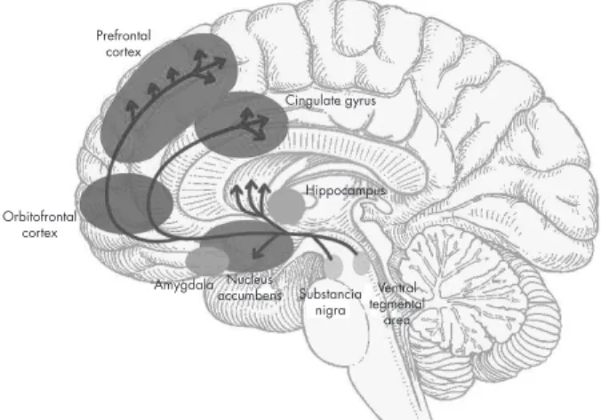
The ventral and dorsal areas of the cingulate cortex play a role in autonomic and a variety of rational cognitive functions, such as reward anticipation, decision making, empathy, and emotional regulation 11 (figure 1).
Reward and aversion
In 1954, James Olds and Peter Milner reported the results of
what became a milestone in the research on reward mech-anisms. They inserted electrodes into the brains of rats and then placed the rats in operant chambers equipped with a lever that, when depressed, would deliver current to the electrodes. Under these conditions, when an electrode was implanted in certain regions of the brain, notably the sep-tal area, the rats would press the lever “to stimulate [them-selves] in these places frequently and regularly for long
pe-riods of time if permitted to do so”.12 Not only will animals
work for food when they were hungry or for water when they were thirsty, but even when sated rats would work for electrical stimulation of their brains.12,13
The seminal work of Olds and Milner unleashed a
barrage of research studies into the brain mechanisms of reward, most immediately, a spate of studies on electrical brain reward.13
The biological basis of mood-related states, such as reward and aversion, is not yet fully understood, albeit it is recognized as a functional neuronal network. Classical
Figure 1. Brain reward pathways. Prefrontal
cortex
Hippocampus Orbitofrontal
cortex
Substancia nigra Nucleus
accumbens tegmentalVentral area Amygdala
formulations of these states implicate the mesocorticolim-bic system, comprising brain areas that include the nucleus
accumbens (NAc), the ventral tegmental area (VTA) and the
prefrontal cortex, implicated reward;14-17 and the amygdala
(AMG), periaquaductal gray (PAG), and the locus coeruleus (LC), often implicated in aversion.14,18
However, the notion that certain brain areas narrowly and rigidly mediate reward or aversion are becoming archa-ic in regard with the development of sophistarcha-icated tools and methodologies that provide new evidence.17
The NAc, especially its outer region, called the shell, has been shown to play a necessary role in assigning moti-vational properties on rewards and to the stimuli that pre-dict them. Thus, for example, in rat and mouse models, an intact NAc is required if the animal is to learn to work (e.g.
lever pressing) to obtain natural rewards, such as palatable foods, or to learn how to self-administer drugs. Once an an -imal learns how to obtain a reward, and the relevant behav-iors become ingrained, reward seeking no longer depends on the NAc and is supported by the dorsal striatum (the
caudate and putamen in humans), the brain structure that
underlies well-learned behaviors and habits.7,19
Under natural conditions, speed and efficiency in
gaining food, water and shelter improve the probability of survival. Thus, a critical role of reward circuitry is to facili-tate the rapid learning of cues that predict the proximity of reward and of the behaviors that maximize the chances of
successfully obtaining it. Once learned, predictive cues au -tomatically activate cognitive, physiologic and behavioral responses aimed at obtaining the predicted reward.20,21
VTA dopamine neurons are the primary source of do
-pamine (DA) in target structures such as the medial pre -frontal cortex and the NAc, which play important roles in a broad range of motivating behaviors and neuropsychiatric disorders.22-24
Dopamine, released from VTA neurons in the NAc, plays
the key role in binding rewards and reward associated cues to adaptive reward-seeking responses.25,26 In animals,
im-planted electrodes can record the firing of dopamine neurons;
microdialysis catheters and electrochemical methods can be used to detect dopamine.27 In humans, positron emission
tomography (PET) permits indirect measures of dopamine
release by observing the displacement of a positron-emitting D2 dopamine receptor ligand previously bound to receptors following a stimulus or pharmacologic challenge. Using such methods in multiple paradigms, it has been well-established
that natural rewards cause firing of neurons and dopamine
release in the NAc and other forebrain regions. When
do-pamine action is blocked −whether by lesioning dodo-pamine
neurons, blocking post-synaptic dopamine receptors or
in-hibiting dopamine synthesis− rewards no longer motivate
the behaviors necessary to obtain them.27-29
Much evidence suggests that the precise pattern of do
-pamine neuron firing, and the resulting synaptic release of
dopamine in forebrain circuits, act to shape behavior so as to maximize future reward.19,30
In a basal state, dopamine neurons have a slow tonic
pattern of firing. When a new, unexpected, or greater than
expected reward is encountered, there is a phasic burst of
firing of dopamine neurons causing a transient increase
in synaptic dopamine. When a reward is predicted from known cues and is exactly as expected, there is no change
from the tonic pattern of firing, that is, there is no addition -al dopamine release. When a predicted reward is omitted
or less than expected, dopamine neurons pause their firing
to levels below their tonic rate. Phasic increases in synaptic dopamine signify that the world is better than expected, fa-cilitate learning of new predictive information and bind the newly learned predictive cues to action.20,21
Two distinct patterns of dopamine neural activity occur
in the behaving animal. Midbrain dopamine neurons typi
-cally fire at low frequencies of 1-5 Hz, which are thought to produce a tone on the high-affinity dopamine D2 receptor
in terminal regions of the mesolimbic dopamine system, including the NAc. In contrast, when animals are present-ed with motivationally salient stimuli, such as conditionpresent-ed cues that predict drug availability, midbrain dopamine
neu-rons fire in high frequency bursts (>20 Hz), thereby produc -ing transient increases in NAc DA concentrations that are
sufficiently to occupy low affinity DA D1 receptors.31-33
The pleasure–reward system
Reward involves multiple neuropsychological components: 1. the hedonic sensation of pleasure itself; 2. the motivation
to obtain the reward (incentive component); and 3. the re -ward-related learning.34
Pleasure represents the subjective hedonistic value of rewards; it can indeed be either rewarding when it follows satisfaction, or incentive when it reinforces behaviors.35,36
Pleasure is not merely a sensation. Even a sensory plea-sure such as a sweet taste requires the co-recruitment of ad-ditional specialized pleasure-generating neural circuitry to add the positive hedonic impact to the sweetness that elicits liking reactions.34,37
Different rewarding stimuli may elicit qualitatively and quantitatively different reward values. The pleasure or re-warding value associated with sexual intercourse may not only exceed in magnitude that associated with scratching an itch, but the two pleasures or rewarding values may them-selves be in addition qualitatively distinct.13
There is evidence, in animals, of an increased
dopami-nergic activity in the VTA either from an unexpected reward
Endocannabinoid system
The endogenous cannabinoid system is a signaling system composed of cannabinoid receptors, endogenous ligands for these receptors and proteins involved in the formation and deactivation of these endogenous ligands.38-41
Two types of CB receptors have been identified. CB1 is
highly expressed in the brain and mediate most of the psy-choactive/central effects of cannabis10,42-44 Discovered later,
CB2 are presented predominantly in the periphery and, at low levels, in some areas of the brain.43-47 In the
dopaminer-gic mesolimbic system, the best known circuit involved in motivational processes, average to high concentrations of CB1 receptors are found in the terminal region, the striatum, whereas low concentrations of CB1 receptors are found in
the origin, the ventral tegmental area (VTA).10,48
Neurobiology of addiction
All drugs of abuse increase dopamine concentrations in ter-minal regions of the mesolimbic dopamine system. Increas-es in NAc DA are theorized to mediate the primary positive reinforcing and rewarding properties of all known drugs of abuse.49-52
In addition, when animals are presented with condi-tioned stimuli that predict drug availability, transient dopa-mine events that are theorized to mediate the secondary re-inforcing effects of drugs of abuse and initiate drug seeking are also observed in the NAc.31,53
Specifically, each drug mimics or enhances the actions
of neurotransmitters at receptors for these
neurotransmit-ters. Opioids are presumed to be habit-forming because of
actions at opiate receptors, and so is nicotine because of the action at nicotinic acetylcholine receptors. Phencyclidine
acts at N-methyl-D-aspartate (NMDA) and sigma receptors,
and also blocks dopamine reuptake.5,54
Tetrahydrocannabi-nol (THC) binds to endocannabinoid receptors.55,56 Although
amphetamines and cocaine do not act directly at dopamine
receptors, they are reinforcing because they increase the concentration of dopamine at the dopamine receptors of the nucleus accumbens and frontal cortex.16
It has become clear that the acute administration of most drugs of abuse increases dopamine transmission in the basal ganglia,16 and that dopamine transmission in this
brain region plays a crucial role in mediating the reinforcing effects of these drugs.57-60
The mesolimbic dopamine pathway is made up of
do-paminergic cells in the VTA projecting into the NAc, locat -ed in the ventral striatum, and is consider-ed crucial for drug reward.54 The mesostriatal (dopamine cells of the substantia
nigra projecting to the dorsal striatum) and mesocortical (dopamine cells of the VTA projecting to the frontal cortex)
pathways are also recognized in contributing to predicting
drug reward (anticipation) and addiction.61 The time course
of dopamine signaling is also a key factor, where the fastest time course predominantly has a role in reward and attrib-uting value to predicted outcomes of behavior, while steady activation of dopamine release plays a role in providing an
enabling effect on specific behavior-related systems.24 The
mode of dopamine cell firing (phasic vs. tonic) also differ -ently modulates the rewarding, conditioning effects of drugs
(predominantly phasic) versus the changes in executive func
-tion that occur in addic-tion (predominantly tonic) 62 (table 1).
The aversion system by the amygdala
Pavlovian fear conditioning is an associative learning task in which subjects are presented with a neutral conditioned
stimulus (CS) paired with an innately aversive uncondi
-tioned stimulus (US).63
Pavlovian fear conditioning (and its aversive
reinforce-ment) is severely impaired by disruption of the amygdala,
indicating that the amygdala plays an important role in learn-ing to respond defensively to stimuli that predict aversion.63,64
Fear is an intervening variable between sets of con-text-dependent stimuli and suites of behavioral response.
Table 1. Properties of addictive drugs
Drug Neurotransmitter Drug target Effect after binding
Opiates (morphine, heroin) Endorphins μ and δ opioid (agonist) Activate Gi/Go Activate K+ channels Psycho-stimulants (cocaine,
amphetamines) Dopamine (DA) Dopamine transporter (DAT) (an-tagonist) Increase synaptic DA; stimulate presynaptic and postsynaptic DA receptors Nicotine Acetylcholine Nicotinic acetylcholine receptors
(nAChRs) (agonist) Stimulates cation channel (may desensitize) Alcohol γ-Aminobutyric acid (GABA)
Glutamate GABAA receptor (agonist) Activate Cl− channel Marijuana
(Δ9-tetrahydrocan-nabinol) Anandamide Cannabinoid CB1 (agonist) Activate Gi/Go
Hallucinogens (Phencyclidine,
ketamine) NMDA receptor channel (antag-onist) Inhibit Ca2+ entry
Unlike with reflexes, in the case of an emotion like fear, this link is much more flexible and the state can exist prior
to and after the eliciting stimuli.65,66
Previous studies have shown that electrical stimulation of the amygdala evokes fear-associated responses such as car-diovascular changes,67,68 potentiated startle,69 and freezing.70
Contemporary fear models posit that the basolateral
amygdalar (basal and lateral nuclei) complex is intercon
-nected with the central nucleus (CeA), which is thought to
be the main amygdaloid output structure sending efferent
fibers to various autonomic and somatomotor centers in
-volved in mediating specific fear responses.71
Neural activity in the amygdala appears to be required for expression of both conditioned and unconditioned re-sponses to aversive stimuli, whereas synaptic plasticity appears to be required only for acquisition of conditioned responses, and not for expression of conditioned or uncon-ditioned responses.63
After fear memory consolidation, which requires 4–6 h, fear memory becomes remarkably resistant to perturbation, giving way only to numerous unreinforced CS presenta-tions, which lead to the extinction of learned fear respons-es. However, substantial remnants of the originally learned fear survive even after extensive extinction and causes the reappearance of fear-related behavior in a variety of circum-stances, such as fear renewal and facilitated reacquisition.72
The lateral amygdala is essential in the acquisition and consolidation of auditory cued-fear conditioning. Thalamic and cortical auditory inputs to the LA are potentiated after
fear conditioning, which are relayed to the basal and central amygdala so as to evoke aversive behavior.72
It seems that the fear-conditioning circuitry of the amygdala is functionally lateralized according to which side
of the body (left or right) a predicted threat is anticipated
on.73
The LA neuronal population displayed increased
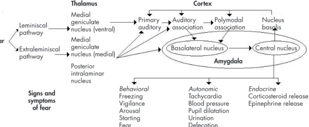
aver-age CS-evoked firing after conditioning, decreased respons -es after extinction, and potentiat-es r-espons-es after recondi-tioning, in tight correlation with the changes in the freezing responses 72,74 (figure 2).
CONCLUSION
The reward and aversion systems are important mecha-nisms for individual and species survival (which is present
in all mammalian animals and perhaps in all animals). The
mesolimbic cortical dopaminergic system is the main sub-strate for the reward system, and the main neurotransmitter is dopamine, although serotonin, noradrenaline, endoge-nous opiates and cannabinoids, and perhaps other transmit-ters, also play a role.
The original function of the reward/aversion system is most likely the survival of the individual and the species, by promoting defense and/or pleasure mechanisms. However, the reward system is also important for sexual behavior, nurs-ing, eating and drinking behavior. Potent psychoactive drugs
stimulate in a non-natural way (massive stimuli) the reward/
Figure 2. Auditory conditioned fear pathways. Sensory information is transmitted to the thalamus via the leminiscal pathway involving the ventral medial geniculate nucleus, which projects only to the primary auditory cortex. Pathways thought to be involved in emotional learning involve extraleminis-cal pathways, which project to the medial division of the medial geniculate nucleus, and the posterior intralaminar nucleus, which project to primary auditory cortex, auditory association cortex, and the basolateral nucleus of the amygdala, which also receives inputs via the auditory association and polymodal association cortex. The basolateral nucleus projects to the central nucleus of the amyg-dala, a major output of the amygamyg-dala, which projects to brain areas that produce the behavioral, autonomic, and endocrine manifestations of fear. The central nucleus also feeds back to the cortex via the nucleus basalis.
Adapted from: Heninger GR. Neuroscience, Molecular Medicine, and New Approaches to the Treatment of Depres-sion and Anxiety. In: Waxman S, editor. From Neuroscience To Neurology. San Diego: Elsevier Academic Press; 2005. Chapter 12.
Signs and symptoms of fear
Thalamus Cortex
Amygdala Ear
Extraleminiscal pathway Leminiscal pathway
Behavioral Freezing Vigilance Arousal Starting Fear Medial
geniculate nucleus (ventral) Medial geniculate nucleus (medial) Posterior intralaminar nucleus
Autonomic Tachycardia Blood pressure Pupil dilatation Urination Defecation
Endocrine
Corticosteroid release Epinephrine release Primary
auditory Auditoryassociation Polymodalassociation Nucleusbasalis
aversion system causing a disproportionate and unnatural response, and thereby inducing addiction and sensitization.
The aversive system may help the animal inducing a
fight/flight behavior or avoiding some food. The amygdala
is probably the main anatomical structure subserving aver-sive behavior, although the inhibition of the reward systems may also trigger aversive behavior.
Activation and sensitization (tachyphilaxia) of the
aversive system is likely to be involved in anxiety disorders, panic attacks, post-traumatic stress disorders, phobias and abstinence syndromes
The reward/aversion system is closely related to mem-ory, emotions and the autonomic and endocrine systems, and it is also related to cognitive functions through its con-nections to the prefrontal lobe. There is a heuristic value in considering the reward/aversion system as a functional unit. Both systems are crucial for survival and this common function should not be underestimated, particularly from an evolutionary viewpoint. Both systems share, albeit not all, anatomical structures and many neurotransmitters
sub-stances. It is perhaps an oversimplification to state that the
withdrawal of one system leads to the overaction of the oth-er but this hypothesis should be explored in the future in clinical and animal research.
Funding
None.
Conflict of interest
No author of this paper has a conflict of interest, including specific financial interest, relationships, and/or affiliations relevant to the
subject matter included in this manuscript.
REFERENCES
1. Schultz W. Multiple dopamine functions at different time courses. Annu Rev Neurosci 2007;30:259–288.
2. Herman JP, Ostrander MM, Mueller NK, Figueiredo H. Limbic system mechanisms of stress regulation: hypothalamo-pituitary-adrenocor-tical axis. Prog Neuropsychopharmacol Biol Psychiatry 2005;29:1201– 1213.
3. Phillips PE, Stuber GD, Heien ML, Wightman RM et al. Subsecond dopamine release promotes cocaine seeking. Nature 2003;422:614–618. 4. Happaney K, Zelazo PD, Stuss DT. Development of orbitofrontal
function: current themes and future directions. Brain Cogn 2004;55:1– 10.
5. Mohanty A, Engels S, Herrington JD et al. Differential engagement of anterior cingulate cortex subdivisions for cognitive and emotional function. Psychophysiology 2007;44:343–351.
6. Hyman SE. Chapter 30: Biology of addiction. En: Goldman L, Scha-fer AI (24 Ed.) Goldman´s Cecil medicine. Philadelphia, PA: Elseviere Saunder; 2012.
7. Tsou K, Brown S, Sanudo-Pena MC, Mackie K et al. Immunohisto-chemical distribution of cannabinoid CB1 receptors in the rat central nervous system. Neuroscience 1998;83:393–411.
8. Dreyer JK, Herrik KF, Berg RW, Hounsgaard JD. Influence of pha -sic and tonic dopamine release on receptor activation. J Neurosci 2010;30:14273–14283.
9. Sesack SR, Grace AA. Cortico-basal ganglia reward network: microcir-cuitry. Neuropsychopharmacol 2010;35:27–47.
10. Bjorklund A, Dunnett SB. Dopamine neuron systems in the brain: an update. Tr Neurosci 2007;30:194–202.
11. Bush G. Cingulate, frontal, and parietal cortical dysfunction in
atten-tion-deficit/ hyperactivity disorder. Biol Psychiatry 2011;69(12):1160–1167.
12. Olds J, Milner P. Positive reinforcement produced by electrical stim-ulation of septal area and other regions of rat brain. J Comp Physiol Psychol 1954;47(6):419-427.
13. Marks LE. A brief history of sensation and reward. In: Gottfried JA (ed.). Neurobiology of sensation and reward. Boca Raton (FL): Chapter 2: CRC Press; 2011.
14. McEwen BS, Magarinos AM. Stress effects on morphology and func-tion of the hippocampus. Ann N Y Acad Sci 1997;821:271–284. 15. William C, Mark, T. Biological substrates of reward and aversion:
a nucleus accumbens activity hypothesis. Neuropharmacology 2009;56(Suppl 1):122–132.
16. Grace AA. The tonic/phasic model of dopamine system regulation and
its implications for understanding alcohol and psychostimulant crav-ing. Addiction 2000;95(Suppl 2):S119–S128.
17. Robinson TE, Berridge KC. Addiction. Annu Rev Psychol 2003;54:25– 53.
18. Fride E. Endocannabinoids in the central nervous system: from neu-ronal networks to behavior. Curr Drug Targets CNS Neurol Disord 2005;4:633–642.
19. Oleson EB, Cheer, JF. A brain on cannabinoids: The role of dopa-mine release in reward seeking. Cold Spring Harb Perspect Med 2012;2:a012229.
20. Matsumoto M, Hikosaka O. Two types of dopamine neuron dis-tinctly convey positive and negative motivational signals. Nature 2009;459:837–841.
21. Cohen JY, Haesler S, Vong L, Lowell BB et al. Neuron-type-specific
signals for reward and punishment in the ventral tegmental area. Na-ture 2012;482:85–88.
22. Bock J, Braun K. The impact of perinatal stress on the functional mat-uration of prefronto-cortical synaptic circuits: implications for the pathophysiology of ADHD? Prog Brain Res 2011;189:155–169. 23. Carlezon WA Jr, Wise RA. Rewarding actions of phencyclidine and
re-lated drugs in nucleus accumbens shell and frontal cortex. J Neurosci 1996;16(9):3112–3122.
24. Onaivi ES, Ishiguro H, Gong JP, Patel S Et al. Discovery of the pres-ence and functional expression of cannabinoid CB2 receptors in brain. Ann N Y Acad Sci 2006;1074:514–536.
25. Bush G, Luu P, Posner MI. Cognitive and emotional influences in an -terior cingulate cortex. Trends Cogn Sci 2000;4(6):215–222.
26. Urban NB, Martínez, D. Neurobiology of addiction. Insight from neu-rochemical imaging. Psychiatr Clin N Am 2012;35:521–541.
27. Roberts DC, Corcoran ME, Fibiger HC. On the role of ascending cate-cholaminergic systems in intravenous self- administration of cocaine. Pharmacol Biochem Behav 1977;6:615–620.
28. Kim Y, Wood J, Moghaddam B. Coordinated activity of ventral teg-mental neurons adapts to appetitive and aversive learning. PLoS ONE 2012;7(1):e29766.
29. Vogel Z, Barg J, Levy R, Saya D et al. Anandamide, a brain
endoge-nous compound, interacts specifically with cannabinoid receptors and
inhibits adenylate cyclase. J Neurochem 1993;61(1):352–355.
30. Wise RA. The role of reward pathways in the development of drug dependence. Pharmacol Ther 1987;35(1–2):227–263.
31. Solinas M, Goldberg SR, Piomelli D. The endocannabinoid system in brain reward processes. British J Pharmacology 2008;154:369–383. 32. Bozarth MA, Wise R. Intracranial self-administration of morphine
into the ventral tegmental area in rats. Life Sci 1981;28:551–555.
33. Tsai HC, Zhang F, Stuber GD, Bonci A et al. Phasic firing in do
-paminergic neurons is sufficient for behavioral conditioning. Science
34. Berridge KC, Kringelbach ML. Neuroscience of affect: Brain mechanisms of pleasure and displeasure. Curr Opin Neurobiol 2013;23(3):294–303.
35. Leknes S, Tracey I. A common neurobiology for pain and pleasure. Nat Rev Neurosci 2008;9(4):314-320.
36. Chenu A, Tassin JP. Pleasure: Neurobiological conception and Freud-ian conception. Encephale 2014;40(2):100-107.
37. Berridge KC. Pleasures of the brain. Brain Cogn 2003;52(1):106-128. 38. Wise RA, Rompré PP. Brain dopamine and reward. Annu Rev Psychol
1989;40:191–225.
39. Herkenham M, Lynn AB, Johnson MR, Melvin LS et al. Characteriza-tion and localizaCharacteriza-tion of cannabinoid receptors in rat brain: a quantita-tive in vitro autoradiographic study. J Neurosci 1991;11:563–583. 40. Braun K. The brefrontal-limbic system: Development, neuroanatomy,
function, and implications for socioemotional development. Clin Peri-natol 2011;38:685–702.
41. Goeders NE, Smith JE. Cortical dopaminergic involvement in cocaine reinforcement. Science 1983;221:773–775.
42. Ritz MC, Lamb RJ, Goldberg SR, Kuhar MJ. Cocaine receptors on do-pamine transporters are related to selfadministration of cocaine. Sci-ence 1987;237:1219–1223.
43. Bromberg-Martin ES, Matsumoto M, Hikosaka O. Dopamine in motivational control: rewarding, aversive, and alerting. Neuron 2010;68:815–834.
44. Grace AA. Phasic versus tonic dopamine release and the modulation of dopamine system responsivity: A hypothesis for the etiology of schizophrenia. Neuroscience 1991;41:1–24.
45. Devane WA, Axelrod J. Enzymatic synthesis of anandamide, an en-dogenous ligand for the cannabinoid receptor, by brain membranes. Proc Natl Acad Sci U S A 1994;91(14):6698–6701.
46. Phillips AG, LePiane G. Disruption of conditioned taste aversion in the rat by stimulation of amygdale: a conditioning effect, not amnesia. J Comp Physiol Psychol 1980;94:664–674.
47. Piomelli D. The molecular logic of endocannabinoid signalling. Nat Rev Neurosci 2003;4:873–884.
48. Owesson-White CA, Ariansen J, Stuber GD, Cleaveland NA et al. Neural encoding of cocaine-seeking behavior is coincident with pha-sic dopamine release in the accumbens core and shell. Eur J Neurosci 2009;30:1117–1127.
49. Munro S, Thomas KL, Abu-Shaar M. Molecular characterization of a peripheral receptor for cannabinoids. Nature 1993;365:61–65.
50. Van Sickle MD, Duncan M, Kingsley PJ, Mouihate A et al. Identifi -cation and functional characterization of brainstem cannabinoid CB2 receptors. Science 2005;310:329–332.
51. Wise RA. Roles for nigrostriatal—not just mesocorticolimbic—dopa-mine in reward and addiction. Trends Neurosci 2009;32(10):517–524. 52. Freund TF, Katona I, Piomelli D. Role of endogenous cannabinoids in
synaptic signaling. Physiol Rev 2003;83:1017–1066.
53. Di Marzo V, Bifulco M, De Petrocellis L. The endocannabinoid system and its therapeutic exploitation. Nat Rev Drug Discov 2004;3:771–784. 54. Witten IB, Steinberg EE, Lee SY, Davidson TJ et al. Recombinase-driv-er rat lines: tools, techniques, and optogenetic application to dopa-mine-mediated reinforcement. Neuron 2011;72:721–733.
55. Herkenham M, Lynn AB, Little MD, Johnson MR et al. Cannabinoid receptor localization in brain. Proc Natl Acad Sci USA 87:1932–1936. 56. Koob GF, Le Moal M. Drug addiction, dysregulation of reward, and
allostasis. Neuropsychopharmacology 2001;24(2):97–129.
57. Van Zessen R, Phillips JL, Budygin EA, Stuber GD. Activation of VTA GABA neurons disrupts reward consumption. Neuron 2012;73:1184– 1194.
58. Gong JP, Onaivi ES, Ishiguro H, Liu QR et al. Cannabinoid CB2 re-ceptors: immunohistochemical localization in rat brain. Brain Res 2006;1071:10–23.
59. Lammel S, Lim BK, Ran C, Huang KW et al. Input-specific con -trol of reward and aversion in the ventral tegmental area. Nature 2012;491(7423):212–217.
60. Blair HT, Sotres-Bayon F, Moita MA, Ledoux JE. The Lateral Amygda-la Processes The Value of Conditioned and Unconditioned Aversive Stimuli. Neuroscience 2005;133(2):561–569.
61. Koob GF. Neural mechanisms of drug reinforcement. Ann N Y Acad Sci 1992;654:171–191.
62. Hyman SE, Malenka RC, Nestlr EJ. Neural mechanisms of addiction: the role of reward-related learning and memory. Annu Rev Neurosci 2006;29:565-598.
63. Blanchard DC, Blanchard RJ. Innate and conditioned reactions to threat in rats with amygdaloid lesions. J Comp Physiol Psychol 1972;81:281–290.
64. Rosen JB, Davis M. Enhancement of acoustic startle by electrical stim-ulation of the amygdala. Behav Neurosci 1988;102(2):195–202. 65. Wise RA, Bozarth MA. Brain mechanisms of drug reward and
eupho-ria. Psychiatr Med 1985;3:445–460.
66. Applegate CD, Kapp BS, Underwood MD, McNall CL. Autonomic and somatomotor effects of amygdala central N. stimulation in awake rabbits. Physiol Behav 1983;31(3):353–360.
67. Weingarten H, White N. Exploration evoked by electrical stimulation of the amygdala of rats. Physiol Psychol 1978;6(2):229–235.
68. Joo Kima E, Horovitzb O, Pellmana B, Mimi Tana L et al. Dorsal peri-aqueductal gray-amygdala pathway conveys both innate and learned fear responses in rats. PNAS 2013;110(36):14795–14800.
69. Bobae An, Hong I, Choi S. Long-term neural correlates of reversible fear learning in the lateral amygdala. J Neuroscience 2012;32(47):16845–16856. 70. Blair H, Huynh V, Vaz V, Van J et al. Unilateral storage of fear
memo-ries by the amygdala. J Neuroscience 2005;25(16):4198–4205.
71. Iwata J, Chida K, LeDoux JE. Cardiovascular responses elicited by stimulation of neurons in the central amygdaloid nucleus in awake but not anesthetized rats resemble conditioned emotional responses. Brain Res 1987;418(1):183–188.
72. Tan KR, Yvon C, Turiault M, Mirzabekov JJ Et al. GABA neurons of the VTA drive conditioned place aversion. Neuron 2012;73:1173–1183. 73. Adolphs, R. The biology of fear. Curr Biol 2013;23(2):R79–R93. 74. Heninger GR. Neuroscience, molecular medicine, and new