R E S E A R C H
Open Access
The fetomaternal interface in the placenta of
three species of armadillos (Eutheria, Xenarthra,
Dasypodidae)
Lorenna C Rezende
1, Claudio G Barbeito
2, Phelipe O Favaron
1, Andrea Mess
1*and Maria A Miglino
1Abstract
Background:Placental characters vary among Xenarthra, one of four supraordinal clades of Eutheria. Armadillos are known for villous, haemochorial placentas similar to humans. Only the nine-banded armadillo has been well studied so far.
Methods:Placentas of three species of armadillos were investigated by means of histology, immunohistochemistry including proliferation marker, and transmission and scanning electron microscopy.
Results:The gross anatomy differed:Euphractus sexcinctusandChaetophractus villosushad extended, zonary placentas, whereasChaetophractus vellerosushad a disk. All taxa had complex villous areas within the maternal blood sinuses of the endometrium. Immunohistochemistry indicated the validity of former interpretations that the endothelium of the sinuses was largely intact. Tips of the villi and the columns entering the maternal tissue possessed trophoblast cell clusters with proliferation activity. Elsewhere, the feto-maternal barrier was syncytial haemochorial with fetal vessels near the surface.
Conclusions:Differences among armadillos occurred in regard to the extension of the placenta, whereas the fine structure was similar. Parallels to the human suggest that armadillos are likely to be useful animal models for human placentation.
Background
Molecular cladistics divides eutherian mammals into four supraordinal clades. Xenarthra likely has a basal position and may influence interpretations of character transformations [1-4]. Phylogenetics have stimulated evolutionary studies as on the placental system [5-9]. Distinct placental types have been recognized with re-gard to invasiveness, shape and internal organization [6,8]. However, only about 1% of eutherian species have been investigated, varying on ordinal and family levels [10]. Attention to rather exotic species such as
armadil-los were drawn from the middle of 20th century on
[11,12], indicating that placental characters vary within Xenarthra. Armadillos are known for haemochorial, vil-lous placentas [11-14] similar to the human [15],
indicating that they could be used as animal models [16].
Only the nine-banded armadillo Dasypus novemcinctus
from North America is well studied in regard to placen-tal development [11-14,17-20]. Data show a peculiar condition in that the developing villi entered pre-existing maternal blood sinuses and enlarged them while leaving
the maternal vessels endothelium largely intact
[11,13,14]. The sinuses were supplied by derivatives of the spiral arteries that were passing in endometrial sep-tae and opened into the intervillous space [11,13,14]. However, data for other armadillo species are sparse [21-23]. We investigated armadillos from Brazil and Argen-tina, the large hairy armadillo Chaetophractus villosus,
DESMAREST, 1904, the small hairy armadillo
Chaeto-phractus vellerosus, GRAY, 1865, and the six-banded
armadilloEuphractus sexcinctus, LINNAEUS, 1758. The
species studied here exhibit litter sizes of 1 to 3 young and gestation periods of 60 to 70 days and inhabit pam-pas, cerrado and chaco vegetations [24-28].
* Correspondence:[email protected]
1Department of Surgery, Faculty of Veterinary Medicine, University of Sao
Paulo, Av. Prof. Dr. Orlando Marques de Paiva, no. 87, Cidade Universitária, São Paulo, SPCEP 05508-000, Brazil
Full list of author information is available at the end of the article
were fixed in 2.5% glutaraldehyde, post-fixed in 2% phos-phate-buffered osmium tetroxide, critical point dried and
gold sputtered. TEM samples were embedded in Spurr’s
[image:2.595.59.538.288.659.2]PCNA (proliferation cell nuclear antigen, clone PC10; 1:300; SIGMA, St Louis, USA); then a biotinylated sec-ondary antibody and streptavidin-HRP (Dako) were
applied and detection was done by Envision and a DAB and substrate chromogen system (DAKO). Negative con-trols were performed using bovine serum albumin to substitute the primary antibody. The research was approved by the Ethical Committee at the Faculty of Vet-erinary Medicine and Animal Science of the University of Sao Paulo.
Results
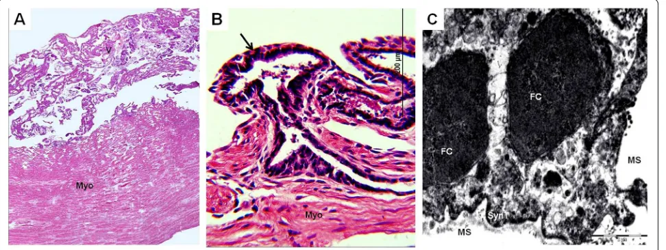
The gross anatomy of the chorioallantoic placenta differed in the three armadillos:Chaetophractus villosus(Figure 1A) andEuphractus sexcinctushad extended, zonary placentas and Chaetophractus vellerosus had a disk (Figure 2A). Chaetophractus villosus had 3 fetuses within a single
chorionic sac (Figure 1A); whereas Chaetophractus
[image:3.595.59.540.90.236.2]vellerosus (Figure 2A) and E. sexcinctus had two fetuses. The placentas of different embryos were not fully sepa-rated (Figure 2A). The placenta was established at the fun-dic end of the uterus. Independent from the macroscopic organization, all three taxa had complex villous areas within the maternal blood sinuses of the endometrium (Figures 1B,C,2B,3A) that reached the myometric region. Indicated by the application of vimentin and cytokeratin in Chaetophractus villosus, the blood sinuses retained parts of the endothelial border (Figures 1D,E). In all three species, this regions were supplied by derivatives of the spiral arteries that had been invaded by the trophoblast (Figures 1D,E,3B). These vessels run within connective tis-sue that was derived from the endometrium (Figure 2B). Tips of the villi and columns entering the maternal tissue Figure 2Chaetophractus vellerosus. (A) Macroscopic anatomy. Chorioallantoic placenta disk (CP) of two embryos (E) with separate umbilical cords (UC). (B) Hematoxylin and eosin. Montage of the villous area (V) with fetal blood vessels inside (BV). Magnification 4*. (C) Hematoxylin and eosin. Villi in the intervillous space with capillaries near the surface.
[image:3.595.58.544.506.689.2]centas occurred in Chaetophractus villosus, Euphractus sexcinctus as well as inCabassous chacoensis,Tolypeutes matacusandDasypus hybridus[22].Dasypus novemcinc-tuspossessed a restricted zonary placenta [11,13]. A disk occurred inChaetophractus vellerosus. For Chaetophrac-tus villosus,Chaetophractus vellerosusand the other spe-cies this represented the term state, whereas for Euphractus sexcinctuswe did not have material from late gestation. All armadillo species studied so far had a com-plex villous area [11-14,17-23]. The application of
immu-nohistochemistry in Chaetophractus villosus indicated
the validity of former interpretations derived from Dasy-pus novemcinctus, that the developing villi entered ma-ternal blood sinuses without fully destroying and replacing the vessels endothelium [11,13,14]. In addition, our results proved the expression of a proliferation mar-ker in the trophoblast cell clusters especially occurring at the tips of the villi [11-13,19-23]. Thus, proliferation can be assumed as general activity of these cells, as was sug-gested by former work on morphology only [11-13]. In the human, the syncytiotrophoblast is formed from pro-liferative cytotrophoblast cells [14,32,33]. We suggest that these cytotrophoblast cells in armadillos may have a similar function. Elsewhere in the villi, the interhaemal barrier was haemomonochorial, syncytial and thin in places; these areas were associated with hypertrophied mesenchymal cells and capillaries that were near the sur-face [11-14,19-23]. These features exhibit parallels to humans [15]. The way in which the villous areas were established was different in primates and armadillos. In humans invasive trophoblast cells migrate deeply into the maternal tissues and remove the endothelium of the arteries [32-35]. However, armadillos are among the very few mammals with villous and invasive placentas. Thus, they may play a role as additional animal models for human placentation [16].
Conclusions
Principal differences between three armadillo species have been revealed in regard to the extension of the pla-centa, whereas the fine structure was mostly similar. Major parallels occurred compared to the human,
Author details
1Department of Surgery, Faculty of Veterinary Medicine, University of Sao
Paulo, Av. Prof. Dr. Orlando Marques de Paiva, no. 87, Cidade Universitária, São Paulo, SPCEP 05508-000, Brazil.2Faculty of Veterinary Science, La Plata
University, 118 La Plata, Buenos Aires, Argentina.
Authors' contributions
MAM devised the study and participated in its design. LCR established the procedure of the material and performed the analysis, helped by CGB, SASK and POF. AM wrote the manuscript. All authors read and approved the final manuscript.
Received: 28 February 2012 Accepted: 24 April 2012 Published: 4 May 2012
References
1. Murphy WJ, Eizirik E, Johnson WE, Zhang YP, Ryder OA, O'Brien SJ:
Molecular phylogenetics and the origins of placental mammals.Nature
2001,409:614–618.
2. Springer MS, Stanhope MJ, Madsen O, Jong WW:Molecules consolidate the placental mammal tree.Trends Ecol Evol2004,19:430–438.
3. Springer MS, Murphy WJ:Mammalian evolution and biomedicine: new views from phylogeny.Biol Rev2007,82:375–392.
4. Delsuc F, Scally M, Madsen O, Stanhope MJ, Jong WW, Catzeflis FM, Springer MS, Douzery EJP:Molecular phylogeny of living Xenarthrans and the impact of character and taxon sampling on the placental tree rooting. Mol Biol Evol2002,19:1656–1671.
5. Mess A, Carter AM:Evolutionary transformations of fetal membrane characters in Eutheria with special reference to Afrotheria.J Exp Zool Part B: Mol Dev Evol2006,306B:140–163.
6. Mess A, Carter AM:Evolution of the placenta during the early radiation of placental mammals.Comp Biochem Physiol2007,148:769–779. Part A.
7. Wildman DE, Chen C, Erez O, Grossman LI, Goodman M, Romero R:
Evolution of the mammalian placenta revealed by phylogenetic analysis. PNAS2006,103:3203–3208.
8. Elliot MG, Crespi BJ:Phylogenetic evidence for early hemochorial placentation in Eutheria.Placenta2009,30:49–67.
9. Martin RD:Evolution of placentation in primates: implications of mammalian phylogeny.Evol Biol2008,35:125–145.
10. Thornburg LL, Hunt JS:Contemparary comparative placenta research - an interview with Allen Enders.Int J Dev Biol2010,54:231–236.
11. Enders AC:Development and structure of the villous haemochorial placenta of the nine-banded armadillo (Dasypus novemcinctus).J Anat
1960,94:34–45.
12. Enders AC:Electron microscopic observations on the villous haemochorial placenta of the nine-banded armadillo (Dasypus novemcinctus).J Anat1960,94:205–215.
13. Enders AC:Placentation in armadillos, with emphasis on development of the placenta in polyembryonic species.InThe biology of the Xenarthra.
Edited by Vizcaíno SFL, W. J. Gainesville: University Press of Florida; 2008: 172–180.
14. Carter AM, Enders AC:The evolving placenta: Different developmental paths to a hemochorial relationship.Placenta2012,33:S92–S98.
16. Carter AM:Animal models of human placentation - a review.Placenta
2007,28:S41–S47.
17. Strahl VH:Über den Bau der Placenta vonDasypus novemcinctus.Anat Anz1913,44:440–447.
18. Fernández M:Zur Anordnung der Embryonen und Form der Placenta bei Tatusia novemcincta.Anat Anz1914,46:253–258.
19. Enders AC, Welsh AO:Structural interactions of trophoblast and uterus during hemochorial placenta formation.J Exp Zool1993,266:578–587.
20. Benirschke K:Nine-banded ArmadilloDasypus novemcinctus.Comparative placentation2005 [http://medicine.ucsd.edu/cpa].
21. Fernández M:Über einige Entwickelungsstadien des Peludo (Dasypus villosus) und ihre Beziehung zum Problem der spezifischen Polyembryonie des GenusTatusia.Anat Anz1915,48:305–327.
22. Adamoli VC, Cetica PD, Merani MS, Solari AJ:Comparative morphologic placental types in Dasypodidae (Chaetophractus villosus,Cabassous chacoensis,Tolypeutes matacusandDasypus hybridus).Biocell2001,
25:17–22.
23. Benirschke K:Three-banded armadilloTolypeutes matacus.Comparative placentation2010 [http://medicine.ucsd.edu/cpa].
24. Redford KH, Eisenberg JF:Mammals of the Neotropics. The Southern Cone. Chile, Argentinia, Uruguay, Paraguay. Illinois: University of Chicago Press; 1992:440.
25. Redford KH:The Edentates of the cerrado.Edentata1994,1:4–10.
26. Cuellar E:Biology and ecology of armadillos in the Bolivian chaco.InThe biology of the Xenarthra.Edited by Vizcaíno SF, Loughry WJ. Gainesville: University Press of Florida; 2008
27. García SV:Especies locales, mercado y transporte en las investigaciones embriológicas: el estudio de la poliembrionía en armadillos a principios del siglo XX.Hist Ciênc Saúde-Manguinhos2008,15:697–717.
28. Bonato Vc, Martins EG, Machado G, da-Silva CQ, dos Reis SrF:Ecology of the ArmadillosCabassous unicinctusandEuphractus sexcinctus(Cingulata: Dasypodidae) in a Brazilian Cerrado.J Mammal2009,89:168–174.
29. Miglino MA, Carter AM, Ambrosio CE, Bonatelli M, De Oliveira MF, Dos Santos, Ferraz RH, Rodrigues RF, Santos TC:Vascular organization of the hystricomorph placenta: a comparative study in the agouti, capybara, Guinea pig, paca and Rock cavy.Placenta2004,25:438–448.
30. Kanashiro C, Santos TC, Miglino MA, Mess AM, Carter AM:Growth and development of the placenta in the capybara (Hydrochaeris hydrochaeris). Reprod Biol Endocrinol2009,7:57.
31. Oliveira MF, Favaron PO, Ambrosio CE, Miglino MA, Mess A:Chorioallantoic and yolk sac placentation inThrichomys laurentinus(Echimyidae) and the evolution of hystricognath rodents.J Exp Zool B (Mol Dev Evol)2012,
318:13–25.
32. Vicovac LJ, Jones CJP, Aplin JD:Trophoblast differentiation during formation of anchoring villi in a model of the early human placenta in vitro.Placenta1995,16:41–56.
33. Huppertz B, Frank HG, Kingdom JCP, Reister F, Kaufmann P:Villous cytotrophoblast regulation of the syncytial apoptotic cascade in the human placenta.Histochem Cell Biol1998,110:495–508.
34. Pijnenborg R, Robertson WB, Brosens I, Dixon G:Trophoblast invasion and the establishment of haemochorial placentation in man and laboratory animals.Placenta1981,2:71–92.
35. Kaufmann P, Castellucci M:Extravillous trophoblast in the human placenta.Placenta1997,18:21–65.
doi:10.1186/1477-7827-10-38
Cite this article as:Rezendeet al.:The fetomaternal interface in the placenta of three species of armadillos (Eutheria, Xenarthra,
Dasypodidae).Reproductive Biology and Endocrinology20121:38. Submit your next manuscript to BioMed Central
and take full advantage of:
• Convenient online submission
• Thorough peer review
• No space constraints or color figure charges
• Immediate publication on acceptance
• Inclusion in PubMed, CAS, Scopus and Google Scholar
• Research which is freely available for redistribution