Biotechnological approaches to cardiac differentiation
of human induced pluripotent stem cells
Claudia Di Guglielmo
ADVERTIMENT. La consulta d’aquesta tesi queda condicionada a l’acceptació de les següents condicions d'ús: La difusió d’aquesta tesi per mitjà del servei TDX (www.tdx.cat) i a través del Dipòsit Digital de la UB (diposit.ub.edu) ha estat autoritzada pels titulars dels drets de propietat intel·lectual únicament per a usos privats emmarcats en activitats d’investigació i docència. No s’autoritza la seva reproducció amb finalitats de lucre ni la seva difusió i posada a disposició des d’un lloc aliè al servei TDX ni al Dipòsit Digital de la UB. No s’autoritza la presentació del seu contingut en una finestra o marc aliè a TDX o al Dipòsit Digital de la UB (framing). Aquesta reserva de drets afecta tant al resum de presentació de la tesi com als seus continguts. En la utilització o cita de parts de la tesi és obligat indicar el nom de la persona autora.
ADVERTENCIA. La consulta de esta tesis queda condicionada a la aceptación de las siguientes condiciones de uso: La difusión de esta tesis por medio del servicio TDR (www.tdx.cat) y a través del Repositorio Digital de la UB (diposit.ub.edu) ha sido autorizada por los titulares de los derechos de propiedad intelectual únicamente para usos privados enmarcados en actividades de investigación y docencia. No se autoriza su reproducción con finalidades de lucro ni su difusión y puesta a disposición desde un sitio ajeno al servicio TDR o al Repositorio Digital de la UB. No se autoriza la presentación de su contenido en una ventana o marco ajeno a TDR o al Repositorio Digital de la UB (framing). Esta reserva de derechos afecta tanto al resumen de presentación de la tesis como a sus contenidos. En la utilización o cita de partes de la tesis es obligado indicar el nombre de la persona autora.
Doctoral Thesis:
Biotechnological approaches to cardiac differentiation
of human induced pluripotent stem cells
PhD Candidate:
Claudia Di Guglielmo
University of Barcelona Biomedicine PhD Program
Institute for bioengineering of Catalonia
(IBEC)
Control of Stem Cell Potency Group
Director: Prof. Dr. Ángel Raya
TABLE OF CONTENTS
I-ACKNOWLEDGEMENTS Pag. I
II-LIST OF ABBREVIATIONS III
III-ABSTRACT V
IV-RESUMEN VII
1-INTRODUCTION Pag. 1
1.1 Induced pluripotent stem cells (iPSC) 1
1.1.1 Embryonic Stem Cells (ESC) and Induced Pluripotent Stem Cells (iPSC) 1
1.1.2 Reprogramming methods 4
1.1.2.1 Integrative methods 4
1.1.2.2 Non Integrative methods 4
1.1.3 Stem Cell Culture 5
1.1.4 Differentiation Potential of iPSC and hESC 6
1.2 Cardiac development and cardiac stem cell differentiation 7
1.2.1 Cardiac development in vivo and differentiation markers 7
1.2.2 Cardiac development in vitro 9
1.2.2.1 Bone morphogenetic protein and mesoderm formation 11
1.2.2.2 Activin signaling pathway 12
1.2.2.3 Wnt family 12
1.2.2.4 Fibroblast Growth Factors 13
1.2.3 Protocols for cardiac differentiation of human pluripotent stem cells 15
1.2.4 Enrichment of differentiated cardiomyocytes 18
1.2.5 Characterization of differentiated cardiomyocytes 19
1.2.5.1 Functional and structural analysis 19
1.2.5.2 Expression of cardiac markers 20
1.2.5.3 Electrophysiology and excitation-contraction coupling 21
1.2.6 Fetal vs. post-natal cardiomyocytes 22
1.2.7.1 Functional and structural analysis 23
1.2.7.2 Pathophysiology of cardiac diseases 23
1.2.7.3 Safety pharmacology and drug discovery 24
1.2.8 Regenerative medicine 25
1.3 Cardiac Tissue Engineering 26
1.3.1 Clinical motivation of cardiac tissue engineering 26
1.3.2 Anatomy and properties of human heart 29
1.3.2.1 Cellular composition of the heart 32
1.3.2.2 Extracellular Matrix 33
1.3.2.2.1 Collagen 34
1.3.2.2.2 Elastin and other ECM proteins 35
1.3.2.3 Contractile apparatus 37
1.3.2.4 Physiological and electrical properties 38
1.3.3 Functional cardiac tissue engineering approaches 39
1.3.3.1 Cell sources 40
1.3.3.2 Cardiac Patches 41
1.3.3.3 Bioreactors 44
2-OBJECTIVES 51
3-METHODS 53
3.1. Maintenance of human ESC and iPSC cells on Feeder Layer 53
3.2. Maintenance of human ESC and iPSC cells on Matrigel 53
3.3. Cardiac Differentiation via ActivinA (Protocol 1) 54
3.4. Cardiac Differentiation via EBs using ActivinA and BMP4 (Protocol 2) 54
3.4.1 Home-made tools fabrication 54
3.4.2 EBs’ formation 55
3.4.3 EBs’ differentiation 55
3.5 Cardiac differentiation via small molecules (Protocol 3) 56
3.6 RNA extraction 57
3.8 PCR 58
3.9 Primers’ List 59
3.10 Immunofluorescence for standard 2D cell cultures 59
3.11 Immunofluorescence “in toto” for 3D cell cultures 60
3.12 Genomic DNA extraction 61
3.13 Plasmids and lentiviral vectors 61
3.14 Lipofectamine transfection of KiPS 3F.7 61
3.15 Lentivirus production and infection of KiPS 3F.7 62
3.16 Flow cytometry and cell sorting 62
3.17 Primary culture of neonatal rat CM and CM Cell culture 63 3.18 Perfusion bioreactor, scaffold and seeding loop 65
4-RESULTS 67
4.1 Cardiac differentiation protocol optimization 67
4.1.1 Cardiac differentiation via Activin A (Protocol 1) 68
4.1.2 Cardiac differentiation via Activin A, BMP4 and DKK1 (Protocol 2) 71
4.1.2.1 Protocol 2 optimization for iPSC 76
4.1.3 Cardiac Differentiation of iPSC with GSK3 inhibitor and IWP4 (Protocol 3) 78
4.2 Transgenic Cell lines 82
4.2.1 KiPS 3F.7 NKX2.5:GFP:Neor cell line 83
4.2.2 KiPS 3F.7 T/Bra: GFP_Rex1:Neor cell line 85
4.2.3 KiPS 3F.7 MHC: GFP_Rex1:Neor cell line 87
4.3 Bioreactor for human iPSC derived cardiomyocytes culture 91
4.3.1 Bioreactor structure 91
4.3.2 Scaffold 93
4.3.3 Seeding optimization and cells viability 94
4.3.4 Rat cardiomyocytes growth 96
4.3.5 Cardiac differentiation of human iPSC-derived cardiac progenitors in 3D 97
4.3.6 Cardiac differentiation of human iPSC-derived early mesodermal cells in 3D 99
4.3.7 Sorted cardiac GFP: MHC population 103
5-DISCUSSION 107
6-CONCLUSION 115
7-ANNEXES 117
8-REFERENCES 135
I-ACKNOWLEDGEMENTS
II-LIST OF ABBREVIATIONS
2D two-Dimensional 3D Three-Dimensional AAS Alpha Sarcomeric Actinin ALKs Activin receptor-like kinases AP Action Potential
APD Action Potential Delay ASA Alpha Sarcomeric Actin AV Atrioventricular
BEHM Bioengineered heart muscle BMP Bone Morphogenetic Protein BTCs Bio-artificial cardiac tissues
CBFHH Calcium and Bicarbonate-Free Hank’s Balanced Salt Solution with HEPES
CMs Cardiomyocytes cTNI Cardiac Troponin I cTNT Cardiac Troponin T Dkk-1 Dickkopf-1
dNTP Deoxynucleotide
ECC Engineered cardiac construct ECM Extracellular Matrix
EDTA Ethylenediaminetetraacetic acid EBs Embryoid Bodies
EHT Engineered heart tissue ESC Embryonic Stem Cells FBMEs Fibrin-based mini-EHTs FBs Fibroblasts
FGF Fibroblast Growth Factor FN Fibronectin
GAG Glycosaminoglycans GFP Green Fluorescent Protein GSK3 Glycogen Synthase Kinase 3 hESC Human Stem Cells
HFF Human Foreskin Fibroblasts
hiPSC Human Induced Pluripotent Stem Cells hPSC Human Pluripotent Stem Cells
iPSC Induced Pluripotent Stem Cells
KiPS Keratinocytes Induced Pluripotent Stem Cells
LM Laminin
LV Lentivirus
MI Myocardial Infarction mRNA Messenger Ribonucleic Acid Neo Neomycin
NKM Non-cardiomyocytes medium O.N. Over night
PBS Phosphate-Buffered Saline PDMS Polydimethylsiloxane PGA Polyglycolic acid PLA Polylactic acid PU Polyurethane RA Retinoic Acid
Rcf Relative centrifugal force RNA Ribonucleic Acid
RT Room Temperature
RT-qPCR Reverse transcription quantitative polymerase chain reaction SCs Stem Cells
SEM Scanning Electron Microscopy SMC Smooth Muscle Cell
SMs Skeletal Myoblasts SN Supernatant
SR Sarcoplasmic Reticulum TBS Tris-Buffered Saline TdP Torsade de pointes TE Tissue Engineering
TGF Transforming Growth Factor
III-ABSTRACT
The heart can be considered the most important organ of our body, as it supplies nutrients to all the cells. When affected from injuries or diseases, the heart function is hampered, as the damaged area is substituted by a fibrotic scar instead of functional tissue. Understanding the mechanisms leading to heart failure and finding a cure for
cardiac diseases represents a major challenge of modern medicine, since they are the leading cause of death and disability in Western world. Being the heart a vital organ it is difficult to have access to its cells, especially in humans. In order to model it or find therapeutic strategies many approaches and cell sources have been studied. For example cardiac stem cells, skeletal myoblasts, bone marrow-derived cells and peripheral blood mononuclear cells have been tested in pre-clinical and clinical trials, without significant tissue regeneration. Human pluripotent stem cells (hPSC) are thought to be the most promising cell type in the field, thanks to their unlimited capacity of self-renewal and retention of differentiation potency. Induced pluripotent stem cells (iPSC) are pluripotent cells derived through reprogramming from adult cells, easily accessible from patients, like keratinocytes. iPSC can be differentiated to cardiac cells, through stage-specific protocols that reproduce embryonic development, offering a very useful platform for modelling diseases of patients with heart failure, for testing new drugs, and for cellular therapy in the future. However, properly mimicking cardiac tissue is very complex, since not only the correct cardiac cell type has to be reproduced, but also its overall cellular composition, architecture and biophysical functions.
follows the expression of different genes specific for stages of differentiation, such as T
(Brachyury) for mesoderm; NKX2.5 for cardiac progenitors; and MHC for cardiomyocytes. Moreover, cardiomyocytes obtained from hPSC using currently available differentiation protocols are typically immature, mostly resembling embryonic or fetal cardiomyocytes, arguably because of the lack of mechanical and electrical
stimuli that only a 3D environment can provide. In order to create a piece of tissue in 3D we used a collagen and elastin-based scaffold, to mimic the structural proteins of endogenous extracellular matrix. We also built a perfusion bioreactor to culture the construct. After initial validation with primary cultures of rat neonatal cardiomyocytes, we tested iPSC-derived cardiac cells at different stages of differentiation. While early mesoderm or cardiac progenitors could not survive in our system, iPSC differentiated to cardiomyocytes, could be retained and maintained alive within the scaffold for at least 4 days.
In conclusion, in this work we combined biotechnological tools in order to obtain a test platform for studying the mechanisms underlying cardiac differentiation, maturation, as well as providing valuable in vitro systems for disease modelling, drug screening of patient-specific heart muscle cells and cell therapy.
IV-RESUMEN
El corazón es el órgano mas importante del cuerpo: impulsando la sangre, aporta
oxigeno y nutrientes a cada célula del organismo. En caso de fallo cardiaco la función
del corazón no puede recuperarse, ya que los cardiomiocitos son reemplazados por una
cicatriz fibrosa no funcional. Las enfermedades cardiacas representan la mayor causa de
muerte y enfermedad en el mundo occidental y entender los mecanismos de las
patologías cardiacas, así como encontrar curas para ellas, es un desafío de primaria
importancia para la medicina moderna. Siendo el corazón un órgano vital y difícilmente
accesible, resulta imprescindible encontrar una fuente celular alternativa. Las células
madre humanas con pluripotencia inducida (iPSC – induced pluripotent stem cells)
parecen óptimas, porque se derivan de simples biopsias de piel de pacientes y se pueden
diferenciar a cualquier tipo celular, cardiomiocitos incluidos. Aún así, diferenciar el
tejido cardiaco es muy complejo: no solamente se debe de reproducir el tipo celular,
sino también su composición celular, su arquitectura y sus funciones biofísicas. Para
estudiar estos aspectos, por un lado obtuvimos tres líneas celulares de iPSC reporteras
de genes específicos de diferentes estadios de diferenciación cardiaca (T para
mesodermo, NKX2.5 para progenitores cardiacos y alpha-MHC para cardiomiocitos), y
por otro desarrollamos un biorreactor adecuado para el cultivo de células cardiacas en
3D. Utilizamos las líneas transgénicas como herramienta para seleccionar células en
diferentes estadios de diferenciación y las co-cultivamos con fibroblastos en un andamio
compuesto de colágeno y elastina (imitando la matriz extracelular cardiaca y la
composición celular del corazón). En conjunto, este estudio revela que las iPSC pueden
ser retenidas y cultivadas en nuestro sistema 3D. Mientras células de mesodermo
temprano y progenitores cardiacos no completaron la diferenciación cardiaca, los
cardiomiocitos derivados de iPSC con cultivo convencional y cultivados en el
biorreactor pudieron ser mantenidos viables en el mismo al menos 4 días. La
aproximación experimental aquí presentada representa una base para desarrollar
plataformas de estudio in vitro paciente-especificas para modelar enfermedades cardiacas humanas y estudios de fármacos, así como ofrecer una herramienta de estudio
de los mecanismos de la diferenciación y maduración cardiacas.
1-
INTRODUCTION
1.1 Induced pluripotent stem cells (iPSC)
1.1.1 Embryonic Stem Cells (ESC) and Induced Pluripotent Stem Cells (iPSC)
Stem cells are cells that, together with the ability to self-renew through mitotic cell
division and to remain undifferentiated, have the capacity to give rise to cell types
different from its own. Proper stem cell maintenance and differentiation is critically
dependent on the surrounding biochemical/biophysical environment.
The ability of a cell to differentiate into other types is called potency.
The potency of a cell specifies its differentiation potential and it is progressively
restricted during development. According to this, cells are classified into (Figure 1.1):
- Totipotent, if they can give rise to any cell type, embryonic and extra-embryonic,
like spores or zygotes.
- Pluripotent, if they can give rise to any cell type that forms an embryo.
- Multipotent, when they can differentiate only into cells of a closely related family,
like progenitor cells.
- Oligopotent, if their differentiation potential is restricted to a few cells, like
lymphoid or myeloid stem cells.
- Unipotent, when they can only produce one cell type, but still have the property of
Figure 1.1: Stem Cell Potency
Stem cell potency is the ability of a cell to differentiate into other cell types. The zygote (fusion of female and male gamete) is a totipotent cell, and can give rise to a whole organism. Pluripotent cells arise from totipotent cells, and differentiate to the cells that constitute the entire organism, which during development restrict their differentiation potential from multipotent to unipotent.
Being ESC cells and iPSC cells pluripotent, they can be used in vitro to obtain different cell types.
Pluripotent cells are normally found in early embryos, where they constitute the inner
cell mass of the blastocyst. In the early 80’s and in the late 90’s, embryonic stem cells
(ESC) lines were derived from mouse and human embryos, respectively (Evans &
Kaufman, 1981), (Thomson et al., 1998). ESC are capable of unlimited proliferation and
allowed to work in vitro with those cells. As well as native pluripotent cells, ESC can be
differentiated into any cell type of the organism and they could be used in the treatment
of diseases like diabetes or Parkinson’s diseases (Thomson et al., 1998).
ESC cells opened great possibilities in research, but they imply the use of live embryos,
which raises ethical concerns. Moreover in case of transplantation in patients there
would be problems of tissue rejection.
The discovery of induced pluripotent stem cells (iPSC) circumvented these issues, in
addition to opening new views on personalized medicine and disease modelling. iPSC
are pluripotent stem cells derived from adult cells through reprogramming. The strategy
was discovered and developed in 2006 by the Japanese laboratory of Yamanaka (K.
To reprogram a cell to pluripotency means to reset an adult cell type to its pluripotent
state by inserting a set of defined factors (Oct-4, Sox2, Klf4 and optionally cMyc),
which was selected among an array of 24 genes highly expressed during pluripotency
by the pioneers of the technique (K. Takahashi & Yamanaka, 2006).
This discovery was made with mouse fibroblasts, then iPSC have also been derived
from other species, proving that the mechanism is evolutionary conserved: examples are
rats (W. Li et al., 2009), pigs (Z. Wu et al., 2009), rhesus monkeys (H. Liu et al., 2008),
and importantly humans (K. Takahashi et al., 2007). Induced pluripotency can be
achieved from all adult tissues, in fact beyond fibroblasts, other cell types that have
been reprogrammed are keratinocytes (Aasen et al., 2008), neural progenitor cells
(Eminli, Utikal, Arnold, Jaenisch, & Hochedlinger, 2008), stomach and liver cells (Aoi
et al., 2008), melanocytes (Utikal, Maherali, Kulalert, & Hochedlinger, 2009) and
lymphocytes (Hanna et al., 2008).
Differences and similarities between iPSC and ESC have to be considered for their
research and clinical applications. iPSC and ESC are very similar in their morphology,
feeder dependence (see 1.1.3 “Stem cells culture” paragraph), surface markers
expression, and in vivo teratoma formation capacity (J. Yu et al., 2007) (K. Takahashi et
al., 2007). Nevertheless iPSC demonstrated different in vitro differentiation potential in
comparison with ESC. For example, they showed reduced and more variable efficiency
in neural (Hu et al., 2010) and cardiovascular (Narsinh et al., 2011) differentiation,
raising the question if iPSC are indeed functionally and molecularly equivalent to ESC.
In fact, iPSC genome expression profile is affected by residual transgene expression
(Soldner et al., 2009), but this issue can be solved using transgene-free iPSC. Another
important difference between iPSC and ESC is that the formers retain an “epigenetic
memory” from the somatic cell of origin: epigenetic marks persist after reprogramming
of fibroblasts, adipose tissue and keratinocytes (Polo et al., 2010) and affect the
resulting iPSC gene expression. Indeed, even if DNA methylation patterns between
human iPSC and ESC are similar, differentially methylated regions were identified
(Lister et al., 2011). Nearly one half are due to a failure to reprogram the somatic cell
epigenome (epigenetic memory), and the remaining half are specific to iPSC, meaning
1.1.2 Reprogramming methods
Since its discovery in 2006, the iPSC cell technology has evolved, being improved in
terms of safety (integrative versus non-integrative methods) and efficiency (Rais et al.,
2013).
1.1.2.1 Integrative methods
Integrative methods of reprogramming are those methods that rely on the integration of
the foreign trans-genes into the host genome. They were applied during the derivation
of the very first iPSC cell lines, when retroviruses and lentiviruses were used to
introduce foreign genes into the host genome of cells (K. Takahashi et al., 2007). Also
the iPSC used in the experiments of this thesis are obtained through retroviruses
infection. The integration of the Yamanaka factors into host genome leads to the
overexpression of these exogenous genes. However, those viral strategies carry the
major drawback of possible mutations and silencing of indispensable genes and/or the
induction of tumorigenicity (Maherali & Hochedlinger, 2008). It has also been shown
that the integrated provirus can alter expression of neighbouring host genes (Hanley,
Rastegarlari, & Nathwani, 2010). Despite these limitations, lots of developments have
been suggested and successfully proven to be viable. Examples are drug-inducible
transgenic systems (Brambrink et al., 2008) (Sommer et al., 2009) (Stadtfeld, Nagaya,
Utikal, Weir, & Hochedlinger, 2008) (Wernig et al., 2008) and the usage of
reprogramming cassettes that can also be LoxP-flanked (Soldner et al., 2009) (Sommer
et al., 2010). Other additional gene delivery systems have been described, like the
piggyback (PB) transposon/transposase and the Sendai virus, a non-viral and viral
system, respectively (Woltjen et al., 2009) (Yusa, Rad, Takeda, & Bradley, 2009) (Ban
et al., 2011). They are useful strategies to deliver large genetic elements and to increase
reprogramming efficiencies in mammalian cells (Kaji et al., 2009).
1.1.2.2 Non-integrative methods
With the potential to impact clinical applications, iPSC cell derivation strategies shifted
chemical compounds (Hou et al., 2013), proteins (D. Kim et al., 2009) (Zhou et al.,
2009) and plasmids (J. Yu et al., 2009) (Okita, Hong, Takahashi, & Yamanaka, 2010).
In fact, somatic cell reprogramming does not require genomic integration or the
continued presence of exogenous reprogramming factors. A fourth method for
integration-free iPSC cell derivation was developed in 2010 (Warren et al., 2010) and
then further optimized. It consists in applying synthetic modified mRNA molecules,
each encoding the Yamanaka factors, to differentiated cells over an extended period of
time. This novel method allows controlling stoichiometry of the Yamanaka factors in a
timely manner. In addition, it opens up novel directed differentiation and
trans-differentiation procedures and addresses a critical safety concern for its possible use in
regenerative medicine.
1.1.3 Stem Cells Culture
hESC and iPSC need special culture conditions to maintain their pluripotency, stable
karyotype and phenotype. Apart from proper culture media, they can need feeder cells
for attachment, nourishment and pluripotency maintenance. Originally hESC were
derived and cultured on top of mouse embryonic fibroblast (MEF) feeder cells in a
culture media containing fetal bovine serum (FBS) (Thomson et al., 1998) (Reubinoff,
Pera, Fong, Trounson, & Bongso, 2000). Later on, human based feeders (Human
Foreskin Fibroblasts, HFF) have been used to replace MEFs (Hovatta et al., 2003)
(Inzunza et al., 2005) (Skottman & Hovatta, 2006) and KnockOut Serum Replacement
(SR) (Invitrogen, Carlsbad, CA, USA) has replaced FBS in hESC culture medium. FBS
contains unknown components and different serum batches vary in their capability to
maintain pluripotency or even differentiate hESC, being a tricky reagent to be used.
Although SR still contains animal-based components, it is more defined than FBS and
has also beneficial effects on hESC proliferation (Koivisto et al., 2004). In addition to
feeder-dependent methods, hESC and iPSC can be cultured on feeder-free culture
systems (International Stem Cell Initiative et al., 2010) (Thomas et al., 2009) with
commercially available Matrigel (BD Biosciences), laminin and fibronectin (Amit,
Shariki, Margulets, & Itskovitz-Eldor, 2004) (Rosler et al., 2004) or similar components
chemically defined (Ludwig et al., 2006) (G. Chen et al., 2011).
Passaging of hESC or iPSC is another challenging step in ESC maintenance in vitro.
Pluripotent stem cells grow in colonies (Figure 1.2) and these colonies have to be
broken either mechanically or enzymatically for passaging. Mechanical cutting of the
colonies into smaller pieces is much more laborious but does not expose the cells to
xenogeneic enzymes (e.g. trypsin or Accutase), which dissociate them in a more
uniform way but at the same time disrupt their cell surface adhesion molecules and
communication with other cells. Moreover, dissociation of hESC into single cells may
lead to karyotype abnormalities (Brimble et al., 2004).
Figure 1.2: Colonies of human iPSC
Human embryonic stem cells grow as cell colonies. A: Colonies of iPSC growing on a Matrigel layer. B: Colony of iPSC cultured on a layer of HFF. (Scale Bar: 500 um)
1.1.4 Differentiation Potential of iPSC and hESC
The possibility to obtain, in vitro, a defined cell population from pluripotent stem cells
in a clinically relevant number, offered exciting prospects for modelling mammalian
development and regenerative medicine. Nevertheless, the other side of the coin of
hESC plasticity is that it is regulated by fine mechanisms, which are extremely difficult
to study and to reproduce or tune in experimental conditions.
Many articles have been published describing differentiation protocols to defined cell
types, such as cardiomyocytes, neurons, hematopoietic cells, hepatocyte-like cells,
endothelial cells, retina, etc. But evaluating the effective differentiation potential of
iPSC cell lines and if they are comparable to hESC is hard. In fact many variables have
First of all, each laboratory adopts its differentiation protocol and uses different cell
lines and culture conditions, so it is difficult to compare lines between different works.
Then, each cell line behaves differently, making hard to compare results (Osafune et al.,
2008) (Cao et al., 2008). Finally, different approaches can be applied to evaluate the
outcome of differentiation: counting the quantity of cells that express one desired
differentiation marker to score the number of differentiated cells (like cardiac troponin
T or I, alpha-sarcomeric actin or actinin) or proving the quality of the differentiation
through fine functional testing of the cells obtained (such as evaluating the beating
capacity of cardiac cells through visual analysis of spontaneously contracting
outgrowths (Lian et al., 2012)). So, in order to establish a standard of differentiation it
is necessary to adapt all these variables to the cell line in use. Also, hESC response to
differentiation stimuli might be different from that of iPSC. In addition to the extent of
reprogramming, this could also be due to the fact that the majority of differentiation
protocols have been established with hESC, and to the epigenetic memory of iPSC
(Bilic & Izpisua Belmonte, 2012) (Hu et al., 2010).
1.2 Cardiac development and cardiac stem cell differentiation
1.2.1 Cardiac development in vivo and differentiation markers
Since human life without heart is not possible, the ontogeny and phylogeny of this
organ have attracted exceptional interest in research. Of the two large conduction
systems that allow life in vertebrate bodies, the nervous system employs electric signals
for its traffic to and from the brain, while the vascular system uses vessels to aid blood
flow to and from the heart. And whereas the vertebrate brain presents variety of sizes,
shapes, and forms, the scheme of cardiovascular system is uniform in all vertebrates and
it is recapitulated equally during embryogenesis. The evolution of multi-cellularity and
of complex body reflected into the evolution of the development of a complex
cardiovascular system, in order to provide cellular nutrition and general physiological
homeostasis. In vertebrates, the heart is the first organ to form and its circulatory
function is essential for the viability of the embryo (Buckingham, Meilhac, & Zaffran,
third week of gestation in humans. At the very beginning of embryo development, after
the hollow cell cluster (blastula) begins the migration and invagination of cells
(gastrulation), unspecialized cells create 3 germ layers: the endoderm, that forms lung,
gut, liver and pancreas, the ectoderm (skin and nervous system), and mesoderm
(muscles – cardiac, smooth and skeletal; bone; tendon and hematopoietic system). So,
myocardial cells are derived from mesoderm, one of the three germ layers that form
during gastrulation from the primitive streak. The initial form of the heart is the heart
tube, shaped as a vessel. It then undergoes multi-phased looping and finally forms the
four-chambered heart (Buckingham et al., 2005). Studies of early stages of heart
differentiation are hampered by the lack of early stage cardiac cell markers (Lough &
Sugi, 2000). Transient expression of T(Brachyury) is widely used to mark mesoderm
and furthermore cardiac lineage formation (Kispert & Herrmann, 1994). Two cardiac
progenitor cell lineages, called the heart fields, contribute to the formation of the heart
(Buckingham et al., 2005). One population contributes to the formation of the left
ventricle, partly the right ventricle, the atrio-ventricular canal and atria. The other
lineage (named the second or secondary heart field) forms the outflow tract and the
whole right ventricle and atria. It is marked by Islet-1 (Isl-1, Insulin Gene Enhancer
protein gene) which expression plays an important role in the embryogenesis of
pancreatic islets of Langerhans. The secondary field forms two thirds of the embryonic
heart, including the cardiac muscle, smooth muscle and endothelial cells (Cai et al.,
2003). (The developmental steps in heart formation are illustrated in Figure 1.3). Other
early markers for cardiac progenitors are mesoderm posterior 1 and 2 (MESP1 and
MESP2), which are transiently expressed in newly formed mesoderm at the primitive
streak (Kitajima, Takagi, Inoue, & Saga, 2000). In mammals, bone morphogenic
proteins (BMPs), transforming growth factor betha superfamily (TGF-betha) and the
fibroblast growth factors (FGFs) have been found to be essential for heart development.
These factors regulate the activation of myocardial regulatory genes such as NK2
transcription factor related gene, locus 5 (NKX2.5) and GATA binding protein-4
Figure 1.3: Heart Development
Scheme of developmental steps in heart formation. Embryonic stem cells in the epiblast can differentiate into cell types of all three germ layers: endoderm, mesoderm or ectoderm. Mesoderm is the origin of cardiac cells. Two cardiac progenitor cell populations, the heart fields, contribute to the formation of the heart. The left ventricle is formed only from the primary heart field, whereas the atria and the right ventricle are formed from both of the progenitor cell populations. The cartoon shows addition of second heart field progenitor cells (blue) to the arterial and venous poles of the heart tube, showing its contribution to heart development.
1.2.2 Cardiac development in vitro
Cardiac differentiation is possible nowadays thanks to the pioneer works with murine
model, since they have generated a large part of the known factors and issues about
human cardiomyogenesis (Doetschman, Eistetter, Katz, Schmidt, & Kemler, 1985;
Gepstein, 2002). In vivo developmental stages are recapitulated in vitro in the murine
and human models as described in Figure 1.3. The improvement of the protocols has
differentiating cells on a dish that resulted in nearly pure cardiomyocytes (CMs) culture.
Data collected from experiments using both the Embryoid Bodies (EBs) and monolayer
methodologies ( see paragraph 1.2.3 “Protocols for cardiac differentiation of human
pluripotent stem cells”) revealed the major signalling pathways (families of proteins and
growth-factors), which are thought to control early stages of mesoderm formation and
cardiomyogenesis. Two key molecules that are added to experiments for in mesoderm
induction in vitro, are the defined growth factors Activin A and BMP4. They are
members of the transforming growth factor (TGF) betha superfamily and they act
through downstream effectors (Smad proteins) depending on their function, referred as
receptor regulated R Smads, co-mediator Co- Smads and inhibitory I-Smads. An
alternative molecular pathway to follow in order to enhance cardiomyocytes generation,
is to inhibit glycogen synthase kinase 3 (GSK3) pathway with small molecules
inhibitors like CHIR99021, since it prepare for mesoderm specification (Lian, Zhang,
Azarin, et al., 2013).
Later during differentiation, Dickkopf-1 (DKK-1) or IWP4 can further enhance CMs
differentiation by terminating the canonical wingless/INT proteins (Wnt) signal and
direct mesoderm to cardiac mesoderm (Yang et al., 2008). During the subsequent period
also, fibroblast growth factor (FGF) 2 and vascular endothelial growth factor (VEGF)
are important mitogens (Noseda, Peterkin, Simoes, Patient, & Schneider, 2011).
Nevertheless, optimal concentrations of those cytokines must be optimized individually
for each cell line to induce efficient cardiac development (Lian et al., 2012) (Paige et al.,
2010). The signalling pathways have to be followed strictly: proteins of TGF-betha,
Wnt inhibition and the FGFs all have highly specific temporal windows for
effectiveness and genetic disruption of their signalling has dramatic effects on cardiac
development (reviewed by Olson (Olson & Schneider, 2003)).
Cardiac progenitor cells already form an extremely organized primary and secondary
heart field, ready to form the heart tube and start the looping procedure. This 3D
organization is important to follow the endothelial and CMs commitment of the cells,
which is dependent on VEGF and FGF signalling along with the Wnt inhibition from
the previous stage of development (S. M. Wu, Chien, & Mummery, 2008) (Yang et al.,
2008). Mesodermal specification has a very close contact to the hematopoietic and
immature and, in the early fetal stages of development; they exhibit spontaneous
contraction, fetal-type ion channel expression (Beqqali, Kloots, Ward-van Oostwaard,
Mummery, & Passier, 2006), and fetal-type electrophysiological signals (Davis, van den
Berg, Casini, Braam, & Mummery, 2011).
In the following paragraphs, molecular pathways involved in key steps of cardiac
differentiation will be described.
1.2.2.1 Bone morphogenetic protein and mesoderm formation
The BMPs form a large part of the TGF-betha superfamily. In particular, BMP4 is a
critical signalling molecule required for the early differentiation of the embryo and
establishing the dorsal-ventral axis (D. Chen, Zhao, & Mundy, 2004). It is an inducing
osteogenesis polypeptide that was originally identified in cartilage and bone
development and it is one of the paracrine signals from endoderm cells (S. M. Wu et al.,
2008). Further on in the development, it is essential for the establishment of primary
mesoderm, and thus of the cardiac differentiation pathway (P. Zhang et al., 2008).
BMP4’ s downstream effectors (Smad1/5) play an essential role in human embryonic
development; a simplified pathway is illustrated in Figure 1.4A. The signalling receptor
has a serine–threonine kinase domain. Among other receptor subtypes, there are many
type I receptors (Activin receptor-like kinases [ALKs]) which mediate BMP binding
(Shi & Massague, 2003). The activated type I receptors directly phosphorylate Smad
transcription factors (Smad1/5/8 for BMPs (Miyazono, Kamiya, & Morikawa, 2010);
Smad2/3 for TGF-betha and Activin A), which then form a complex with a common
partner or co-Smad (Smad4). They translocate to the nucleus, where they interact with
other transcription factors (D. Chen et al., 2004). The aforementioned classes can be
inhibited by inhibitory Smads (Smad6/7): accumulating in the nucleus, they regulate the
transcriptional activity of their target genes. The inhibitory Smads are natural inhibitors
of TGF-betha-induced Smad-mediated signalling (ten Dijke & Hill, 2004). Despite the
origin of BMP4, its in vitro application in the cardiac differentiation protocols described,
activate signalling pathways that induce formation of pre-cardiac mesoderm with the
typical early cardiac marker NKX 2.5. Inhibition of the BMP pathway affects
positive (cTNT) cells in a concentration-dependent manner in Danio (Hao et al., 2010).
Furthermore, blocking BMP activity in serum with BMP antagonist enhances neural
differentiation (Pera et al., 2004).
1.2.2.2 Activin signalling pathway
Activin and Nodal, like BMPs, are members of the TGF-betha family. The TGF-betha
pathway integrates signalling from TGF-betha, BMP, nodal and Activin. Activin has
three isoforms: Activin A, Activin B, and Activin AB. The Activin to Smad pathway is
essential for normal embryonic and extra embryonic growth and differentiation (Noseda
et al., 2011). A simplified pathway is illustrated in Figure 1.4B. Activin acts through
Activin receptor-like transmembrane serine-threonine kinases, to specific intracellular
kinase, in a complex with type I receptor (ALK4). The pathway is restricted to
Smad-dependent signalling. After ligand binding, activated ALK4 transduces the signal by
phosphorylating Smad2 and Smad3. Smads interact with Smad4 and the
R-Smad/Smad4 complex then undergoes nuclear translocation and stimulates various
target gene expression; it also promotes cooperative DNA binding by the fork head
winged-helix protein FoxH1, which mediates many effects of the pathway (Kitisin et al.,
2007). Inibition of Activin A receptor-like kinase ALK5, and its relatives ALK4 and
ALK7 block CMs specification (Inman et al., 2002). Activin was initially reported as a
cardiomyocytes differentiation actor together with BMP4, with an important role in
mesoderm formation (Sugi & Lough, 1995).
1.2.2.3 Wnt family
The Wingless-type Integration gene (Wnt) family takes its name from the Drosophila
gene wingless (wg) and the orthologous int-1, an oncogene in mammary tumours
activated by proviral insertion (Nusse et al., 1991). Cellular responses to these proteins
are often categorized based on their utilization of betha-catenin, a co-activator of
transcriptional effectors. Activity of the Wnt/betha-catenin “canonical” pathway
maintains transcriptional programs that enable stem cells to remain multipotent (Cole,
simplified pathways are illustrated in Figure 1.4C. Non-canonical Wnt signalling
pathways in vertebrates are less well understood, but appear to have no certain function
in heart development. The pathway is started upon the binding of the Wnt protein to the
frizzled receptor at the cell surface. In the absence of ligand, betha-catenin is targeted
for proteasome degradation via its phosphorylation by glycogen synthase kinase 3-betha
(GSK3-betha) and CK1 (casein kinase 1), in a destruction complex also including
Axin1 and the adenomatous polyposis coli complex (APC axin) tumour suppressor
protein. Inhibitors specific for canonical Wnts include the extracellular protein dickkopf
homologue (Bao, Zheng, & Wu, 2012) and small molecule inhibitor of Wnt response,
like IWP4. Wnt Inhibitor IWP-4 was identified in a high throughput screen for
antagonists of the Wnt/betha-catenin pathway. Germ line mutations in the Wnt pathway
cause several hereditary diseases, and somatic mutations are associated with cancer of
the intestine and a variety of other tissues. Despite the pathway being active in the
initial period of mesoderm differentiation, Wnt signalling inhibition is essential for
cardiogenic activity in the phase of cardiac progenitor cells (Lanier et al., 2012).
1.2.2.4 Fibroblast Growth Factors
Fibroblast growth factors (FGFs) constitute a large family of signalling polypeptides
that are expressed in various cell types from early embryos to adults. Across vertebrates,
FGFs are highly conserved and promiscuously bind to four high-affinity trans-
membrane receptor tyrosine kinases, known as FGFR1, 2, 3, and 4. During embryonic
development, FGFs act in multiple biological processes like cell proliferation,
differentiation, and migration (Bottcher & Niehrs, 2005). FGFR1 plays an essential role
in early development, mainly in mesoderm patterning and cell migration during
gastrulation, and it is also involved in cell differentiation at several levels during early
stages of organogenesis (Dvorak et al., 2005) (Figure 1.4D). The signal cascade
downstream of FGFRs involves tyrosine phosphorylation of the docking protein FRS2
followed by recruitment of several Grb2 molecules. Activated Grb2 molecules bound to
FRS2 recruit the nucleotide exchange factor SOS. The formation of FRS2-Grb2-SOS
complexes results in activation of the Ras-Raf-p38 mitogen activated kinases (MAPK)
signalling pathway followed by cell response. In parallel, phosphorylated FGFRs also
docking protein Gab1 that activates the phosphatidyl-inositol 3 kinase (PI3K)-Akt cell
survival pathway (Dvorak & Hampl, 2005). Human ESC express substantial levels of
mRNAs coding for FGF2 and its three high-affinity receptors, FGFR1, FGFR3, and
FGFR4. In addition, hESC express mRNA for the docking protein FRS2 that represents
a major target of activated FGFRs (see Figure 1.4). The mitogen-activated protein
kinase (MAPK) pathway was described as relevant for cardiac differentiation in later
stages (Yook et al., 2011). Through the MEK-ERK pathway, the FGF-2 signalling
pathway switches the outcome of the BMP4-induced differentiation of hESC by
maintaining Nanog levels (P. Yu, Pan, Yu, & Thomson, 2011). FGF2 maintains
pluripotency in hESC alone (Dvorak et al., 2005), or in combination with Activin A
(Vallier, Alexander, & Pedersen, 2005). FGF2 with BMP4 cooperate to promote
mesoderm induction during the initial part of the differentiation (Barron, Gao, & Lough,
2000). Combined use of Activin A, FGF2, BMP4, and VEGF specifically promotes the
formation of multipotent mesoderm-committed progenitor cells, able to generate all
mesoderm cell types, including cardiomyocytes, smooth muscle cells, and endothelial
cells (Evseenko et al., 2010). FGF2 has a complex role in the mesoderm specification
along with FGF8 (Reifers, Walsh, Leger, Stainier, & Brand, 2000), being part of the
highly conserved FGF pathway.
Figure 1.4: Signal pathways that underlie cardiac differentiation of stem cells
A: BMP Pathway; B: Actin and Nodal Pathway; C: Wnt Pathway; D: FGF Pathway. Circle indicates ligand; oval transcription factors; rectangle receptor ligand-binding and signalling domains (light and dark, respectively), square represents protein kinases; triangle G-proteins; hexagon scaffold protein. Adapted from Noseda et al.
1.2.3 Protocols for cardiac differentiation of human pluripotent stem cells
The efficiency of the outcome of a differentiation protocol to obtain cardiomyocytes
from hESC or iPSC depends, in part, on the individual cell line used and, also, on the
methods used for propagation prior to differentiation. The differentiation methods that
have been developed are based on three main techniques: spontaneous differentiation in
EBs, differentiation in mouse visceral-endoderm-like cell (END2) co-culture and
differentiation with defined factors. Apart from being differentiated from pluripotent
cells, cardiomyocytes have been trans-differentiated directly from adult cell types.
In 2001 the group of Kehat published the possibility of getting CMs from human ESC
cells (Kehat et al., 2001). Human ESC (Wisconsin line H9.2) were dissociated using
collagenase IV into small clumps (less than 20 cells) and grown for 7 – 10 days in
suspension to form EBs. Then they were plated onto gelatin-coated culture dishes.
Those started spontaneous contractions after 11–14 days (four days after plating). A
maximum in the number of beating areas was observed at 27–30 days of differentiation,
with 8.1% EBs scored as contracting. Spontaneous differentiation to CMs in aggregates
was soon after observed by others, claiming better efficacy and using different cell lines
(R. H. Xu et al., 2002) (Q. He, Li, Bettiol, & Jaconi, 2003). Counting beating EBs may
not be accurate, as individual EBs may contain significantly different numbers of
cardiac cells, and the temperature of the microscope counting stage may also vary, but
still it is considered a method for evaluating the outcome of a protocol.
First, a directed method for the derivation of CMs from the hESC cell lines has been
described by Mummery et al. (Mummery et al., 2002) (Mummery et al., 2003) and
(Passier et al., 2005). Beating areas were cultured in co-culture of hES[2] cells with
END2. Endoderm secreted signals play an important role in the differentiation of
cardiogenic precursor cells that are in the adjacent mesoderm during the embryonic
development. Earlier co-culture of END2 cells with murine ESC had shown that beating
areas appeared in aggregated cells and that culture medium conditioned by the END2
cells contained cardiomyogenic activity (van den Eijnden-van Raaij et al., 1991).
Mitotically inactivated END2 cells were co-cultured with the human ESC cell line hES2
and gave beating areas after 12 days in co-culture. Later on, a third main way of
differentiation was introduced in monolayer form, using the Matrigel-coated plates. The
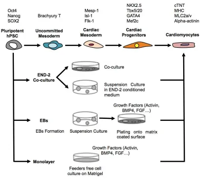
Figure 1.5: Cardiac differentiation cascade and differentiation methods
From the top: Markers of different stages of cardiac differentiation, steps in cardiac differentiation and the differentiation methods. END-2 differentiation has two variables, hESC are either plated on top of END-2 cell layer or are cultured as EBs in suspension in END-2 conditioned medium. In EBs method, differentiation can be performed spontaneously or with differentiation inducing growth factors. Monolayer differentiation is initiated with feeder free hESC cultures. Culturing and differentiation of hESC are preformed on top of Matrigel.
Different cytokine approaches were tested, in agreement with differentiation pathways
discussed in the previous session. After initial spontaneous indirect pathways (Kehat et
al., 2001) and non-selective endoderm-inducing conditions of END2 cells (Mummery et
al., 2002), mesodermal induction with Activin A and BMP4 were approached. Early
differentiation was accomplished by those two factors alone. But shortly after initial 48
Oct4 and Nanog. This strategy resulted in the low amount of contracting colonies / EBs
and various levels of cardio specific proteins in the samples. In order to specify more
the mesodermal differentiation pathway, Wnt inhibitors (DKK1), VEGF and bFGF
were studied and used as cardiac progenitor selective agents. So-called “small
molecules” have various effects on cardiac differentiation. Among others, retinoic acid
(RA) has been shown to accelerate hESC differentiation into CMs (Wobus et al., 1997).
Enhancement was also seen using a retinoid X receptor agonist (Honda et al., 2005). RA
is regulating the expression of NKX2.5, and has an effect on spatial organization of the
primitive cardiac cells, as well as atria and ventricles organization in the animal model
(Yutzey, Rhee, & Bader, 1994). Similarly, ascorbic acid (AA) has shown positive
effects especially on EB based cultures (T. Takahashi et al., 2003), but the specificity of
the AA and its possible pathway is discussed.
Until 2008, serum has been present in the culture medium of all differentiation
protocols. Serum was reported to be un-favourable to maintenance of primary CMs
(Piper, Jacobson, & Schwartz, 1988). The switch to serum free methods eliminated the
“batch bias” and was also required for possible clinical applications. A switch to a
serum-free differentiation medium in mouse EBs resulted in a 4.5-fold increase in the
percentage of beating clusters (Sachinidis et al., 2003). Similarly, insulin (originally in
the differentiation media) was omitted without losing CMs yield and was further shown
to have even a negative effect on cardiac differentiation (X. Q. Xu, Graichen, et al.,
2008).
In addition to classical cardiac differentiation of pluripotent stem cells, cardiac cells
have been obtained directly from other adult cell types. The direct trans-differentiation
idea came from the observation that the generation of hiPSC had shown that a relatively
small set of defined genes could epigenetically alter the global gene expression of a cell.
If a somatic cell could be reprogrammed into a stem/progenitor cell, probably could
undergo also direct cardiac reprogramming. A combination of three developmental
transcription factors (referred as GMT) was shown to reprogram murine post-natal
cardiac and dermal fibroblasts directly into differentiated cardiomyocyte-like cells
[zinc-finger transcription factor recognizing the GATA motif – Gata4, myocyte
enhancer factor 2C (Mef2c) and T-box transcription factor 5 (Tbx5)] (Szabo et al.,
2010) (Qian et al., 2012). This attitude stresses that cardiac fibroblasts comprise over
were also able to generate CMs (the same way as murine cardiac fibroblasts) (Szabo et
al., 2010). More recently, direct reprogramming of human fibroblasts has also been
achieved (Islas et al., 2012). Several studies showed that the murine direct
reprogramming factors Gata4, Mef2c, and Tbx5 (GMT), or GMT plus Hand2 (GHMT),
were insufficient to transform human fibroblasts into CMs (Nam et al., 2013) (Wada et
al., 2013), indicating that the differences between mouse and human cardiovascular
development need to be considered for optimal transdifferentiation to human CMs.
Better results were obtained adding to the cocktail small-non coding RNA miR-1, and
miR-133m that converted human adult dermal fibroblasts into induced
cardiomyocyte-like cells . The transduction of these factors promoted substantial cardiac troponin T
expression in at least 9% of the source population (Nam et al., 2013). Shortly afterward,
introduction of GMT plus Mesp1 and myocardin (GMTMM) was also shown to
successfully convert human fibroblasts to iCMLs (Wada et al., 2013). Since then,
alternative approaches have succeeded in generating human iCMs with gene expression
profiles and functional characteristics similar to those detected in ESC-CMs (Fu et al.,
2013).
1.2.4 Enrichment of differentiated cardiomyocytes
Due to the inefficient differentiation, the resulting cell populations that arise from a
differentiation protocol, are a mixture of different cell types and the yield of
hES/iPSC-CM cultures can be very low. EB differentiation in serum containing medium yields
<1% and the more defined ActivinA/BMP-4 protocol yielded > 30% of cardiomyocytes
(Laflamme et al., 2007). Therefore differentiation methods need considerable up-scaling
and effective enrichment and purification methods should be developed before
differentiated cardiomyocytes can undergo testing and clinical use in the future.
For certain research purposes, it is adequate to enrich the hES/iPSC-CM by
mechanically dissecting beating areas from the differentiation cultures (Kehat et al.,
2001) (Mummery et al., 2003).
PercollTM gradient separation based on density gradient separation has been used in combination with the generation and maintenance of cardiac bodies (C. Xu, Police,
Hassanipour, & Gold, 2006). After separation and 7 days of suspension maintenance,
difficult for others to reproduce (van Laake, Hassink, Doevendans, & Mummery, 2006),
and also in our hands.
Transgenic selection is one technique to enrich cardiomyocytes from hESC
differentiation cultures. This method utilizes transgenic hESC/iPSC lines where a gene
of green fluorescent protein (GFP) or an antibiotic resistance gene is located under the
control of a cardiac specific promoter (D. Anderson et al., 2007; Kolossov et al., 2005)
(Huber et al., 2007) (X. Q. Xu, Zweigerdt, et al., 2008) (Kita-Matsuo et al., 2009).
Although this method is efficient, genetic modification is neither easy for hESC or
human iPSC cell lines nor suitable for possible future clinical use (Mummery, 2010)
(Vidarsson, Hyllner, & Sartipy, 2010). In a study from 2009, they sorted
cardiomyocytes from mixed cell populations by using the endogenously expressed
surface marker activated leukocyte cell-adhesion molecule, ALCAM (CD166) (Rust,
Balakrishnan, & Zweigerdt, 2009). And in 2011 SIRPA (CD172a) was found to be a
marker for cardiac cells from a screening of 370 known CD antibodies (Dubois et al.,
2011). However, there is a lack of cardiac specific surface proteins and therefore a lack
of antibodies to make sorting possible (Mummery, 2010). Nevertheless,
fluorescence-activated cell sorting (FACS) was successfully used in selection by utilizing the high
mitochondria content of cardiomyocytes (Hattori et al., 2010).
1.2.5 Characterization of differentiated cardiomyocytes
1.2.5.1Functional and structural analysis
hES/iPSC-CM have the capacity to beat spontaneously (Kehat et al., 2001) (Mummery et
al., 2003). Beating cells are at an early stage relatively small and round and situated in
circular accumulations in the EBs. At later stages, EBs gradually develop to be larger and
the cells turn to be more elongated in shape and tend to accumulate in strands. Electron
microscopy studies reveal that cardiomyocytes contain myofibrils, which are first
randomly and in a varying manner distributed throughout the cytoplasm. However,
organized sarcomeric structures occur at later stages of differentiation with A, I and Z
bands (see paragraph “1.3.2.2 Contractile apparatus” and figure 1.7). Mitochondria are
also present close to the sarcomeres and cells have intercalated disks with gap junctions
1.2.5.2Expression of cardiac markers
Gene expression profiles of the hESC/iPSC during cardiac differentiation (Beqqali et al.,
2006) (Synnergren et al., 2008) and the differentiated cardiomyocytes have been studied
by DNA microarray (Cao et al., 2008) (Synnergren et al., 2008) (Kita-Matsuo et al.,
2009) (X. Q. Xu, Soo, Sun, & Zweigerdt, 2009). These studies reveal that the molecular
signature of hESC-CM resembles the cardiomyocytes from the human heart (Vidarsson
et al., 2010). hESC/iPSC-CM differentiation can be predicted by the transient
expression of the early mesodermal marker T (Brachyury). T (Brachyury) expression
peak is detected at the time point of 3 days in END-2 co-cultures (Beqqali et al., 2006),
a day later in EBs (Bettiol et al., 2007) and at day 1 in GSK pathway inhibition based
protocols on iPSC monolayers (Lian et al., 2012; Lian, Zhang, Zhu, Kamp, & Palecek,
2013). T (Brachyury) belongs to the family of transcription factors, which are encoded
by the T-box genes (Showell, Binder, & Conlon, 2004). This protein family plays roles
in many developmental processes and has a sequence similarity with the DNA-binding
domain, the T-domain (Showell et al., 2004). Brachyury T can be nominated as a classic
transcription factor. It is localized in the nucleus and is an endogenous activator of
mesodermal genes (Conlon et al., 1994) (Showell et al., 2004) (Kispert, Koschorz, &
Herrmann, 1995). In the embryo, T (Brachyury) expression is suggested to be induced
by TGF-beta and FGF signalling (Hemmati-Brivanlou & Melton, 1992) (Amaya, Stein,
Musci, & Kirschner, 1993). Overall, very few direct targets for T-box genes have been
identified. However, embryonic FGF (eFGF) (Casey, O'Reilly, Conlon, & Smith, 1998),
has been suggested as downstream target for Brachyury T. Differentiation cascade can
be further followed by the expression of cardiac regulatory transcription factors such as
Islet-1 (Isl-1), Mesp 1, GATA4 and NKX2.5 (Graichen et al., 2008) (Yang et al., 2008).
Cardiac troponin T (cTNT) is encoded by the TNNT2 gene (Thierfelder et al., 1994). It
is the tropomyosin-binding subunit of the troponin complex and can therefore be used
for characterizing hESC/hIPSC-CM. Troponin complex is located on the thin filament
of striated muscles and regulates muscle contraction in response to alterations in
intracellular calcium ion concentrations (Farah & Reinach, 1995) (Tobacman, 1996). In
addition to cTNT, other cardiac specific structural proteins are used for confirming
cardiac phenotype of the beating hES/iPSC-CM such as cardiac troponin I, myosin or
cardiac alpha-actinin (Kehat et al., 2001) (Mummery et al., 2003). Also, proteins of
channels can be used in characterization. Gap junctions are formed from connexin
proteins and have an important role in signal transduction. Connexin 43 (Cx43) is the
most common form in the ventricle, Cx40 predominates in the atria and Cx45 is found
in both atria and ventricle (Gaborit et al., 2007).
1.2.5.3Electrophysiology and excitation-contraction coupling
Human ESC as well as iPSC cell-derived cardiomyocytes exhibit heterogenic action
potential (AP) morphologies which can be divided into nodal, atrial and ventricular
subtypes according to the shape of AP (J. Q. He, Ma, Lee, Thomson, & Kamp, 2003) (J.
Zhang et al., 2009) (Zhang et al., 2009). If compared to the human neonatal or adult
atrial or ventricular cardiomyocytes, hESC-CM have relatively positive maximum
diastolic potential and slow maximum rate of rise of the AP, and are therefore called
embryonic atrial- and ventricular like cells (J. Q. He et al., 2003). Differentiated beating
cells exhibit spontaneous APs and contractile activity and therefore express cardiac
structural proteins and ionic currents (Kehat et al., 2001) (Mummery et al., 2003) (J. Q.
He et al., 2003). During differentiation, the expression of some ion channel genes
increases suggesting that hESC-CM reach a more mature state with time in culture
(Sartiani et al., 2007). Traditionally, patch clamp has been used in analysing the action
potential and also the electrophysiological properties of cardiomyocytes. Microelectrode
array (MEA) technology provides another useful platform to study cell
electrophysiology, especially ESC-derived cardiomyocytes (Hescheler et al., 2004)
(Reppel et al., 2004). In MEA, cells are plated on top of electrodes in a cell culture
well-type platform and can be cultured and measured repeatedly for long periods of time.
Regarding excitation-contraction coupling, hESC/iPSC-CM have functional, even if
immature, calcium handling components when compared to adult cardiomyocytes
(Dolnikov et al., 2006) (J. Liu, Fu, Siu, & Li, 2007) (Satin et al., 2008). For clinical
applications, the calcium system should be functioning properly to hESC-CM to
integrate properly after transplantation since poor integration to the host myocardium
could have serious arrhythmias as a consequence. Nevertheless, a better understanding
1.2.6 Fetal vs. post-natal cardiomyocytes
There are marked phenotypic differences between fetal and post-natal CMs, including
age-dependent changes in protein kinase expression, betha-adrenergic signalling
(Schaffer & Williams, 1986), contractile apparatus maturity, and the hyperplastic
(Rybin & Steinberg, 1994) versus hypertrophic (Pandya, Santani, & Jain, 2005)
responses to mechanical stress. The accumulation of maturation specific isoforms of
sarcomeric proteins including cardiac myosin heavy chain, sarcomeric actin and actinin,
titin, cardiac troponin-T and -I, and the maturation of sarcolemmal and sarcoplasmic
reticulum (SR) ion channels (Hagege & Menasche, 2000) (Tenhunen et al., 2006)
(Assmus et al., 2002) allow the functional characterization of cardiomyocytes identity
and maturation. These characteristics determine the overall phenotypic appearance of
CMs, which can differ depending on their location.
Regarding stem cell derived-cardiomycytes, there is currently no standard to what a
stem cell derived-CM is. Many groups use the expression of early cardiac genes:
NKX2.5 or GATA4 and the presence of rhythmic beating. This is not necessarily
sufficient because skeletal and smooth muscle also contracts. Also, it has been widely
accepted that terminally differentiated mature cardiac muscle does not express proteins
that are specific to skeletal muscle. However, studies have shown that several skeletal
muscle specific proteins, such as skeletal muscle specific troponins or ion channels, are
transiently present in the developing heart (Haufe et al., 2005)(Saggin, Gorza, Ausoni,
& Schiaffino, 1989). Similarly, “cardiac” and “skeletal” excitation-contraction coupling
mechanisms co-exist in the developing skeletal muscle with the “cardiac” type
dominant in the early phases of myogenesis and the “skeletal” dominating in more
mature muscle (Cognard, Rivet-Bastide, Constantin, & Raymond, 1992). These studies
suggest the co-existence of many cardiac and skeletal muscle specific proteins (MHCs,
troponins, etc.) as well as excitation-contraction coupling mechanisms within the
developing tissue, but also within cultured cells, especially those that are considered to
be immature. Thus, the functional characterization of CMs for their research and
1.2.7 Applications for hESC or iPSC derived cardiomyocytes
1.2.7.1Functional and structural analysis
Since the establishment of the first permanent hESC line (Thomson et al., 1998) there
has been a great hope of replacing damaged heart tissue by hESC derived
cardiomyocytes. However, many major problems need to be solved before hESC-CMs
are usable in clinics. Before clinical use becomes a reality, it is likely that the derived
cardiomyocytes will be applicable for drug discovery and safety pharmacology
applications (Braam, Passier, & Mummery, 2009). Nevertheless, cardiac differentiation
and the beating cells are already a useful tool for developmental biology and to study
the pathophysiology of human cardiac diseases. In addition, iPSC technology enables
the production of patient specific cell lines, which extends the potential use even further.
1.2.7.2Pathophysiology of cardiac diseases
Many cardiac diseases are caused by gene mutations or gene-environment interactions.
So far, these severe diseases have been studied in animal models, especially with
transgenic mice. Even though mouse models can provide valuable information,
differences between human and mouse physiology limit the applicability of the results,
for example the much faster beating rate of the mouse may override the effects of
arrhythmias which would be severe for humans (Freund & Mummery, 2009).
Cardiomyocytes derived from genetically modified hiPSC could be used as a disease
model. To derive a mutated hiPSC line and the disease model, it needs to be genetically
manipulated. However, genetic manipulation of hESC has proven to be more
challenging if compared to mouse ESC cells and only a few reports of successful gene
targeting and manipulation have been published (Braam et al., 2008) (Giudice &
Trounson, 2008). To obtain disease specific lines, the genetic manipulation step can be
circumvented by deriving iPSC cell lines from patients with genetic diseases (Ebert et
al., 2009; Freund et al. 2010; Park et al., 2008). The differentiation of these model iPSC
cells to the desired cell type makes it possible to study the development and the
pathophysiology of the disease. In addition, the factors affecting the development and