and molecular mechanisms in
Williams-Beuren syndrome
María Gabriela Palacios Verdú
DOCTORAL THESIS UPF / 2015
THESIS SUPERVISORS
Prof. Luis Alberto Pérez Jurado
Dr. Miguel del Campo Casanelles
“Let me tell you the secret that has led me to my goal. My strength lies solely in my tenacity”.
A mis papis, a Andy, a Titi,
Parece que fue ayer cuando caminaba por las calles de Barcelona y me topé con un hermoso edificio en primera línea de mar. Cuál fue mi sorpresa cuando leí su letrero “Parc de Rercerca Biomèdica de Barcelona”, en ese momento decidí que ahí era donde quería hacer mi tesis doctoral. Desde entonces han pasado unos cuantos años, sin duda, años que quedarán por siempre en la memoria. Hay muchas personas a las que quiero agradecer por esta magnífica temporada. Espero no olvidar a nadie, y si es así, ruego disculpen mi frágil memoria que como muchas de ustedes saben, a los casi cuarenta años de edad la memoria suele comenzar a fallar.
Primero que nada quisiera agradecer a mi director de tesis, Luis, por haberme dado la oportunidad de entrar al laboratorio para completar una etapa más de mi formación. Por haberme enseñado, que todo, absolutamente todo tiene una base genética. Por tus consejos y enseñanzas de laboratorio y de clínica. Por las conversaciones de política, local e internacional.
A todos los que son o han formado parte del Lab 422: Cris, Maria, Clara, Mariví, Ivon, Raquel, Tina, Marta, Aïda, Fátima, Judith, Armand, Roser, Debora; por todas las horas compartidas, las reuniones, las cenas de inicio y fin de curso, los consejos, los “berenars” y muchas cosas más.
A Mariví, gracias por dejarme ser parte del “team mouse”, por enseñarme a pelar patas, por las discusiones científicas y no tan científicas, por tu apoyo y consejos.
volley e, inclusive, una barbacoa jugando al “Just Dance”.
A Maria, mi vecina y compañera de lab, de master, de volley y de basket! Gracias por estar ahí siempre, a pesar de que ahora nos separan unos cuantos kilómetros de distancia. Y, last but not least, gracias por ser mi profesora de catalán.
A Clara, por tus consejos, por contestar mis dudas, por momentos vitales compartidos (bodas, embarazos), por SEAGen y el máster. Gracias por aquellas tardes de merienda en los sofás que alegraban el día.
A Aïda, porque no se que hubiera hecho sin ti durante estos últimos meses.. Por tu paciencia, por tu forma de ver las cosas, por tu sinceridad y apoyo. Por el volley y, en pocas ocasiones, running. Muchísima suerte en esta nueva etapa de tu vida!
A Tina, por ser mi compañera de poyata, si comenzara a enumerar todas nuestras experiencias no acabaría jamás! Gracias por escucharme, por compartir el Northern, por tu apoyo, por tus correcciones.. Por cada temporada de volley, por el combo Capitina y Gabitana.
A Marta, por tus consejos, por tu apoyo en momentos difíciles, por compartir aficiones (chocolates), por nuestros trayectos en bus que incluía, en ocasiones, discusiones filosóficas. Y por haber continuado con mi formación en catalán (“fiyoll”) y refranes locales (“Es broma, poma!”).
inclusive deportivos.
A Miguel, por tus críticas constructivas y por hacerme ver el lado bueno de los trabajos que he realizado.
A mi queridísimo Doc, por ser el primero en formarme en este mundo tan interesante de la genética, por estar siempre pendiente de mis pasos académicos, deportivos y personales, y por siempre tener las puertas abiertas por si un día decido volver a la llacta.
A QGenomics; Manel, Olaya, Benja, Sonia, Cristina, Xavi, Lluis, María Jesús, y sus nuevos integrantes; por compartir desayunos, actividades, conversaciones y momentos especiales. Por compartir la centrífuga, el termociclador y pipetas en momentos de necesidad absoluta.
A Gens Bojos, Sondas de Array, PhD-4 años de esclavitud, Game of Clones… Por cinco magníficas temporadas de volley playa, por darlo todo en cada encuentro, por cuatro copas (espero que este año no sea la excepción) y por aguantar mi espíritu competitivo.
A las Cabritas, al equipo, a Tatay y a Neus, gracias por las horas compartidas dentro y fuera de pista, por estar ahí, por su apoyo en momentos difíciles y por creer en mi. Tatay gracias por enseñarme más allá de técnica individual y táctica de juego, porque siempre podemos dar más.
A la junta de SEAGen; Anna, Núria, Estela, Mar y Clara; por sus actividades, reuniones, jornadas, charlas y, aunque pocas, actividades lúdicas.. Seguiremos trabajando para difundir el asesoramiento genético.
A Edgar, María, Vale, Jorge, Maty y Gastón; por sus llamadas que nos acortan la distancia, por las deliciosas parrilladas, por nuestros desayunos en Jürgen, por sus frases de apoyo en momentos complicados, porque están presentes en cada momento especial.
A mi familia, porque siempre han estado ahí aún a la distancia. Por nuestras películas nocturnas y viajes larguísimos! Por nuestras noches de bohemia, por escucharme horas (aún cuando se quedaban dormidos), por nuestros cafés de los domingos, por las farras locas en UIO o BCN… En fin, por todas las vivencias que hemos podido compartir y las que están por llegar. Les extraño!
A Andy y a Titi, porque siempre han estado presentes en los buenos y no tan buenos momentos. Por escucharme, por aconsejarme, por enseñarme, por tener paciencia y apoyarme en cada paso que he dado.
A mis papis, por haberme brindado todas las oportunidades para llegar a donde he llegado y por ser lo que soy, por creer en mi en todo momento, por siempre estar ahí y por ser un gran ejemplo de lo que quiero llegar a ser.
A Mauri, mi compañero, mi amigo, my better half. Porque sin ti este camino no habría sido posible, por suplir en lo que a mi me falta, por estar ahí incondicionalmente, por creer en mi y no dejarme caer cuando el camino se complicaba. Por todas nuestras cenas de los viernes que ayudaban a aclarar la mente y hermosos momentos en nuestro “Happy place”.
Gracias Totales!
"No solo no hubiéramos sido nada sin ustedes, sino con toda la gente que estuvo a nuestro alrededor desde el comienzo; algunos siguen hasta hoy. ¡Gracias totales!"
Williams-Beuren Syndrome (WBS, MIM 194050) is a neurodevelopmental disorder with multisystemic manifestations of variable expressivity, including dysmorphic features, vascular stenoses, and intellectual disability with an uneven neurocognitive profile. It is caused by a recurrent 1.55-1.83 Mb deletion secondary to non-allelic homologous recombination (NAHR) at chromosome band 7q11.23 that includes 26 to 28 genes. A genotype-phenotype correlation has only been established for some of the genes involved in this deletion, the most studied is the elastin gene associated with the cardiovascular phenotype. In this thesis project, we have aimed to further elucidate the molecular mechanisms involved in NAHR, as well as possible cis
and trans-acting mechanisms. We have also described novel clinical characteristics (metabolic phenotype), described in greater detail the cardiovascular phenotype present in patients with WBS, as well as determining possible candidate genes associated with both of these phenotypes.
RESUMEN
Numerous researchers have studied Williams-Beuren syndrome since it was first described in the 1960’s with the objectives of determining the phenotype and the natural history of the syndrome, elucidating the causal molecular mechanisms involved and the genotype-phenotype correlations. The final objective of the effort of the scientific community is to improve the medical management of patients, by establishing follow-up guidelines, medical treatment and therapies to improve their physical and psychological symptomatology.
The global aims of this thesis project are to improve the definition of the WBS phenotype, the natural history of this disorder, to characterize the deletion and the molecular mechanisms involved, establish clinical-molecular correlations to identify possible candidate genes associated with specific phenotypes.
The introduction focuses on the clinical characteristics and management of the WBS, an overview of the molecular mechanisms involved and previously reported chromosomal rearrangements, and previously known genotype-phenotype correlations.
The thesis body is divided in three chapters that present in detail the methodology used and the results obtained. The first chapter is about the molecular mechanisms involved in WBS. The second chapter describes the metabolic phenotype. The third chapter describes cardiovascular disease and hypertension in WBS.
CONTENTS
ABSTRACT………..xi
PROLOGUE………..xiii
LIST OF FIGURES……….………..xix
LIST OF TABLES……….………..…xxiii
INTRODUCTION…………...1
1. WILLIAMS BEUREN SYNDROME CLINICAL CHARACTERISTICS ... 3
1.1 Facial Phenotype ... 3
1.2 Growth and Puberty ... 4
1.3 Cardiovascular Phenotype ... 4
1.4 Endocrinological Phenotype ... 9
1.5 Gastrointestinal Manifestations ... 10
1.6 Neurological Phenotype ... 11
1.7 Cognitive and Behavioral Phenotype ... 11
1.8 Auditory Manifestations ... 15
1.9 Ophthalmologic Manifestations ... 15
1.10 Dental Manifestations ... 15
1.11 Genitourinary Manifestations ... 16
1.12 Musculoskeletal Manifestations ... 16
1.13 Integumentary system Manifestations ... 16
2 WILLIAMS-BEUREN SYNDROME: CRITICAL REGION
(WBSCR) ... 20
2.4 7q11.23 Genomic Architecture ... 20
2.5 Mutational Mechanisms ... 22
2.5.1 Non-Allelic Homologous Recombination ... 22
2.5.2 Paracentric Inversion ... 25
2.5.3 Deletion ... 26
2.5.4 Duplication ... 30
2.5.5 Triplication ... 31
2.5.6 Copy-Number Variants ... 31
2.5.7 Novel technologies used to refine breakpoints ... 32
3 GENOTYPE – PHENOTYPE CORRELATION ... 35
3.4 Cardiovascular phenotype ... 39
3.4.1 Elastin ... 40
3.4.2 Neutrophilic cytosolic factor 1 ... 41
3.5 Neurocognitive and craniofacial phenotype ... 42
3.5.1 Transcription Factor II-I Family ... 42
3.5.2 LIM Domain Kinase 1 ... 44
3.5.3 CAP-GLY Domain containing linker protein 2 ... 45
3.5.4 Bromodomain adjacent to zinc finger domain 1B ... 46
3.5.5 Frizzled 9 ... 46
3.5.6 Syntaxin 1A ... 47
3.5.7 Eukaryotic translation initiation factor 4 ... 47
3.6 Metabolic phenotype ... 48
3.6.1 Mlx-interacting protein like ... 48
3.6.2 Syntaxin 1A ... 49
OBJECTIVES………...53
Hotspots for recurrent meiotic rearrangements mediated by segmental duplications at 7q11.23
CHAPTER 2……….…95
Metabolic abnormalities in Williams-Beuren syndrome CHAPTER 3………...…121
Cardiovascular manifestations and evaluation in Williams-Beuren syndrome DISCUSSION………....….165
CONCLUSIONS……….…...…183
REFERENCES………...…….……..187
LIST OF ACRONYMS………..……215
LIST OF FIGURES
INTRODUCTION
Figure 1. Typical facies of three unrelated patients with WBS... 4 Figure 2. Left ventriculography of a patient with WBS…………...……... 5 Figure 3. Relative strengths and weakness of WBS cognitive
phenotype……….... 12
Figure 4. Atypically focused attention of a child with WBS to the
experimenter´s face during a cognitive task………... 13
Figure 5. Stellate iris pattern of a patient with WBS………... 15 Figure 6. Schematic representation of the WBS Critical Region
(WBSCR)………. 22
Figure 7. Schematic representation of non-allelic homologous
recombination………... 23 Figure 8. Mechanisms for non-allelic homologous recombination
(NAHR)………... 24
Figure 9. Schematic representation of the origin of 7q11.23 paracentric
inversion………... 26
Figure 10. Schematic representation of the WBS deletion………... 27 Figure 11. Schematic representation of the meiotic misalignment and
crossing-over event between an inverted and a normal chromosome……... 28 Figure 12. Schematic representation of the 1.55 Mb and 1.83 Mb deletions and breakpoint characterization………... 29 Figure 13. Schematic representation of novel technologies used to refine
breakpoint location……….. 34
Figure 14. Schematic representation of typical and atypical 7q11.23
deletions………... 36
Figure 15. Schematic representation of WBS critical region at
chromosomal band 7q11.23 in humans and in chromosomal band 5G2 in
mice………...……….…….. 37
Figure 16. Schematic representation of WBS critical region at
chromosomal band 7q11.23………...…...…... 39
CHAPTER 1
Figure 1. Schematic representation of SSNs distributed along the B-block and the frequency of deletions at each interval for 1.55 Mb deletion and
normal and inverted 7q.11.23 genomic region……….. 63 Figure 3. Schematic representation of the architectural structure of a
deletion with a breakpoint located within GTF2IRD2……….. 65
Figure 4. Genome location of genotyped SNPs at 7q11.23………... 69 Figure 5. Multidimensional approach to discriminate between inversion
and no-inversion carriers………... 71
CHAPTER 2
Figure 1. A). Histogram of triglyceride plasma levels in centiles in individuals with WBS. B). Histogram of the distribution of triglyceride
z-score values……….……….……….... 101
Figure 2. A). Histogram of total bilirubin plasma levels in centiles in individuals with WBS. B). Histogram of the distribution of total bilirubin
z-score values………...……….... 102
Figure 3. TB, DB and IB z-score values with respect to UGT1A1
phenotype………..………... 102
Figure 4. A). Intraperitoneal glucose tolerance test. B). Langerhans islets
area analysis………. 104 Figure S1. Schematic representation of WBS deletion and the WBS
murine models used in this study. …...………....………. 112 Figure S2. A). Histogram of TSH plasma levels in centiles in individuals
with WBS. B). Histogram of the distribution of TSH z-score values……… 112 Figure S3. A). Histogram of glucose plasma levels in centiles in individuals with WBS. B). Histogram of the distribution of glucose z-score values…… 113 Figure S4. A). Histogram of total cholesterol plasma levels in centiles in
individuals with WBS. B). Histogram of the distribution of total cholesterol z-score values………... 113 Figure S5. Relative expression of Ugt1a1 studied in liver of WBS mouse
models……….… 114
CHAPTER 3
Figure 1. Percentage of patients with HTN compared to BMI
classification………..………... 131 Figure 2. Mean, systolic, diastolic compared by BMI classification.………. 134 Figure 3. Percentage of patients with LV geometry alterations separated
by BMI….. ……….. 135 Figure 4. Malondialdehyde concentration in normotensive and
Figure 5. Mean, systolic, diastolic and nocturnal BPI and BPL at 0 and 12
months after losartan treatment………... 136
Figure 6. Plasma renin activity and aldosterone plasma concentration at 0 and 12 months after losartan treatment………..…….. 137 Figure 7. Malondialdehyde concentration at 0 and 12 months after
losartan treatment,………...………. 137 Figure 8. Mean, systolic, diastolic and nocturnal BPI and LV geometry….. 139 Figure 9. NCF1 copy number and percentage of office hypertension
subdivided by gender……….... 139 Figure 10. Association of SBPI and DBPI with PLCE1 genotype………... 141 Figure S1. Histogram ofplasma renin activity and aldosterone plasma
levels……… 160 Figure S2. Histogram of malondialdehyde plasma values in 32 individuals with WBS………. 160 Figure S3. Comparison of MDA plasma concentration with NCF1 copy
LIST OF TABLES
INTRODUCTION
Table 1. Cardiovascular findings in WBS individuals ………... 7 Table 2. Recommendations for medical monitoring of individuals with
WBS……… 18
CHAPTER 1
Table 1. PRDM9 allelic frequencies in the transmitter, non-transmitter
group and in Europeans……… ………... 68 Table 2. Allelic frequency of SNPs located at 7q11.23 in CEU HapMap,
no-inversion carriers and inversion carriers………..…… 103
Table S1. Polymorphic block CNVs in transmitter and non-transmitter
group………..……….……….………. 92
Table S2. SSNs used for mapping the deletion breakpoint in patients with 1.55 Mb deletion. ……..……….………..……….………... 92 Table S3. Site-specific PCR primers to amplify across Bt/Bm
interchromosomal junction fragments. ……….………... 93 Table S4. PCR primers used to amplify and Sanger-sequence GTF2IRD2
paralogous sequence variants………... 93
CHAPTER 2
Table 1. Biochemical parameters studied in individuals with WBS………... 101 Table 2. Metabolic parameters analyzed in WBS mice models at 25 weeks
of age..……….……….… ..……….……… 103
Table S1. Crude values of biochemical parameters studied in 154 WBS
individuals………..……….……….……… 115 Table S2. Primer sequences for genotyping and qRT-PCR. ……..……….. 109 Table S3. Alterations in biochemical parameters in children and adults
with WBS……….………….………….………….…...…………... 110 Table S4. Candidate SNPs genotyped in WBS individuals………... 110 Table S5. UGT1A1 genotype frequency and bilirubin concentrations in
healthy controls and WBS individuals……….……….….……... 110
Table S6. Metabolic parameters analyzed in WBS mice models at 4.5
CHAPTER 3
Table 1. Genotyped SNPs associated with hypertension and
cardiovascular risk………...……….……….… ..………... 128 Table 2. Cardiovascular lesions reported in the cohort of 136 patients in
the 47 patients who underwent an echocardiographic study……….… 130
Table 3. ABPM variables studied in a cohort of 49 patients with WBS.…... 132 Table 4. Frequency of vascular stenosis detected by Doppler US and CT
angiogram in normotensive and hypertensive individuals with
WBS……… 138 Table 5. Association of relevant SNPs with cardiovascular stenosis and
vascular disease………...……….……... 140 Table 6. Association of PLCE1 gene with systolic and diastolic HTN…… 141 Table S1. Ambulatory hypertension in pediatric and adult patients with
WBS……… 162 Table S2. Oxidative stress parameters levels compared with mean,
systolic, diastolic and nocturnal hypertension………...……… 162 Table S3. ABPM variables at 0 and 12 months after losartan treatment in hypertensive patients with WBS………... 163 Table S4. Oxidative stress parameters at 0 and 12 months after losartan
1.
WILLIAMS BEUREN SYNDROME CLINICAL
CHARACTERISTICS
Williams Beuren syndrome (WBS, OMIM 194050) is a neurocognitive disorder characterized by multisystemic manifestations of variable expressivity. It has an incidence of 1/7,500 – 1/10,000 individuals [1], almost always with a sporadic occurrence, although it can be transmitted in an autosomal dominant manner [2].
WBS was first described in 1952 in a report encompassing the clinical characteristics of children who presented infantile hypercalcemia, short stature and congenital malformations [3]. It was several years later that Dr. John Cyprian Phipps Williams and Dr. Alois Beuren described the main characteristics of this disorder; supravalvular aortic stenosis, intellectual deficit and common facial characteristics [4, 5].
1.1 Facial Phenotype
The facial gestalt of WBS is unique and distinct from other neurodevelopmental disorders. In infants and young children it is characterized by a broad forehead, bitemporal narrowing, low nasal root, bulbous nasal tip, strabismus, an iris stellate pattern, periorbital fullness, malar flattening, long philtrum, wide mouth, full lips, full cheeks and widely spaced teeth with malocclusion [6] (Figure 1.A).
1.2 Growth and Puberty
The incidence of prenatal growth deficiency is approximately 50-70% [7, 8]. During the first two years of age WBS individuals usually have poor growth and follow the 3rd percentile curve until 9 and 11 years of age in females and males, respectively[8]. The rate of linear growth during childhood is 75% of normal [9]. WBS individuals present a premature and brief pubertal growth spurt. Menarche usually occurs at an earlier age [8]. Mean adult height is at the 3rd percentile or lower for 70% of WBS individuals [8].
Several reports have shown that between 25 to 65% of adults with WBS have a body mass index (BMI) compatible with an overweight to obesity range [10, 11].
1.3 Cardiovascular Phenotype
One of the main hallmarks of WBS phenotype is cardiovascular (CV) disease, which is characterized by stenoses of medium and large arteries [12]. The reduction of elastin content in the media of arteries
Figure 1.Typical fancies of three unrelated patients with WBS.A. The young
child has a flat nose bridge, upturned tip of the nose, long philtrum and full cheeks.
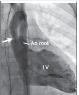
may lead to recurrent injury and fibrosis; as well as an increase in hemodynamic stress to the vascular endothelium, which ultimately leads to proliferation of smooth muscle cells and fibroblasts, fibrosis and a subsequent intimal hyperplasia [13–15]. Vascular stenoses can occur in any artery of the body; however the most frequent locations are the supravalvular aortic (Figure 2) and peripheral pulmonic stenosis [12].
Supravalvular aortic stenosis (SVAS) occurs in approximately 70% of WBS individuals, with a severity that ranges from mild to severe [16– 18]. This cardiovascular involvement tends to progress with time, especially during the first five years of life [19–21]. However, a previous study reported that 16% of patients presented an unexpected spontaneous resolution, suggesting that when SVAS is mild it is more likely to improve than to progress [22]. Peripheral pulmonic stenosis (PPS) occurs in approximately 30% of patients and tends to improve with age in the majority of patients [21, 22].
Systemic arteriopathy is present in approximately 20% of WBS patients and the affection of the elastic arteries can be a local or diffuse narrowing [23, 24]. The most frequent is the stenosis of the thoracic aorta (STA), which is a separate entity from coarctation of the aorta [25]. A study in a cohort of 270 patients with WBS found that STA was present in 14%, long-segment STA was more frequent than
Figure 2. Left ventriculography shows
[image:29.499.86.214.202.355.2]discrete STA [25]. Many patients with STA presented other vascular stenoses; including neck vessels, renal artery, abdominal aorta, mesenteric and celiac artery [25]. It is important to diagnose systemic stenoses before a surgical intervention to avoid hypoperfusion of the organs irrigated by the stenotic arteries and because it can change the surgical technique used [23, 25]. Abdominal aortogram studies in WBS patients showed that the morphology of the abdominal aorta is characterized by segmental changes in the diameter, hypoplastic at the diaphragm level, suprarenal narrowing, renal artery stenosis (RAS) and with an increase in infrarenal aorta diameter [26]. Middle aortic syndrome (MAS); consisting of stenoses of the thoracic and abdominal aorta, mesenteric and renal arteries, has been described in 2-70% of cases [27, 28]. The higher incidence of other arterial stenoses in patients with STA might indicate that STA is a marker for worsened generalized arteriopathy [29]. RAS is present in 7-58% of patients [29], when associated with hypertension its etiology be considered renovascular [26]. CT or RM angiogram are good imaging techniques to study the vascular complications in WBS patients, diagnose MAS and control it after its surgical intervention [28].
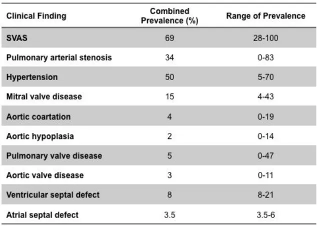
Other frequent cardiovascular alterations reported in WBS, that can occur either isolated or accompanied by SVAS or PPS, are mitral valve disease, aortic coarctation, pulmonary or aortic valve disease or ventricular septal defect (Table 1)[12, 18, 21, 22].
Arterial hypertension (HTN) can occur in approximately 50% of WBS individuals, although, as seen on Table I, this percentage is highly variable depending on the criteria and the methods used for blood pressure measurement [12, 16, 31–33]. A study using ambulatory blood pressure measurements (ABPM) found an increased incidence of HTN in WBS compared to the control group (40% vs. 14%); the difference was more striking when comparing children (46% vs. 6%). The authors also reported that WBS increased by 10 mmHg the mean daytime and nighttime blood pressures [33]. Another study using ABPM found a similar frequency of HTN, with a higher frequency of systolic HTN compared to diastolic HTN, and a blunted nocturnal decrease in normotensive individuals [34]. This finding was corroborated in a study evaluating exercise tolerance and ABPM, in which 47% of WBS patients did not have an adequate nocturnal blood pressure decrease [35]. A blunted nocturnal decrease is associated with increased end-organ damage and cardiovascular events [36, 37]. Some
Table 1.Cardiovascular findings in WBS individuals. Adapted from [12, 18, 21,
of the causative mechanisms include an increase in adrenergic activity and decrease in vagal activity during sleep, increased sympathetic activity due to sleep disturbances or activation of the renin-angiotensin-aldosterone axis with volume excess [38]. A recent study using ABPM studied the behavior of central and peripheral blood pressures and arterial stiffness parameters in children with WBS using ABPM and found a higher nighttime heart rate and an impaired physiological reduction in the day-night shift suggesting an abnormal sympathetic cardiovascular control and an increase in small arteries resistance [39]. Regarding the pharmacological treatment of HTN, at the moment there is no information about which should be the first line of treatment for HTN in WBS patients [12, 32].
The high variability of the cardiovascular phenotype in WBS patients has been attributed to the presence of modifying factors [12], for instance gender as described previously [30]. A possible genetic modifying factor is NCF1 copy number [31]. A study found that HTN was significantly less prevalent in patients whose deletion included NCF1 [31]. Patients with only one copy of NCF1 were protected against HTN by a reduction of angiotensin-II mediated oxidative stress [31], as it will be explained in greater detail in the final part of the introduction.
Left ventricular function and textural properties of WBS patients has only been analyzed in one study. The authors performed an echocardiography in a cohort of 16 patients with WBS that had SVAS or HTN and found left ventricular hypertrophy (LVH) in 56% of patients and abnormal left ventricular geometry in 62% [40]. WBS patients with LVH presented diastolic dysfunction compared to patients with a normal left ventricular mass [40].
myocardial ischemia [42]. Pham et al reviewed cardiovascular data from 242 individuals from the Pediatric Cardiac Care Consortium with WBS and found a 6% mortality rate (15/242) after cardiac surgery or catheterization. Patients presenting both SVAS and PPS had the highest mortality rate [43].
1.4 Endocrinological Phenotype
Endocrinological alterations described in WBS individuals include transient hypercalcemia of infancy, subclinical hypothyroidism and glucose intolerance.
Transient hypercalcemia in infancy has a frequency of approximately 15%, although its frequency ranges from 5-50% depending on the criteria and methods used to diagnose it [44, 45]. The episodes are usually asymptomatic, although patients can also present nonspecific symptoms such as hypotonia, irritability, decreased appetite and constipation[16]. In some instances, it can be accompanied by hypercalciuria that often persists during the first year of life [24, 45]. Nephrocalcinosis can be found in less than 5-10% of WBS patients undergoing a renal ultrasonography [16, 45–47].
Abnormal glucose metabolism was observed in 75% of adult and in 63.6% of young adults with WBS after a 2-hour glucose tolerance test [51, 52]. Glucose metabolism alterations ranged from glucose intolerance to diabetes mellitus (DM). The authors of this study recommended the glucose tolerance test as the gold standard to diagnose glucose alterations in WBS individuals, since they did not find significant differences with respect to controls in fasting glucose, median insulin levels or hemoglobin A1C [51]. A recent study in young adults with WBS evaluated β-cell function and insulin sensitivity and found an association with impaired insulin sensitivity but not with β-cell function, islet autoimmunity or other traditional risk factors for type 2 DM (age, BMI or family history) [52].
1.5 Gastrointestinal Manifestations
Gastrointestinal manifestations are frequent findings at all ages. During infancy and childhood, frequent manifestations include feeding difficulties, gastrointestinal reflux, constipation, colic and failure to thrive [24].
Gastrointestinal reflux also occurs in approximately 25% of adults and responds well to medical treatment [44].
Celiac disease is present in 2.2-10% of patients with WBS [54–56], much higher than the mean prevalence estimate in the general population (0.9%) [57]. Antibody screening for celiac disease (anti-endomysium and anti-gliadin) should be performed in WBS with clinical suspicion of celiac disease.
1.6 Neurological Phenotype
Neurological signs are present in 40 to 70% of WBS individuals of all ages [58, 59] and include central hypotonia, hyperreflexia of lower extremities accompanied with peripheral hypertonia [60]. Cerebellar signs include intention tremor, dysmetria, dysdiadokinesis and ataxia [60]
The most common central nervous system structural abnormality reported in WBS individuals is Arnold Chiari malformation type I [16]. Its exact prevalence is unknown since neuroimaging techniques (MRI) are not routinely performed on WBS patients. However, a study found that it was present in approximately 10% of WBS patients [61]. Acute symptoms include headaches, syncope and suboccipital pain secondary to flow obstruction of cerebral spinal fluid (CSF) [62]. If untreated, chronic posterior fossa compression can lead to upper extremity weakness, muscle atrophy and paresthesias [62]. It is recommended that WBS individuals presenting neurological manifestations should be evaluated by a neurologist and with neuroimaging techniques [44].
Neuroanatomical findings in WBS include a reduction of 10-15% compared to controls in overall cerebral volume, with relative preservation of cerebellar size [63, 64]. Cerebral volume reduction is at the expense of white matter; since grey matter and CFS seem to be spared [63, 64].
1.7 Cognitive and Behavioral Phenotype
relative strengths in concrete language, concrete non-verbal reasoning and verbal short-term memory and extreme weaknesses in visual spatial construction [66, 67].
Children with WBS present a delay in language acquisition; they develop strengths in concrete receptive and expressive vocabulary but difficulties in vocabulary for spatial, dimensional and temporal concepts [67], as well with the pragmatics of language, for instance maintaining the topic of conversation [67]. The most severe deficit is in visual spatial construction, evaluated with the use of complex drawings (Rey-Osterrieth Complex Figure) or pattern construction tasks [60]. Figure 3 compares their relative verbal strengths and visual spatial weakness when a 15-year old individual with WBS is asked to describe and draw an elephant [68].
Figure 3. Relative strengths and weakness of WBS cognitive phenotype.
As it can be observed, the boy is able to explain and describe with great detail an elephant. However, when asked to draw it, he draws in detail all the parts of the elephant but is unable draw it as a whole.
Individuals with WBS have a unique social phenotype characterized by an overly friendly, gregarious and empathetic personality [60, 69, 70]. Infants and toddlers first manifestation of extreme sociability is the prolonged time they stare at human faces [60]. As can be observed in Figure 4, when given a task, instead of responding to the stimulus (object), they try to engage with the examiner [68, 71]. Patients with WBS have a propensity to focus at the faces, specifically at the eyes [72, 73].
Even though they have a tendency to engage in social interactions with strangers, a trait that is maintained throughout their lifespan [60, 74], many individuals have difficulties establishing and maintaining friendships [75]. Many adults are socially isolated, without significant relationships with the opposite gender or without stable social relations with peers [76].
Figure 4.Atypically focused attention of a child with WBS to the examiner’s
Adaptive behavior refers to practical, conceptional and social skills that are learned for an individual to function independently in the community and it has been evaluated in WBS individuals by parent rated scales [60]. WBS individuals usually have higher performance scores in socialization and communication than in daily living and motor scales [77, 78]. A study conducted in adults with WBS found that approximately 16% were living independently, 38% had some form of job placement (part time jobs, low paid or voluntary), and few adults could handle their finance [79]. The majority of adults were able to take care of their basic self-hygiene such as dressing and using the lavatory. Approximately half of the adults were able to go alone to local shops or to familiar environments [79].
1.8 Auditory Manifestations
WBS is associated with increased auditory problems. Approximately 90% of WBS individuals present hypersensitivity to sound (hyperacusia) or pain (odynacusis) that persists till adulthood [85]. It has also been described that approximately 50% of children with WBS present chronic otitis media [24]. Mild to moderate high frequency sensorineural or mixed hearing loss has been described in 60 to 70% of school-aged children with WBS possibly secondary to cochlear dysfunction [85–87]. Recurrent ear-wax build-up is common in adults with WBS [11].
1.9 Ophthalmologic Manifestations
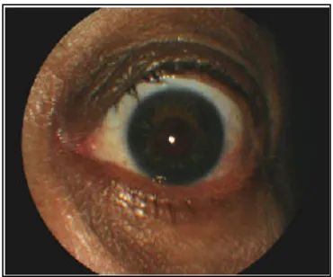
Ophthalmologic manifestations are also frequent among individuals with WBS, including a stellate iris pattern in 70% (Figure 5) [88], high frequency of refractory errors (67% presented hyperopia, 20% astigmatism and 7% myopia) and strabismus in 37% of patients [89, 90].
1.10 Dental Manifestations
[image:39.499.85.270.369.523.2]Individuals with WBS have a high frequency of dental problems; the most frequent are malocclusion (85%), microdontia (95%), hypoplastic enamel defects and abnormal tooth morphology [91]. In
Figure 5.Stellate iris pattern of
a patient with WBS (Modified
adults with WBS a frequent problem is poor dental hygiene which can lead to caries and gum disease [11, 44].
1.11 Genitourinary Manifestations
Genitourinary structural abnormalities are present in 20 to 35% [11, 24, 53] of children with WBS and include renal ectopia, agenesis, hypoplasia, duplication, hydronephrosis and duplicated collecting system [46, 47, 92, 93]. Other frequent genitourinary manifestations are enuresis present in 50% of patients and recurrent urinary tract infections in 30% [24]. Bladder diverticulum has also been reported in WBS adults, and is also thought to be secondary to elastin haploinsufficiency [44].
1.12 Musculoskeletal Manifestations
A significant number of individuals with WBS have musculoskeletal manifestations. Approximately 90% of infants present significant hypotonia and joint laxity, which may contribute to the delay in ambulation [24, 44]. Around 60% of children adopt an awkward gait characterized by a posture of bent knees, flexed-hip stance to improve their stability [24, 44]. Radio-ulnar synostosis is present in approximately 20% of children with WBS [24]. With increasing age, individuals with WBS present joint contractures (50%), which tend to worsen overtime if they are not properly treated [44]. Kyphosis and lordosis are also a frequent finding, occurring in approximately 20 and 40%, respectively [24].
1.13 Integumentary system Manifestations
Approximately 40% of young children have had surgical repairs of inguinal hernias [44] and 15 -20% have umbilical hernias [44, 92].
1.14 Management and Genetic Counseling
Williams Beuren syndrome is a multisystemic disorder with variable expressivity. Patients should be followed-up in multidisciplinary consults to evaluate patients in a global manner, diagnose and treat symptomatology promptly and decrease the number of hospital visits.
At the moment of clinical suspicion a series of evaluations are recommended, including a complete physical and neurological exam, anthropometric data, cardiovascular evaluation (blood pressure and echocardiography), ophthalmological exam, calcium metabolism evaluation, a neuropsychological evaluation and a molecular test to confirm the presence of a 7q11.23 deletion [95]. When giving a diagnosis, it is recommended that it should be a clinical geneticist, who should inform about the sporadic genetic cause, the natural history of the disease, the recurrence risk and the importance of starting early intervention programs and school support [95].
Table 2. Recommendations for medical monitoring of individuals with WBS
[24, 44, 95].
*Periodicity depends on patient’s symptomatology or clinical diagnosis.
although few cases have been reported with an autosomic dominant pattern [96]. The recurrence risk for successive pregnancies for parents not carriers of susceptibility alleles is very low, approximately the same as for the general population, 1/7,500-1/10,000 [1, 95, 97]. If the progenitors are carriers of susceptibility alleles (copy number variants at 7q11.23 or paracentric inversion) the risk increases slightly, approximately 1/1000 [97]. First-degree relatives have the same occurrence risk as the general population.
2
WILLIAMS-BEUREN SYNDROME: CRITICAL
REGION (WBSCR)
2.47q11.23 Genomic Architecture
The 7q11.23 WBS critical region has a complex genomic architecture (Figure 6). The single copy gene region is flanked by three large segmental duplications (SD) or low copy repeats (LCRs). SD or LCRs are large blocks, usually of 10-400 kb of genomic DNA, of highly identical sequence (>97%) generated during primate evolution that can predispose the region to genomic rearrangements [98]. The LCRs are located in the centromeric, medial and telomeric segment of the WBS locus; and each is composed of three blocks called A, B and C. The centromeric and medial duplicons are in the same orientation (tandem repeats) but different order. While the telomeric duplicon is in the opposite transcriptional orientation (inverted repeats) but in the same order as the centromeric block [97, 99, 100]. The three blocks share a very high degree of nucleotide homology (≈98-99%) [97, 99] that can lead to misparing and unequal crossover during meiosis causing deletions, duplications or inversions.
The single copy gene region is located between the medial B and C blocks and span an area of ≈1.2 Mb [97, 99, 100].
The A blocks contain four pseudogenes: STAG3L, PMS2L, GATS-L and a fragment with very high homology to WBSCR19. The medial block A, also contains the gene WBSCR16, which encodes an RCC-1 like G-exchange factor [99]. Its pseudogene is located at the telomeric block A [99].
TF-II, is located in the medial block Bm and its two pseudogenes (GTF2IP1 and GTF2IP2), transcribed as a truncated protein, are located at the centromeric and telomeric blocks [99], respectively. The medial and telomeric copies of another member of the transcription factor family TF-II, GTF2IRD2, are fully transcribed, while the centromeric copy (GTF2IRD2P) is not expressed since it lacks the first two exons. NCF1 encodes for the p47 phox subunit of the nicotinamide adenine dinucleotide phosphate-oxidase complex (NADPH) and the only active copy is located in the medial B block [101]. The centromeric (NCF1B) and telomeric (NCF1C) copies are pseudogenes with a 2-bp GT deletion at the beginning of exon 2 [31].
2.5Mutational Mechanisms
2.5.1 Non-Allelic Homologous Recombination
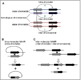
The characteristics of the genomic architecture can predispose to the occurrence of rearrangements and genomic instability [102, 103]. Non-allelic homologous recombination (NAHR) can result in recurrent rearrangements that include deletions, duplications or inversions [104]. Recurrent rearrangements are those that include the same genomic interval in unrelated individuals and the breakpoints usually lie in the LCRs flanking the event [105]. As shown in Figure 7, NAHR that occurs between duplicated sequences in direct orientation can result in deletion or duplication. While NAHR that occur between sequences aligned in an inverted position can result in inversions [106].
Figure 6.Schematic representation of the WBS Critical Region (WBSCR).
Figure 7.Schematic representation of non-allelic homologous recombination,
between: A. Blocks in the same orientation can generate duplication or deletion. B.
Blocks in an inverted position can result in an inversion (Modified from [107]).
Various studies have been done to determine which factors can predispose to the rate of genomic rearrangements [106, 108, 109]. The rate of formation of genomic rearrangements is directly proportional to the degree and length of sequence homology, and indirectly proportional do the distance between them [108]. Cis-acting factors that can influence the recombination rate are the enrichment and formation of secondary structures such as G-quadruplexes, cruciforms, recombination motifs, Alu signal recognition, microsatellites and large A-T repeats [110–114].
Figure 8. Mechanisms for non-allelic homologous recombination (NAHR).A.
[image:48.499.84.367.75.353.2]In 2008, a 13-mer degenerative motif (5’-CCNCCNTNNCCNC-3’) was discovered to be crucial for recruiting crossovers in 40% of all human hotspots [115]. This sequence is likely the binding site of the PR domain-containing 9 gene (PRDM9, OMIM 609760), that is a meiosis-specific histone H3 methyltransferase with a C-terminal tandem-repeat C2H2 zing finger domain encoded by a minisatellite [116]. PRDM9 is likely to act as a trans-modifying factor for some recurrent genomic disorders, such as Charcot-Marie Tooth Type 1A (CMT1A) and hereditary neuropathy with liability to pressure palsies (HNPP)[117].
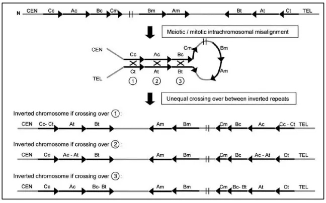
2.5.2 Paracentric Inversion
Figure 9. Schematic representation of the origin of 7q11.23 paracentric
inversion. During meiosis or mitosis an intra-chromosomal misalignment occurs
between inverted low copy repeat blocks (LCRs). Non-allelic homologous recombination (marked in X) can occur between any of the centromeric and telomeric blocks of LCR, originating an inversion of variable size. From [97].
The presence of the inversion can predispose to chromosomal misalignment during meiosis, resulting in reciprocal duplication or deletion [97].
2.5.3 Deletion
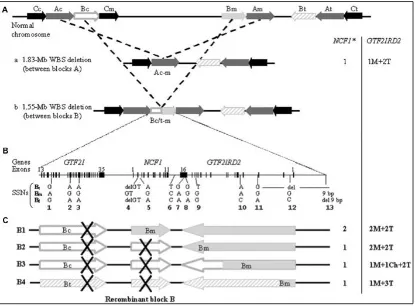
[image:50.499.87.411.61.263.2]Figure 10.Schematic representation of the WBS deletion. (Modified from [97]).
The deletion occurs more frequently between the blocks B because of their higher degree of homology compared to that of the blocks A (99.6% compared to 98.2%) and because of the shorter genomic interval between the centromeric and medial B blocks (1.55 Mb) compared to the centromeric and medial A blocks (1.84 Mb) [97].
Figure 11. Schematic representation of the meiotic misalignment and crossing
over event between an inverted and a normal chromosome. Type 1 unequal
crossing over occurs between the block Bt from the inverted chromosome and Bm of the normal chromosome. Type II unequal crossing over occurs between block Bm of the inverted chromosome and Bt of the normal chromosome (Modified from [97]).
Approximately two-thirds of the deletions arise by interchromosomal rearrangements, while a third of cases are intra-chromosomal [97, 120]. However, all inversion mediated deletions occur after an inter-chromosomal crossover [97].
Figure 12. Schematic representation of the 1.55 Mb and 1.83 Mb deletions and
breakpoint characterization. A. Representation of the 7q11.23 region, in black are
represented the blocks C, in grey the A blocks and in white the B blocks. The top of the figure depicts the 1.83 Mb and 1.55 Mb deletions. B. Representation of the genomic content of the B blocks, as well as the location of the 13 genotyped site-specific nucleotides (SSNs) to refine the breakpoints of the deletion [97]. C. Scheme of the different locations of the 1.55 Mb deletion. In breakpoint 1 (B1) the crossover occurs at GTF2I, therefore NCF1 gene content and the functional copy of GTF2IRD2 are not affected. In B2, the breakpoint is located at NCF1, therefore the patient presents with only one functional copy but GTF2IRD2 remains the same. In B3, the breakpoint occurs at GTF2IRD2 generating a chimeric copy with the final exons belonging to the centromeric block and the initial exons belonging to the medial block. Finally B4, which is the product of the inversion-mediated deletion, has a loss of NCF1 gene and a gain of the telomeric GTF2IRD2 copy. From [31].
gene, the two functional copies of NCF1 and the two medial and telomeric copies of GTF2IRD2 (2T+2M) are kept (Breakpoint 1, B1). When the crossover takes place at NCF1 gene, there is a loss of a functional copy of the gene but GTF2IRD2 telomeric and medial copies are maintained (2T+2M). In the third case, the breakpoint occurs at GTF2IRD2 and there is a loss of NCF1 gene and the creation of a chimeric copy of GTF2IRD2, with the final exons belonging to the centromeric copy and the initial exons belonging to the medial copy. Finally, the fourth scenario is the inversion-mediated deletion, were there is a loss of NCF1 gene and a gain of the telomeric copy of GTF2IRD2 [31, 97]. In the 1.83 Mb deletion, the crossover occur between the centromeric and medial A blocks, result in a loss of NCF1 gene and the medial copy of GTF2IRD2 (1M+2T) [97].
Secondary to gene conversion events between NCF1 gene and its pseudogenes, approximately 15% and 1% of individuals with WBS present three or four copies of NCF1, respectively [97, 121].
2.5.4 Duplication
The NAHR mechanism predicted the existence of the reciprocal 7q11.23 duplication. However, it was not until 2005 that the first case was described [122]. Possible explanations for the relatively recent description are that the phenotype of the reciprocal duplication is very different from the deletion, it is difficult to predict the possible phenotype associated to the duplication based on the knowledge of the gene content of WBS and, as has been observed, there is a great phenotypical variability among individuals [123, 124]. Moreover, detecting adjacent duplications by FISH can be challenging.
cardiovascular manifestations (aortopathy) and other congenital abnormalities [122–127]. In several reported cases, the duplication was transmitted from one of the progenitors and the majority of progenitors had previous history of speech delay and/or learning difficulties [124, 126]. However, there were also few progenitors who presented with normal speech and cognitive abilities [126]. There are two possible explanations for the lack of phenotype in parents, either because the neurocognitive problems might improve over time or because the studied progenitors presented a milder phenotype [126]. The duplication has been found in 0.1 to 0.12% of patients referred for cytogenetic testing for developmental delay, congenital malformations and autism [124, 128, 129] and in 0.076% of patients diagnosed with schizophrenia [130].
2.5.5 Triplication
The first case of a 7q11.23 triplication was recently described in 2010 [131]. The patient presented intellectual disability, severe expressive language delay, behavioral problems and dysmorphisms [131]. The triplication did not encompass the entire WBS critical region; since FZD9 and FKBP6 were not included [131]. Since the breakpoint was not located at the segmental duplications, it is possible that another mechanism different from NAHR could have mediated the triplication [131]. The phenotype of the triplication was more severe than the phenotype spectrum of the duplication, indicating a possible gene-dosage effect [131].
2.5.6 Copy-Number Variants
7q11.23 paracentric inversion acts as a predisposing allele for WBS, but also the type of CNVs present in the region [132].
2.5.7 Novel technologies used to refine breakpoints
Conventional technologies used to refine breakpoints of recurrent genomic disorders required the use of several techniques. Pulsed-field electrophoresis followed by genomic Southern blot hybridization was used to obtain an atypical hybridization band with the breakpoint of interest [133]. This method required the preparation of high molecular-weight DNA, the identification of informative restriction enzymes and the design of probes that flank the deleted interval [134]. Although reliable, this approach was laborious and time-consuming.
Novel technologies available for breakpoint refinement include array comparative genomic hybridization (array CGH) and targeted capture of informative regions using multiple inversion probes (MIPs) followed by next generation sequencing (NGS) [134].
3 GENOTYPE – PHENOTYPE CORRELATION
Figure 14. Schematic representation of typical and atypical 7q11.23 deletions.
On the left, the main clinical characteristics, including craniofacial features, developmental delay, and visual spatial construction deficits (Modified from [150]).
Mlxipl [159]. Partial and complete deletions models that recapitulate most of the features of WBS have also been created [160, 161].
[image:61.499.137.359.123.485.2]The proximal deletion model (PD) involving Gtf2i to Limk1, recapitulates the social phenotype associated to WBS, including
Figure 15. Schematic representation of WBS critical region at chromosomal band
7q11.23 in humans and in chromosomal band 5G2 in mice[151, 152]. There is a high
reduced social fear and gregariousness [162], suggesting that genes telomeric to the elastin locus might me involved in the WBS social phenotype. The distal deletion (DD) model includes Limk1 to Fkbp6 [160, 162] and although it includes a greater number of genes, it was associated with less obvious phenotypes [162]. Mice presented craniofacial abnormalities, including decreased cranial volumes, and also with connective tissue abnormalities such as hernias [162]. This is not unexpected since this deletion includes the elastin locus. With regards to the cardiovascular manifestations, DD mice presented 10-20% increase in mean blood pressure and histological sections showed disorganized and fragmented lamellar units in the blood vessel walls [160]. A double heterozygous mice (D/P) was also studied and it presented similar manifestations as the partial deletion models, however this model presents a homozygous deletion of Limk1 [160]. A complete deletion (CD) mice was created encompassing the entire WBS critical region [161]. CD mice present decreased body weight at birth, decreased brain weight and a milder cardiovascular phenotype compared to DD, including mild increase in mean blood pressure, mild increase in arterial wall thickness and cardiac hypertrophy [161].
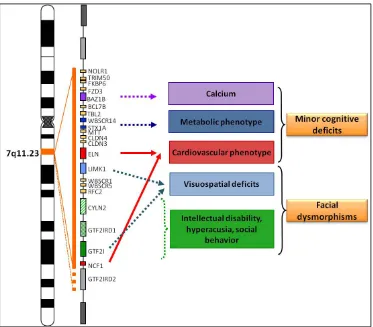
Figure 16. Schematic representation of WBS critical region at chromosomal
band 7q11.23. Genotype-phenotype correlations are marked in colors. Evidence
from atypical deletions are marked in orange, centromeric genes have been associated with minor cognitive deficits while telomeric genes have been associated with facial dysmorphisms.
3.4Cardiovascular phenotype
[image:63.499.68.441.67.394.2]3.4.1 Elastin
Elastin (ELN, OMIM 130160) was the first gene to be linked with a specific phenotype; it is responsible for the cardiovascular and connective tissue abnormalities observed in WBS patients. In 1993, it was found that a t(6;7)(p21.1;q11.23) translocation disrupted the elastin locus and co-segregated with non-syndromic supravalvular aortic stenosis suggesting it as the cause [163]. Since then it has been shown that point mutations, deletions or translocations that disrupt the elastin locus can cause non-syndromic supravalvular aortic stenosis (OMIM 185500) [163–166].
Elastin is the main component of the arterial extracellular matrix [158]. It is mostly expressed during the third trimester of gestation and the first postnatal years. Elastin is synthesized by smooth muscle, it is secreted as a monomer, tropoelastin, and after post-translational modifications it is organized into insoluble polymers that form concentric rings of elastic lamellae around the arterial lumen (Figure 15) [167, 168].
Elastin is a potent autocrine regulator of vascular smooth muscle activity, it induces actin stress fiber organization, inhibits proliferation, regulates migration and signals via a integrin, heterometric G-protein coupled pathway [158].
3.4.2 Neutrophilic cytosolic factor 1
The neutrophilic cytosolic factor 1 gene (NCF1, OMIM 608512), located at the medial B block, encodes the p47phox subunit of the nicotinamide adenine dinucleotide phosphate (NADPH) oxidase complex. Mutations in NCF1 cause granulomatous disease (OMIM 233700)[169].
The NADPH oxidase complex is composed of five subunits, the p47phox, p67phox, p40phox, p22phox and gp91phox[170, 171]. Vascular NADPH oxidase is regulated by humoral growth factors such as cytokines and vasoactive agents, and by physical factors, including stretch and shear stress [172]. The p47phox subunit has a major role in activating the NAPDH oxidase complex [173]. NADPH oxidase enzyme generate oxidative stress by the conversion of oxygen to O2- [171].
The role of oxidative stress in essential hypertension has been studied in great detail and there are indirect evidence supporting its association with increased blood pressure, including increased plasma and urine levels of oxidative stress markers, increased vascular production of superoxide anion, decreased plasma levels of antioxidant vitamins and anti-hypertensive drugs reduce reactive oxygen species production and decrease oxidative stress by inhibiting the action of NADPH oxidase [174].
Later studies in mouse models confirmed previous findings. The p47phox knock-out mouse model did not present hypertension when induced with angiotensin II and had lower oxidative stress production, vascular hypertrophy and endothelial dysfunction when compared to wild type mice [176]. The cardiovascular phenotype for the distal deletion mice were partially rescued when decreasing Ncf1 gene dosage [177]. Double heterozygous mice for the distal deletion and a loss of function of Ncf1 presented normal blood pressure, reduced angiotensin II levels and decreased heart size [177].
3.5Neurocognitive and craniofacial phenotype
The neurocognitive and craniofacial phenotype in WBS has been associated to various genes, including members of the transcription factor II-I family (GTF2I, GTF2IRD1 and GTF2IRD2), LIMK1, CLIP2, BAZ1B, FZD9, STX1A and EIFH4H.
3.5.1 Transcription Factor II-I Family
General transcription factor II-I (GTF2I, OMIM 601679), GTF2I repeat domain-containing protein I (GTF2IRD1, OMIM 604318) and GTF2I repeat domain-containing protein II (GTF2IRD2, OMIM 608899) are members of the transcription factor II-I (TFII-I) family of proteins characterized by the presence of multiple helix-loop-helix – like domains known as I repeats [178]. TFII-I family has been implicated in several important biological functions including cell cycle and proliferation, growth factor (TGF-β), calcium and immune signaling [179].
GTF2I and GTF2IRD1 have been proposed as potential candidates genes involved in the craniofacial, social and neurocognitive phenotype observed in WBS individuals. Individuals with smaller atypical deletions not including GTF2I or GTF2IRD1 do not present the facial gestalt of WBS individuals, nor the social personality and visual spatial deficits [145, 147, 183]. An individual carrying a smaller deletion including all three TFII-I family members and 14 genes in the distal region exhibited autistic traits and the Williams Syndrome Cognitive Profile (WSCP) including visual spatial deficits corroborating their involvement [146]. Deletions involving only GTF2IRD1 have increased our knowledge about its individual contribution [144, 184]. There are two cases reported in the literature, both present craniofacial dysmorphisms and borderline to normal cognitive abilities. However, they are discrepant in the overly-friendliness and specific visual spatial impairments [144, 184].
There are several Gtf2i murine models. Homozygous mutant mice generated by gene targeting with an inframe deletion of exon 2 presented decreased viability and fertility, and abnormal craniofacial morphology [185]. Heterozygous mice presented milder craniofacial alterations and some neurobehavioral alterations (decreased exploratory activity and anxiety) and a low threshold for sound intolerance [185]. A second model created by mutagenesis with a gene trap insertional vector presented similar characteristics. Homozygous mice died during embryiogenesis and presented neural tube defects. Heterozygous mice did not present relevant dysmorphic traits, but presented altered social behavior and increased social interaction [186]. Finally expression analysis of murine lines with Gtf2i and Gtf2ird1 inactivation showed decreased expression of craniofacial related genes [187].
embryonic lethality, neural tube and vascular defects [155]. With regards to their behavioral phenotype, mice presented decreased fear and aggression, and increased social behaviors [189], a hypoactive-anxious phenotype, with gait and sensory motor abnormalities [153, 190].
GTF2IRD2 is not always deleted in WBS, it depends on the breakpoint and size of the deletion. Although its function is still unknown, it was observed that GTF2IRD2 interacts directly with GTF2I and GTF2IRD1 and that it regulates its activities by direct protein interactions and sequestration of the proteins to a novel nuclear compartment [191]. GTF2IRD2 is expressed in human fetal and adult brain, and with in silico expression analysis it was determined that it is specially expressed in the cerebellum, orbitofrontal cortex and dorsolateral prefrontal cortex [192].
A study comparing cognitive, behavioral and psychological functioning between individuals with a 1.55 Mb deletion and with a 1.83 Mb deletion (including GTF2IRD2) found that individuals with the larger deletion presented increased impairments in spatial functioning, social reasoning and cognitive flexibility[192]. The authors attributed these differences to the haploinsufficiency of GTF2IRD2 [192]. However, further studies are needed to corroborate this finding.
3.5.2 LIM Domain Kinase 1
LIMK1 has been proposed as a contributing factor to impaired visual spatial constructive cognition. However, evidence from smaller atypical deletions has been inconclusive. In 1996, two families with deletions encompassing ELN and LIMK1 were evaluated and the individuals presented with supravalvular aortic stenosis and the characteristic cognitive profile of WBS patients, including poor spatial construction skills and proficient verbal ability [196]. However, a later study reported two individuals with the same partial deletion who did not present the WSCP [197, 198].
The Limk1 knockout model presented significant abnormalities in spine morphology and synaptic function and presented altered fear response and spatial learning. The authors conclude that Limk1 plays a critical role in the morphogenesis and function of dendritic spines [199].
Therefore, it seems that although LIMK1 might contribute to the visual spatial constructive cognition, its haploinsufficiency is not sufficient and other genes in the WBS region might be involved.
3.5.3 CAP-GLY Domain containing linker protein 2
The CAP-GLY domain containing linker protein 2 (CLIP2, OMIM 603432) regulates cytoskeleton by polymerization of the microtubule network [200, 201]. It is abundantly expressed in dendrites and cell bodies of neurons in the brain [202].
The homozygous and heterozygous mouse model of Clip2 present mild growth deficiency, brain abnormalities, hippocampal dysfunction and specific deficits in motor coordination [205].
As in the case of LIMK1, it seems that CLIP2 haploinsufficiency is not the sole cause of the phenotype but that it might contribute to the visual spatial and cognitive impairments along with other genes present in the region.
3.5.4 Bromodomain adjacent to zinc finger domain 1B
Bromodomain adjacent to zinc finger domain IB (BAZ1B, OMIM 605681) is a subunit of a chromatin-remodeling complexes, involved in DNA repair [206]. BAZ1B is highly expressed in cranial neural crest derived mesenchyme [207].
Murine models showed that BAZ1B could be a potential candidate gene involved in craniofacial phenotype of WBS. Random mutagenesis in mice resulted in a heterozygous L733R change in a highly conserved amino acid [207]. Homozygous mice were smaller and had widened bulbous foreheads, shortened snouts and a reduction in parietal and nasal bone [207]. Heterozygous mice also presented similar alterations, including a decrease in cranium width to height ratio and a decrease in palatine bone [207]. Partial deletion mouse model including Baz1B also presents with an abnormal craniofacial appearance [162]
3.5.5 Frizzled 9
Frizzled 9 gene (FZD9, OMIM 601766), first identified in 1997 [208], is selectively expressed throughout life in the hippocampus [209] and it has been postulated that it may act as a Wnt receptor in the canonical Wnt pathway, signaling via β-catenin pathway [210].
facial features and moderate neuropsychological deficits [150]. However, the frizzled 9 murine model pointed towards its potential involvement in the neurocognitive phenotype. Null and heterozygous mice presented with increased apoptotic cell death and increased proliferation of precursors during hippocampal development [211]. Null mice presented severe deficits in learning and memory, which could reflect hippocampus functional deficits [211]. Heterozygous mice presented milder alterations in spatial memory and lower seizure threshold [211]. A second murine model for Fzd9 was analyzed and it did not exhibit the same cognitive alterations, instead it had an abnormal B cell development [212]. Further studies of this model revealed that frizzled 9 null mice also presented low bone mass caused by impaired bone formation, with defects in matrix mineralization [213].
3.5.6 Syntaxin 1A
Syntaxin 1A (STX1A, OMIM 186590) encodes a protein that belongs to the SNARE (soluble N-ethylmaleimide-sensitive factor attachment protein receptors) complex [214] which is involved in vesicle fusion process [215]. The SNARE complex plays a major role in insulin exocytosis, as well as in neurotransmitter release.
Stx1A hemizygous mice did not present any apparent behavioral or cognitive phenotype [216]. Homozygous mice for a truncated form of Stx1a presented altered synaptic plasticity [217] and both, homozygous and heterozygous mice, showed abnormal behavior similar to neuropsychological alterations observed in psychiatric patients [218].
3.5.7 Eukaryotic translation initiation factor 4

![Figure 14. Schematic representation of typical and atypical 7q11.23 deletions.On the left, the main clinical characteristics, including craniofacial features, developmental delay, and visual spatial construction deficits (Modified from [150])](https://thumb-us.123doks.com/thumbv2/123dok_es/5199935.94239/60.499.52.460.71.395/schematic-representation-characteristics-including-craniofacial-developmental-construction-modified.webp)
![Figure 15. Schematic representation of WBS critical region at chromosomal band 7q11.23 in humans and in chromosomal band 5G2 in micedegree of conserved sinteny, in mice the WBS region is in inverse orientation with [151, 152]](https://thumb-us.123doks.com/thumbv2/123dok_es/5199935.94239/61.499.137.359.123.485/schematic-representation-critical-chromosomal-chromosomal-micedegree-conserved-orientation.webp)