Assessment of prepubertal sheep oocyte competence for in vitro embryo production by the Brilliant Cresyl Blue test.
106
0
0
Texto completo
(2)
(3) La Dra Maria Teresa Paramio Nieto, Catedrática del Departamento de Ciencia Animal y de los Alimentos de la Facultad de Veterinaria de la Universidad Autónoma de Barcelona. CERTIFICA:. Que la tesis titulada “Assessment of prepubertal sheep oocyte competence for in vitro embryo production by the Brilliant Cresyl Blue test.” presentada por María Gracia Catalá para optar al grado de Doctor, se realizó bajo mi dirección y con un financiamiento del Ministerio de Ciencia e Innovación (AGL 2007 60227/GAN y AGL2011-23784) y una beca otorgada por la Universidad Autónoma de Barcelona (UAB-2007FI00193).. I para que conste a los efectos oportunos, firmo la presente en Bellaterra (Cerdanyola del Vallès), el 12 de marzo de 2012. Dra. Maria-Teresa Paramio Nieto.
(4)
(5) A mis padres, A mi hermano, A mi hermana del alma, A Amine,.
(6)
(7) Chapter 1: General Background…………………………………………………………. 10. Chapter 2: Bibliographical Revision…………………………………………………….. 16. 2.1. Current situation of the in vitro embryo production (IVEP) in sheep ……………. 18. 2.1.1. In vitro Maturation (IVM)…………………………………………………….... 18. 2.1.2. IVF and sperm capacitation…………………………………………………….. 18. 2.1.3. Intracytoplasmic Sperm Injection………………………………………………. 19. 2.1.4. Parthenogenetic activation…………………………………………………….... 19. 2.1.5. Embryo culture and blastocyst production….………………………………….. 20. 2.2. Study of the oocyte……………………………………………………………….... 20. 2.2.1. Meiosis: nuclear and cytoplasmic maturation………………………………….. 20. 2.2.3. Maturation promoting factor…………………………………………………… 21 2.2.4. Mitochondria and ATP…………………………………………………………. 22. 2.2.5. Gene expression……………………………………………………………….... 22. 2.2.6. Non invasive oocyte quality assessment: Brilliant Cresyl Blue Strain……….. 23. 2.3. Parameters affecting oocyte quality……………………………………………….. 25 2.3.1. Age of donor……………………………………………………………………. 25. 2.3.2. Follicular size…………………………………………………………………... 26 2.3.3. Oocyte size……………………………………………………………………... 26 2.4. Improving oocyte quality using in vitro media…………………………………..... 27. 2.4.1. Insulin Transferrin Selenium (ITS)…………………………………………….. 28. 2.4.2. Ascorbic acid ………………………………………………………………….. 29. Chapter 3. Objectives……………………………………………………………………. 32 Chapter 4. Brilliant Cresyl Blue stain selects largest oocytes with highest mitochondrial activity, maturation-promoting factor activity and embryo developmental competence in prepubertal sheep…………………………………………...... 36. Chapter 5. Effect of insulin transferrin and selenium and Ascorbic Acid in maturation media on embryo development, MPF activity and ATP content of prepubertal sheep oocytes selected by brilliant cresyl blue test…………………………………………………... 50. Chapter 6. Effect of oocyte quality on blastocyst development after in vitro fertilization (IVF) and intracytoplasmic sperm injection (ICSI) in a sheep model……………………... 72. Chapter 7. General Discussion…………………………………………………………... 80. Chapter 8. Conclusions………………………………………………………………….. 86. Chapter 11. Bibliography ……………………………………………………………….. 90. Thanks to, ………………………………………………………………………………. 104.
(8)
(9) Chapter 1:. General background. 10.
(10) 11.
(11) General background Assisted reproductive technologies (ART) have been one of the major tools towards increasing productivity in livestock industry. In this field, artificial insemination (AI) is the earliest and most powerful among the reproductive technologies because it is easy to perform, costeffective, and highly successful (Vishwanath. 2003). For over the years AI has been used to obtain offspring from genetically superior and from sub-fertile animals. Moreover, by the 1960´s, significant improvements in cryopreservation and storage of semen made AI even more accessible to livestock producers (Vishwanath. 2003). In the modern dairy industry, new ART techniques were developed as cryopreservation not only of semen but also of gametes and embryos, induction of multiple ovulations, embryo transfer, in vitro fertilization (IVF), sex determination of sperm or embryos, nuclear transfer, cloning, etc. In the small ruminants as sheep and goat, these techniques had a lower development. Despite the improvement in ART protocols, the pregnancy rates are still relatively low and early embryonic mortality has been reported after embryo transfer. It has been shown that 40% of total embryonic losses occur between day 8 and day 17 of pregnancy (Thatcher et al. 1994) and one of the reasons could be due to the oocyte quality (Snijders et al. 2000). The oocyte quality is used as synonymous of oocyte competence, defined as the ability of an oocyte to resume meiosis, cleave following fertilization, develop to the blastocyst stage, induce a pregnancy and bring the offspring to term in good health (Sirard et al. 2006). The oocyte competence is acquired gradually during the course of folliculogenesis as the oocyte grows and its somatic cells cohort differentiates (Eppig et al. 1994). Many factors have been shown to affect the oocyte competence as: follicle size (Lonergan et al. 1994; Romaguera et al. 2011), phase of follicular wave (Machatkovaa et al. 2004), hormonal stimulation (Sirard et al. 2006), maturation environment (Warzych et al. 2007), season (Sartori et al. 2002), nutrition (Fouladi-Nashta et al. 2007) and donor’s age (Rizos et al. 2005). The assessment of oocyte quality is one of the major objectives in ART, especially in human where the use of the best quality oocytes to be inseminated may improve significantly the embryo production. Consequently, multiple methods of oocyte selection have been proposed. One of the most popular is to select the oocyte by morphology which is relatively quick and simple; however this simple technique leads to identify more frequently the negative than the positive aspects, and overall it is not fully satisfactory (Balaban and Urman. 2006). Other methods have been proposed as the use of polarizing microscopy analysis (Heindryckx et al. 2011), gene expression of the granulosa cells or in the oocyte itself (Patrizio et al. 2007), polar body biopsy to screen oocytes with chromosomal defects deriving from errors in the meiotic division (Dawson et al. 2006). Most of these techniques are quite complicated, require. 12.
(12) General background expensive laboratory equipment and time-consuming procedures, and some of them are invasive and consequently are not currently applicable in the clinical practice. In research, the law is extremely restrictive in the use of human embryos; consequently using animals has become through time a valuable tool. The bovine is the mostly used ruminant in ART research because of its economic importance and its wide geographical distribution, although using small ruminants as oocyte donor could be an interesting alternative. Using the sheep (Ovis aries) can bring great benefits to reproduction research because of their younger age to reach puberty, short gestational periods, the possibility to have more than one offspring in a single gestation and the maintenance cost are lower than in bovine. Moreover, sheep have been domesticated for over 10,000 years and are also widespread across the world, being adapted to many different climatic conditions. During the past 60 years, sheep have been the subject of considerable research starting in 1949 with an experimental super ovulation protocol (Ortavant et al. 1949) until nowadays in which sheep receptors, genes (Leoni et al. 2007; Kyasari et al. 2012) and proteins (Grazul-Bilska et al. 2011) are being studied. In addition, using prepubertal animals as oocyte donor has some additional benefits comparing to the use of adult donors, as for example; reducing the generational interval (Duby et al. 1996), their ovaries produce a major number of oocytes than adult (Koeman et al. 2003) and this oocytes could serve as a model of low quality oocyte in research as they are characterized of having an abnormal cytoplasmic maturation and lower ability to achieve the blastocyst stage (Revel et al. 1995; O'Brien et al. 1996; Armstrong. 2001). One of the difficulties of using ovaries coming from prepubertal animals, is to release the complex oocyte cumulus (COCs) by traditional follicular aspiration because of having a high percentage of antral follicles with a smaller diameter than 3 mm (Martino et al. 1994). Consequently, the release of these oocytes is made by slicing the ovary surface obtaining oocytes with heterogeneous diameter, different COC morphology and stage of atresia. The Brilliant Cresyl Blue (BCB) test has been successfully used as a non invasive methodology to select oocytes with a higher diameter and more competent to develop up to the blastocyst stage in cow (Pujol et al. 2004; Alm et al. 2005; Bhojwani et al. 2007; Torner et al. 2008; Opiela et al. 2010), pig (Ericsson et al. 1993; Roca et al. 1998; El Shourbagy et al. 2006; Egerszegi et al. 2010), goats (Rodriguez-Gonzalez et al. 2002; Rodriguez-Gonzalez et al. 2003; Kątska-Książkiewicz et al. 2007), mouse (Wu et al. 2007) and buffalo (Manjunatha et al. 2007). To our knowledge there are no previous reports using this stain to select oocytes by their competence in the sheep. The aim of this study is to test the ability of the BCB staining to select the more competent sheep oocytes for in vitro embryo production. Also, in this work we pretend to improve the. 13.
(13) General background knowledge about oocyte competence related to their cytoplasmic and molecular performances, their responses to different techniques of fertilization and the in vitro culture media needed to improve the blastocyst production.. 14.
(14) 15.
(15) Chapter 2.. Bibliographical Revision. 16.
(16) 17.
(17) Bibliographical Revision 2.1. Current situation of the in vitro embryo production (IVEP) in sheep 2.1.1. In vitro maturation (IVM) A correct maturation of the cumulus-oocyte complex (COC) is one of the most important factors which determine the entry of the oocyte into metaphase II (MII), subsequent successful fertilization, as well as the ability of an embryo to undergo an appropriate growth and development. Performing the COC maturation under in vitro conditions provides an excellent opportunity for having cheap and abundant oocytes for carrying out basic research and for the application of emerging biotechnologies like cloning and transgenesis. Several aspects of the IVM of sheep oocytes have been studied (Wani et al. 2000; Rao et al. 2002). Sheep COC’s are most commonly matured in Tissue Culture Medium (TCM199) containing Earle’s salts. The supplementation of the IVM medium with epidermal growth factor (EGF) (Guler et al. 2000), mare serum (Motlagh et al. 2008), fetal calf serum (FCS) (Ghasemzadeh Nava and Tajik. 2000), estrous sheep serum (ESS) (Ghasemzadeh Nava and Tajik. 2000), insulin –like growth factor (IGF-I) (Guler et al. 2000) and cysteamine (de Matos et al. 2002) among others stimulate sheep oocyte nuclear and cytoplasmic maturation. After the improvements in the IVM media, currently the most commonly media used to in vitro mature sheep oocytes is the TCM199 supplemented 2 mM glutamine, 100 µM cysteamine, 0.3 mM sodium pyruvate, 10% fetal bovine serum (FBS), 5 µg/mL FSH, 5 µg/mL LH and 1 µg/mL estradiol (Loi et al. 2008). Moreover supplementing the IVM media with ESS instead of FBS has also shown good results (Bebbere et al. 2010). 2.1.2. IVF and sperm capacitation. The first reports of IVF in sheep used heparin to capacitate the ram spermatozoa reaching an 80% of fertilization but only 15% were viable embryos (Slavik et al. 1992). However, the use of ESS instead of heparin rapidly gained importance by showing a fertilization rate of 85% (Huneau et al. 1994) and 56% reached the blastocyst stage (Walker et al. 1994). The ESS works through the binding-protein albumin that may facilitate the sperm capacitation by contributing to the depletion of the sperm cholesterol membrane (Huneau et al. 1994). Ccurrently most of the authors use the ESS to capacitate fresh and frozen semen using a concentration of 2 to 20% of ESS (Walker et al. 1994). Bebbere et al ( 2010) using frozen ram semen capacitated with 2% ESS reached 54% of blastocyst using adult sheep oocytes and Shirazi ( 2009) with fresh semen and 20% ESS reached a 34% of blastocyst.. 18.
(18) Bibliographical Revision 2.1.3. Intracytoplasmic Sperm Injection. Intracytoplasmic sperm injection (ICSI) consists in fertilizing a MII oocyte by the injection of a single spermatozoon into the cytoplasm with both the acrosome and sperm membrane intact. This technique was reported with success for the first time in hamsters 30 years ago (Uehara and Yanagimachi. 1976). Since then, the ICSI has become the most commonly used procedure to overcome male infertility problems in human reproduction. In animals, the ICSI has been in general used for research purposes, in which studies with sex-sorted semen (Wilson et al. 2006), sperm mediated gene transfer (Lavitrano et al. 2006; Pereyra-Bonnet et al. 2011) and cryopreserved oocytes (Matson et al. 1997; Pope et al. 2012) are the most commonly reported. The first lamb born after oocyte maturation, sperm sexing by flow cytometry and ICSI was published by Catt et al. ( 1996). For the ICSI procedure they have directly injected the spermatozoa into the cytoplasm without any chemical activation obtaining a low efficiency of the technique that was traduced in 251 oocytes injected and transferred but only one lamb arrived to term. Later, this same group reported that the manipulation by itself was not enough to cause proper oocyte activation and that the addition of calcium in the culture media increased the efficiency of the technique (Gomez et al. 1998). More recently, Shirazi et al. ( 2009) tried to determine the need of an activation protocol after sheep sperm injection, concluding that the chemical activation of oocytes must be considered as an essential part of ICSI in this specie. 2.1.4. Parthenogenetic activation After the entry of the sperm, mammalian oocytes exhibit an increase of the intracellular calcium induced by the same sperm. These transient calcium peaks are propagated throughout the fertilized oocyte in the form of a wave and initiate both the cortical granule exocytosis and escape from the MII arrest to become a zygote [revised by (Loi et al. 1998; Nakada and Mizuno. 1998)]. Oocyte activation protocols have been developed to induce artificially the intracellular calcium levels in the oocyte cytoplasm. This is achieved by exposing the oocyte to a calcium ionomicyn or ionophore and subsequently culturing it with a persistent kinase inhibitor such as 6-DMAP (6-dimethyl amino purine). The treatment with ionomycin 19.
(19) Bibliographical Revision alone caused the resumption of meiosis but no pronuclear formation and the 6-DMAP alone did not cause any resumption of meiosis or pronuclear formation. So, it is important the combination of the two compounds to reach the pronuclear stage (SuskoParrish et al. 1994). In sheep, Alexander et al. ( 2006) using the combination of these two compounds produced 21 % of blastocysts; he also showed that using cycloheximide instead of the 6-DMAP it is also possible to produce blastocyst but in a lower percentage (15%). Loi et al. ( 1998), using the combination of ionomicyn and 6-DMAP to activate sheep nuclear transfer oocytes, reached an efficiency of 83% of blastocyst compared to 25% with no activation protocol. 2.1.5. Embryo culture and blastocyst production. The most common media used during in vitro culture (IVC) of embryos is the Synthetical Oviductal Fluid (SOF: (Tervit et al. 1972). From the beginning, this media showed good results in culturing embryos under in vitro conditions obtaining 25 lambs born after 6 days of IVC (Tervit and Rowson. 1974). In addition, supplementing the SOF media with serum (20% vs. 40%) (Thompson et al. 1998) and BSA (18% vs. 28%) (Carolan et al. 1995) increased blastocyst percentage significantly. Furthermore, the addition of amino acids (aa) to this media appears to be beneficial in sheep producing 58% of blastocyst versus 22% when the aa were not added to the media (Walker et al. 1996). 2.2. Study of the oocyte 2.2.1. Meiosis: nuclear and cytoplasmic maturation. In mammals, oocytes are arrested for several weeks, months or years in prophase of the first meiotic division. During this long period, oocytes accumulate molecules of mRNA, proteins, lipids and sugars as well as they gradually increase in size. The accumulation of all necessary sources of energy and information during oocyte growth is essential for the final step of oogenesis: the oocyte maturation. Maturation consists of two interlinked and mutually dependent processes: cytoplasmic and nuclear maturation. The cytoplasmic maturation of the oocyte includes cytoplasmic changes as organelle redistribution, micro and macro molecular changes that occur during oocyte maturation. These modifications contribute to the oocyte’s ability to undergo: nuclear. 20.
(20) Bibliographical Revision maturation, successful fertilization, cleavage and the development at least until the activation of the embryonic genome. Nuclear maturation includes chromatin changes during the oocyte maturation starting from germinal vesicle (GV) breakdown (GVBD) through Meiosis I and Meiosis II when the oocyte is finally arrested in the MII stage. At this moment the oocyte is physiologically prepared to complete the second meiotic division upon fertilization. Under in vivo conditions, only fully grown oocytes can resume meiosis which implies that cytoplasmic changes that occur before maturation are essential for the acquisition of the developmental competence [Revised by Marteil et al ( 2009)]. However, when oocytes are removed manually before ovulation from an antral follicle, the separation triggers a pseudo-maturation event leading in general to the completion of the first meiotic division and the arrest at the MII stage. This process has been called spontaneous maturation and is believed to be induced by the removal of the oocyte maturation inhibitor (OMI) present in the follicle where cAMP is involved [Revised by Sirard ( 2011)]. A comparison between oocytes that were removed from the follicular environment and in vitro matured compared to in vivo matured oocytes, showed the same rates of nuclear maturation, fertilization and cleavage, but the percentage of blastocyst was significantly lower on in vitro matured group [30% vs. 60%, revised by (Sirard and Blondin. 1996)] indicating that the cytoplasmic competence must be different between the in vitro and the in vivo matured oocytes. 2.2.3. Maturation Promoting Factor. Meiosis is regulated by the maturation-promoting factor (MPF). This universal cell cycle regulator is a heterodimer protein composed of two subunits, the catalytic subunit p34cdc2 (serine-threonine kinase activity) and the regulatory subunit cyclin B1. The association of these two subunits is a requirement for the activation of the protein kinase activity; also the phosphorilation of p34cdc2 on threonine 161 by the protein kinase CAK (Cdc2 activation kinase) and dephosphorylation on threonine 14 and tyrosine 15 by Cdc25 phosphate is necessary. The MPF activity appears just before GVBD increasing until metaphase I (MI); its activity decreases in the anaphase-telophase while its maximum level is reached at the MII stage (Figure 1). Incompetent goat oocytes have a limited amount of Cyclin B1 (Hue et al. 1997) and p34cdc2 (Anguita et al. 2007). In calf and lamb oocytes the MPF activity is significantly lower than in cow and ewe oocytes (Ledda et al. 2001; Salamone et al. 2001) whereas Han D et al. ( 2010), showed that the MPF activity of prepubertal mice oocytes was significantly higher than adult mice oocytes, suggesting a difference in the mechanisms according to species. In prepubertal. 21.
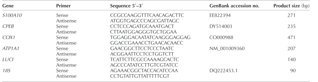
(21) Bibliographical Revision goats, Anguita et al. ( 2007) showed higher MPF activity and competence in oocytes with a diameter larger than 135 µm compared to the smaller diameters. Figure 1: Schematic representation of MPF activity during oocyte maturation.. GV-Intact. MPF. MPF. GVBD. Metaphase I. MPF. MPF MetaphaseII. 2.2.4. Mitochondria and ATP Mitochondria are maternally inherited organelles that use oxidative phosphorylation to supply energy as adenosine triphosphate (ATP) to the cell (Stojkovic et al. 2001). This source of ATP, has a central role in the establishment of the developmental competence (Van Blerkom. 2004; Van Blerkom et al. 2008). Even though mitochondria are the most abundant organelles in the oocyte, little is known about their different functions. The mitochondria distribution and activity change during oocyte maturation and fertilization with the aim of bringing mitochondria to the region of the cell where a higher level of ATP (Van Blerkom and Runner. 1984) or calcium (Sousa et al. 1997) are required. Energy in the form of ATP is crucial; spindle formation and chromosome behavior depend on the expression and activity of motor proteins, which use ATP as their energy source. It has been proposed that mitochondria reorganization and ATP levels are influenced by the oocyte quality (Stojkovic et al. 2001), compactness of the cumulus (Torner et al. 2007) and cumulus apoptosis (Torner et al. 2004), GnRH (Dell'Aquila et al. 2009) and the microtubule cytoplasmic network (Brevini et al. 2005) affecting the early stages of the embryo (Tarazona et al. 2006). Therefore, several authors concluded that better quality oocytes contained significantly higher ATP levels and produced significantly higher blastocyst rates after fertilization (Van Blerkom et al. 1995; Stojkovic et al. 2001; Van Blerkom. 2004). 2.2.5. Gene expression In the last few years, the study of mammalian genes has been the focus of several studies in the belief that a good expression pattern could derive in a successful oogenesis, folliculogenesis, fertilization and early embryonic development. In the course of acquiring the oocyte competencies and a good embryo development a correct mRNA transcription is a crucial process occurring in the cytoplasm (Crozet et al. 1981; Brevini-Gandolfi and Gandolfi. 2001;. 22.
(22) Bibliographical Revision Patel et al. 2007).The mRNA content in oocytes is affected by animal nutrition (Pisani et al. 2008), donor age (Hamatani et al. 2004), follicle diameter (Caixeta et al. 2009), IVM culture media (Saadeldin et al. 2011; Salhab et al. 2011), in vivo and in vitro conditions (Wells and Patrizio. 2008), apoptosis (Li et al. 2009) and the cumulus cells (Adriaenssens et al. 2010) among others. In this thesis we are going to study the expression of four genes in relation with the oocyte quality; two genes involved in metabolism: ATP1A1 (ATPase NaC/KC transporting a 1) and COX1 (cytochrome c oxidase subunit 1), and two genes involved in the constitutive function of the cell: CPEB (cytoplasmic polyadenylation-element-binding protein) and S100A10 (calciumbinding protein). ATP1A1 gene is translated in an enzyme responsible for the transport of Na+ out of and K+ into the cell and that is an important key regulator of bovine blastocyst formation and is necessary for the in vitro production of healthy bovine embryos (Watson et al. 1999). CPEB plays an important role in the regulation of the mRNA translation targets required for oocyte maturation (Cai et al. 2010). COX1 is a gene that produces a mitochondrial energy-transfer enzyme of the respiratory chain. Opiela et al. ( 2010) found a high expression of COX1 in oocytes of better quality versus lower quality oocytes. According to Tingaud-Sequeira et al. ( 2009), S100A10 plays an antiapoptotic role and that a high expression levels of S100A10 in the follicles may have a dual function protecting follicular cells from apoptosis during atresia and acting as chemoattractant for leukocytes and macrophages. After a microarrays of bovine oocytes, Torner et al. ( 2008) showed a higher expression of S100A10 in lower quality than in high quality oocytes. 2.2.6. Noninvasive oocyte quality assessment: Brilliant Cresyl Blue Stain. With the aim of selecting the most competent oocytes, relevant results in goat (RodriguezGonzalez et al. 2002), bovine (Pujol et al. 2004; Alm et al. 2005; Bhojwani et al. 2007; Torner et al. 2008; Opiela et al. 2010), pig (El Shourbagy et al. 2006), mouse (Wu et al. 2007) and buffalo (Manjunatha et al. 2007) were published when the Brilliant Cresyl Blue (BCB) stain was used to select oocytes prior to the IVM. This is a non invasive methodology that allows selecting oocytes with larger diameters among a heterogeneous pool. Brilliant Cresyl Blue is a compound (Figure 2; C17H20N3OCl · 1/2ZnCl2) with a molecular weight of 385.96 g/mol which is used to determine the intracellular activity of glucose-6-phosphate dehydrogenase (G6PDH). The G6PDH is a regulatory enzyme synthesized within the oocyte during oogenesis, and is a component of the pentose phosphate pathway (Figure 3) that controls the flow of carbon through this pathway and produces reducing equivalents in the form of NADPH to meet. 23.
(23) Bibliographical Revision cellular needs for reductive biosynthesis and maintenance of the cellular redox state. The activity of the G6PDH gradually decreases as oocyte reach their growth phase (Mangia and. Epstein. 1975). The BCB dye can be reduced by the G6PDH enzyme activity, thus the oocytes that have reached their growth phase cannot reduce BCB to a colorless compound exhibiting a. blue colored cytoplasm (BCB+). However, the growing oocytes are expected to have a high activity of G6PDH and be able to reduce the blue compound which results in a colorless oocyte. cytoplasm (BCB–). Figure 2: Brilliant Cresyl Blue compound. Figure 3: A reduce scheme of the Pentose Phosphate Pathway. Glucose-6-phosphate NADP+. Glucose-6-phosphate dehydrogenase. NADPH. 6-phosphogluconolactone H2O. Gluconolactonase. H+. 6-phosphogluconate NADP+. 6-phosphogluconate. NADPH CO2. Ribulose-5-phosphate. Non-oxidative reactions (Nucleotides, Co-enzymes, DNA, RNA). 24.
(24) Bibliographical Revision The first studies using the BCB compound appeared in the 80's, when this stain was used as a colorimetric assay to detect X-linked enzymes such as G6PDH to predict the sex of the embryos, resulting in a 64% of efficiency in sex prediction (Williams. 1986). Some years later, Ericsson et al. ( 1993) changed the use of the BCB stain in predicting sex, and used it to select more competent porcine oocytes for IVM and IVF. He reported for the first time a significant increase in maturation (82% vs. 49%) and sperm penetration (51% vs 26%) for the BCB+ respect the BCB- oocytes. In our laboratory we have previously shown the usefulness of the BCB stain to select the larger and more competent oocytes for in vitro blastocyst production. The percentages of total embryos undergoing development to the morula plus blastocyst stages were higher in the group of BCB+ than in BCB− oocytes in prepubertal goat [12% vs. 4%, respectively; (RodriguezGonzalez et al. 2002)] and in heifers [13% vs. 2%, respectively; (Pujol et al. 2004)]. Additional reports reiterate the value of the BCB stain for the selection of developmentally competent oocytes related to bovine nuclear transfer procedures (Bhojwani et al. 2007) and in COC’s collected by OPU (Tagawa et al. 2006). Torner et al. ( 2008) studied in bovine the efficiency of the BCB stain by analyzing the phosphorylation pattern of protein kinases, the cDNA microarray for gene expression profiles, the fluorescence labeling and photometric measurement for chromatin configuration of the nucleus and the mitochondrial activity of the oocytes. They reported a higher phosphorylation levels of Akt and MAP kinases and an increase in transcription of genes that are involved in regulating transcription (SMARCA5), cell cycle (nuclear autoantigenic sperm protein, NASP) and protein biosynthesis (RPS274A and EF1A) in BCB+ oocytes. On the other hand, BCB- oocytes revealed higher mitochondrial activity and an increase in mRNA expression of genes involved in ATP synthesis (ATP5A1), mitochondrial electron transport (FL405), calcium ion binding (S100A10) and growth factor activity (BMP15). Mohammadi-Sangcheshmeh, A. ( 2011b) using equine oocytes, showed a greater proportion of expanded COC (71% vs. 50%), maturation rate (59% vs. 29%), cleavage rate (46% vs. 29%), and blastocyst (9% vs. 1%) percentage in BCB+ compared with BCB- oocytes. Regarding the gene expression, eight genes were significantly higher for BCB+ oocytes (ATPV6E, IF-3, TFAM, DNMT1, STAT3, Aurora-A, ODC1, and CKS2) whereas BCBoocytes showed higher expression of COX1. In porcine, Egerszegi I. et al. ( 2010), observed that BCB+ oocytes were characterized by a high mitochondrial activity with a homogeneous distribution while BCB- oocytes had a low mitochondrial activity with a heterogeneous distribution. In addition, BCB+ oocytes showed an increase in maturation (82% vs. 7%) and fertilization (17% vs. 0%) respect BCB- porcine oocytes.. 25.
(25) Bibliographical Revision 2.3. Parameters affecting oocyte quality 2.3.1. Age of the donor. The age of the female donor is an important issue in ART evidencing a reduced developmental competence when prepubertal donors are used. Armstrong et al.( 1997) suggested that the optimal age to collect oocytes from prepubertal lambs is between 4 and 6 weeks of age as this is the time of most follicular responsiveness. In a sheep study, in which oocytes were in vivo fertilized and flushed from oviducts of prepubertal or adult ewes and transferred to adult recipients, showed that only 33% of the zygotes transferred from prepubertal donors resulted in birth compared to 73% from adult oocyte donors (Quirke and Hanrahan. 1977). Ledda et al.( 2001) reported that although prepubertal sheep oocytes reach the MII stage at the same percentages as adult animals, they show a lower level of MPF compared to adult ones. In addition, lambs produce 16% of cleaved zygotes that reaches the blastocyst stage, significantly lower than the 34% produced by adult sheep donors (O'Brien et al. 1997). Comparable results were found in other species. In cow, Revel et al. ( 1995) reported similar rates of IVM, IVF and cleavage for calf and cow oocytes, but after 7 days of IVC, the blastocyst percentage was significantly lower for calf than for cow oocytes (10% vs. 20%, respectively). In pig, Grupen et al. ( 2003) showed that the rates of cleavage (92% vs. 73%) and blastocyst formation (57%vs. 38%) were higher for adult oocytes than for prepubertal oocytes and that the blastocysts derived from adult oocytes had more trophectoderm cells (43 vs. 30) and total cells (51 vs. 36) than those derived from prepubertal oocytes. This drastic reduction in blastocyst development of prepubertal donors is generally attributed to an incomplete cytoplasmic maturation, traduced in the failure of the sperm to penetrate and decondensate, inability to form normal male pronuclei, failure to block polyspermy, early cleavage failure and failure to reach or survive the transition from maternal to embryonic genome expression among others [reviewed by (Armstrong. 2001)]. 2.3.2. Follicular size. Several authors concluded that there is a correlation between the follicle diameter and the oocyte size and its competence (Martino et al. 1994; Fair et al. 1995; Ledda et al. 1999). In prepubertal goat and ovine the most competent oocytes are the ones coming from follicles bigger than 3mm (Martino et al. 1994; Cognie et al. 1998). In adult goats, Crozet et al. ( 2000) obtained a higher percentage of blastocysts (6% ,12% and 26%) using oocytes from small (2-3 mm), medium (3.1-5 mm), large (> 5 mm) follicles, respectively. Comparing adult and. 26.
(26) Bibliographical Revision prepubertal pig oocytes, Bagg et al. ( 2007) showed that rates of blastocyst development after parthenogenetic activation of adult oocytes from three different follicles sizes (3mm, 4mm, and 5-8 mm) were similar (approximately 55%), whereas rates from prepubertal oocytes increased with the increasing follicle size (17%, 36% and 55%, respectively). They concluded that the low developmental competence in prepubertal porcine oocytes is associated with a greater proportion of 3 mm follicles and not to an effect of the female age. In our laboratory we have previously described in prepubertal goat that most of the follicles present in the ovaries were between 2.5 and 3 mm and only 1.1% of follicles per ovary were larger than 3 mm (Martino et al. 1994). More recently, we have reported a higher oocyte size (128 µm vs. 125 µm), higher percentages of TUNEL positive (43% vs. 24%), higher cleavage (48% vs. 23%) and higher blastocyst rates (20% vs. 4%) in oocytes deriving from follicles with diameter >3 mm than from oocytes deriving from follicles with diameter <3mm. Blastocyst mean cell number did not show significant differences between the different follicular groups (123 vs. 104 blastomeres) (Romaguera et al. 2010). As well, significant differences were found when comparing blastocyst rates of oocytes recovered from follicles with diameter <3 mm of prepubertal goats to those from adult goats (5% vs. 21%, respectively). However, when prepubertal goat follicles of >3mm were used, no differences were found comparing to adult oocytes (18%) (Romaguera et al. 2011). In addition, Kauffold et al. ( 2005), showed an increase in blastocyst production in oocytes coming from calf follicles with diameter > 8mm (47%) than from follicles of < 8 mm (<15%). In addition, they found no differences in blastocyst production when comparing oocytes from calf (47%) and cow (59%) from a follicle diameter bigger than 8mm. 2.3.3. Oocyte size. It has been shown that oocyte size is closely related to the developmental competency. Gandolfi et al. ( 1998) showed differences in oocyte size between cow (123 µm) and calf (118 µm) oocytes. This difference in oocyte diameter was reflected in a significant decrease in protein synthesis after 9 h of IVM in calf oocytes, while in cow adult it was detected after 24 h. In prepubertal goat, oocytes from different diameter (< 110, 110-125, 125-135 and > 135 µm) showed a positive correlation to the percentage of oocytes reaching MII stage after IVM (0%, 21%, 58% and 78%, respectively) and the percentage of blastocysts obtained at 8 days postinsemination (0%, 0%, 2% and 13%, respectively). Also, the protein expression of p34cdc2 and the MPF activity increased in each oocyte category after 27 h of maturation (Anguita et al. 2007). Differences in classification of oocyte size were reported in cattle that could be attributed to differences in cattle breeds and methods of measuring oocyte diameter. Hyttel et al.( 1997) showed that even thought oocytes of 100 µm had full competence for the resumption of meiosis,. 27.
(27) Bibliographical Revision they produce significant lower percentages of blastocysts (30%) in comparison to oocytes with a size larger than 110 µm (60%). Otoi et al. ( 1997) classifying oocytes in six categories (<110, 110-114, 115-119, 120-124, 125-129 and ≥ 130 µm) concluded that bovine oocytes with a diameter larger than 115 µm can reach the meiotic competence, but to acquire fully embryo development competence and reach the blastocyst stage the best diameter of oocytes is from 120 µm (6%, 9%, 16%, 24%, 30%, 0% blastocyst, respectively). Arlotto et al. ( 1996) classified oocytes in 4 categories (95-104, 105-114, 115-124, 125-134 µm) with a diameter average of 122.5 µm, concluded that bigger oocytes produce more blastocyst (10%, 23%, 34%, 39%, respectively). Similar results were found in buffalo in which the mean diameter of oocytes was 146.4 µm (<126, 127-144, 145-162, >163 µm) and the rate of blastocyst production in vitro was significantly higher in oocytes with diameters greater than 145 µm [0%, 1%, 7.3%, 10.4%, respectively; (Raghu et al. 2002a)]. 2.4. Improving oocyte quality using in vitro media. As was previously stated, oocytes acquire developmental competence sequentially during follicle growth, reaching the fully meiotic competence at the early antral stage of the follicle growth when they have accumulated all the regulating molecules in sufficient amounts to enable resumption of meiosis. So, reaching the oocyte competency is closely correlated to the oocyte size which is associated with follicle diameter. Since follicles of juvenile animals are usually smaller than those of adults, it is difficult to separate maternal age effects from those related to follicle diameter [revised by (Armstrong. 2001)]. Juvenile donors produce a high amount of small diameter follicles with incompetent oocytes. Consequently, Wu et al. ( 2006) using a growth medium during the IVM of porcine oocytes from small diameter follicles, showed an increase in oocyte nuclear maturation (55% vs. 36%) and developmental competency (13% vs. 3%) of these oocytes compared to those matured in the conventional direct oocyte maturation system. This media consist in a more growthsupporting and less maturation-promoting environment during the first phase of the oocyte maturation with the addition of Insuline Transferrine Selenium and Ascorbic Acid. 2.4.1. Insuline Transferrine Selenium (ITS). Insulin is a polypeptide hormone that promotes the uptake of glucose and amino acids and may have mitogenic effects. It has also been reported that insulin stimulates the proliferation and steroidogenesis of granulosa and theca cells (Campbell et al. 1995; Spicer and Echternkamp. 1995; Duleba et al. 2001). In the ovarian tissue, insulin stimulates granulosa cell progesterone secretion and granulosa cell luteinization (Channing et al. 1976). Insulin and Insulin Growth. 28.
(28) Bibliographical Revision Factor (IGF) produce an increase in the developmental potential of porcine oocytes and embryos during IVM and IVC (Tsafriri and Channing. 1975). In the mouse, an increase of the protein synthesis was detected in the presence of insulin at the compacted morulae stage of development (Rao et al. 1990) when the insulin receptor appears (Harvey and Kaye. 1988). Transferrin and selenium are essential trace elements and may have antioxidant activity in biological systems (Wu et al. 1973; Gutteridge. 1986). Transferrin is a glycoprotein that binds iron very tightly but reversibly. It has a molecular weight of around 80 kDa and contains 2 specific high-affinity Fe3+ binding sites. The affinity of transferrin for Fe3+ is extremely high but decreases progressively with decreasing pH below neutrality (Crichton and CharloteauxWauters. 1987). Selenium can be found in the body as part of at least 25 selenoproteins (Kryukov et al. 2003). Those selenoproteins are considered to be involved in the regulation of various physiological functions including antioxidant protection, redox regulation of gene expression, thyroid metabolism, and sperm structure integrity maintenance (Surai. 2002).Insulin–transferrin–selenium together could be supplemented in both complex and noncomplex media. In pig, the addition of ITS to the in vitro maturation medium promote nuclear maturation [79% vs. 54%; (Hu et al. 2011)], decreased polyspermy (35% vs. 57%) and increased male pronuclear formation (73% vs. 52%) compared to the non addition (Jeong et al. 2008). In buffalo, the ITS increased blastocyst number (Raghu et al. 2002b). Cordova et al.( 2010) showed that supplementing the calf maturation medium with ITS plus L-ascorbic acid during the first 12 h of IVM improves cytoplasmic maturation and the developmental competence respect oocytes matured without ITS plus L-ascorbic acid (20% vs.12%, respectively). 2.4.2. Ascorbic acid. Vitamins are important nutrients involved in multiple cell functions, including mammalian reproduction, not only as cellular antioxidants, but also as modulators of intracellular and extracellular biochemical processes [revised by (Tao et al. 2004)]. The oxidative stress is detrimental to granulosa cells and oocytes, it is for this reason that the use of chemically defined media containing vitamins such as L-ascorbic acid (vitamin C) and α-tocopherol (vitamin E) could improve oocyte quality (Eppig et al. 2000; Tao et al. 2004). L-ascorbic acid is necessary for collagen synthesis, promotes steroidogenesis and acts as an antioxidant in many biological processes [revised by (Murray et al. 2001)]. In addition, Murray et al ( 2001) showed that even thought L-ascorbic acid had no effect on follicles growth or on estradiol production, it significantly reduced apoptosis. In sheep oocytes, Natarajan et al. ( 2010) showed that the addition of 50 µM L-ascorbic acid to the embryo culture medium significantly increased the rates of morulae (41%), blastocysts (20%) and blastocyst total cell number (108 cell) when. 29.
(29) Bibliographical Revision compared to control (30%, 13%, 92, respectively). However, no effect was found when supplementing the in vitro maturation medium with different concentrations of L-ascorbic acid.. 30.
(30) 31.
(31) Chapter 3:. Objectives. 32.
(32) 33.
(33) Objectives. 1- To develop the methodology of. the Brilliant Cresyl Blue stain as a noninvasive. technique to select more competent oocytes for in vitro blastocyst production in sheep.. 2- To study sheep oocyte quality by assessing mitochondria distribution and activity, genes expression, ATP and MPF activity in selected BCB oocytes.. 3- To increase the in vitro blastocyst production of the prepubertal sheep oocytes by improving the oocyte competence using a growth media during the IVM.. 4- To study the response in blastocyst production of BCB selected oocytes after IVF (in vitro fertilization), ICSI (Intracytoplasmic Sperm Injection) and PA (Parthenogenetic Activation).. 34.
(34) 35.
(35) Chapter 4. Brilliant Cresyl Blue stain selects largest oocytes with highest mitochondrial activity, maturation-promoting factor activity and embryo developmental competence in prepubertal sheep. 36.
(36) 37.
(37) REPRODUCTION RESEARCH. Brilliant Cresyl Blue stain selects largest oocytes with highest mitochondrial activity, maturation-promoting factor activity and embryo developmental competence in prepubertal sheep Maria Gracia Catalá, Dolors Izquierdo, Svetlana Uzbekova1, Roser Morató2, Montserrat Roura, Roser Romaguera, Pascal Papillier1 and Maria Teresa Paramio Departament de Ciència Animal i dels Aliments, Facultat de Veterinària, Universitat Autònoma de Barcelona, Bellaterra, Barcelona, Spain, 1Physiologie de la Reproduction et des Comportements, UMR6175 INRA, CNRS, Université de Tours, Haras Nationaux, Nouzilly, France and 2Departament de Medicina i Cirurgia Animal, Facultat de Veterinària, Universitat Autònoma de Barcelona, Bellaterra, Barcelona, Spain Correspondence should be addressed to M T Paramio; Email: [email protected]. Abstract The aim of this study was to test the Brilliant Cresyl Blue (BCB) stain to select prepubertal sheep oocytes for in vitro blastocyst production. Oocyte diameter, mitochondrial activity, maturation-promoting factor (MPF) activity and mRNA relative expression (RE) of genes related to metabolism (ATPase NaC/KC transporting a 1 (ATP1A1) and cytochrome c oxidase subunit 1 (COX1)) and constitutive function of the cell (cytoplasmic polyadenylation-element-binding protein (CPEB) and S100A10) were assessed. Immature oocytes were exposed to different BCB concentrations (13, 26, 39 and 52 mM) and classified according to their cytoplasm colouration as grown BCBC (blue cytoplasm) and growing BCBK (colourless cytoplasm). Staining oocytes with 13 mM BCB during 60 min allows selection of (BCBC) the largest (123.66 mm) and most competent oocytes to develop to the blastocyst stage (21%) with a higher number of cells (69.71G6.19 S.E.M.) compared with non-stained BCBK oocytes (106.82 mm, 9% and 45.91G3.35 S.E.M. respectively). Mitochondrial activity, assessed by MitoTracker Orange CMTMRos probe, was significantly higher in BCBC than in BCBK oocytes after in vitro maturation (3369 and 1565 AU respectively). MPF activity was assessed by CDC2 kinase activity assay showing significantly higher activity at metaphase II stage in BCBC than in BCBK oocytes (1.479G0.09 and 1.184G0.05 optical density respectively). The genes analysed in this work, ATP1A1, COX1, CPEB and S100A10, did not show significant effect in mRNA RE between BCB selected oocytes. In conclusion, BCB stains larger and more competent oocytes to develop to the blastocyst stage with more active mitochondria and MPF activity and higher blastocyst cell number. Reproduction (2011) 142 517–527. Introduction In vitro embryo production is closely related to oocyte source and quality (Rizos et al. 2002, Cognie et al. 2003). Thus, the efficiency of in vitro techniques is low when using prepubertal animals as oocyte donors. Prepubertal oocytes are characterised as having abnormal cytoplasmic maturation and lower ability to achieve the blastocyst stage than those coming from adult donors (Armstrong 2001). This has been shown in cattle (Revel et al. 1995), sheep (O’Brien et al. 1996) and pigs (Peters et al. 2001). Ovaries from prepubertal animals have a high percentage of antral follicles with a diameter smaller than 3 mm (Martino et al. 1994), making it difficult to release the cumulus–oocyte complexes (COCs) by traditional aspiration. For this reason, oocytes are routinely obtained by slicing the ovary surface, resulting in oocytes with heterogeneous diameter, q 2011 Society for Reproduction and Fertility ISSN 1470–1626 (paper) 1741–7899 (online). different COC morphology and at varying stages of atresia. It is known that there is a direct and positive relationship among follicle size, oocyte diameter and embryo development (Gilchrist et al. 1995, Barnes & Sirard 2000). In prepubertal goats, we have previously shown that oocytes with a diameter larger than 125 mm produced higher percentages of blastocyst after IVF (Anguita et al. 2007) and ICSI (Jimenez-Macedo et al. 2007) and oocytes coming from follicles larger than 3 mm develop to the blastocyst stage in a significantly higher percentage than oocytes from follicles smaller than 3 mm (Romaguera et al. 2010). Brilliant Cresyl Blue (BCB) stain is known to be a non-invasive methodology that allows the selection of oocytes with larger diameters among a heterogeneous pool. The BCB test determines the intracellular activity of glucose-6-phosphate dehydrogenase (G6PDH), a pentose phosphate pathway enzyme that gradually decreases its activity as oocytes DOI: 10.1530/REP-10-0528 Online version via www.reproduction-online.org.
(38) 518. M G Catalá and others. reach their growth phase. BCB dye can be reduced by G6PDH activity, therefore oocytes that have reached their growth phase cannot reduce BCB to a colourless compound and exhibit a blue coloured cytoplasm (BCBC). However, growing oocytes are expected to have a high level of G6PDH activity and be able to reduce the blue compound, resulting in a colourless oocyte cytoplasm (BCBK). In our previous studies in prepubertal goats (Rodriguez-Gonzalez et al. 2002) and cows (Pujol et al. 2004), we have shown the usefulness of the BCB stain to select the larger and more competent oocytes for in vitro blastocyst production. Blastocyst viability is related to the timing of blastocyst formation (Majerus et al. 2000), embryo cryotolerance assessed by blastocyst re-expansion rates post-warming (Leoni et al. 2009) and the number of blastomeres at a given age and their allocation to the inner cell mass (ICM) and the trophectoderm (TE; Papaioannou & Ebert 1988). The blastocyst is composed of two different cell lineages: TE and the ICM. The inside cells develop into the ICM of the blastocyst and the outside cells progressively lose their pluripotency, differentiating into an extraembryonic tissue, the TE. Mitochondria are maternally inherited organelles that use oxidative phosphorylation to supply energy (ATP) to the cell (Stojkovic et al. 2001). The distribution of mitochondria changes during oocyte maturation and fertilisation with the aim of bringing mitochondria to the region of the cell where a higher level of ATP (Van Blerkom & Runner 1984) or calcium (Sousa et al. 1997) is required. It has been demonstrated that mitochondrial function and the cytoplasmic ATP level can affect fertilisation, resulting in a significant increase in blastocyst rates or their total failure after IVF (Van Blerkom et al. 1995, Liu et al. 2000). Mitochondrial distribution and activity are modified during oocyte in vitro maturation (IVM) and this differs among species such as cattle (Stojkovic et al. 2001, Tarazona et al. 2006), dogs (Valentini et al. 2010), goats (Velilla et al. 2006), horses (Torner et al. 2007), humans (Van Blerkom et al. 1995, 2008, Dell’Aquila et al. 2009), mice (Calarco 1995) and pigs (Torner et al. 2004, Brevini et al. 2005). Using the fluorescence probe MitoTracker Green, Sun et al. (2001) concluded that in vitro matured pig oocytes present changes in the distribution of mitochondria causing the incomplete movement of mitochondria into the inner cytoplasm affecting the cytoplasmic maturation. In our laboratory, we found differences in the distribution pattern of mitochondria between adult and prepubertal goat oocytes (Velilla et al. 2006). Meiosis and mitosis are regulated by the activity of the maturation-promoting factor (MPF). This universal cell cycle regulator is a heterodimer protein composed of two subunits, the catalytic subunit p34cdc2 (serine– threonine kinase activity) and the regulatory subunit cyclin B1. The association of these two subunits is a requirement for the activation of the protein kinase Reproduction (2011) 142 517–527. activity; also the phosphorylation of p34cdc2 on threonine 161 by the protein kinase CDC2-activation kinase (CAK) and dephosphorylation on threonine 14 and tyrosine 15 by CDC25 phosphatase is necessary. MPF activity appears just before germinal vesicle breakdown (GVBD) increasing until metaphase I; its activity is decreased in anaphase–telophase while its maximum level is reached at metaphase II (MII). It has been shown that incompetent goat oocytes have a limited amount of cyclin B1 (Hue et al. 1997) and p34cdc2 (Anguita et al. 2007). MPF activity in calf and lamb oocytes were significantly lower than in cow and ewe oocytes (Ledda et al. 2001, Salamone et al. 2001), whereas (Han et al. 2010) showed in mice that the MPF activity of prepubertal oocytes was significantly higher than that of adult oocytes. In prepubertal goats, Anguita et al. (2007) showed higher MPF activity and oocyte competence to develop up to the blastocyst stage in oocytes with a diameter larger than 135 mm. In conclusion, MPF activity could be a useful tool in analysing differences in oocyte quality. Competence is acquired during oocyte growth, when the synthesis and storage of proteins and RNA take place (Crozet et al. 1981, Brevini-Gandolfi & Gandolfi 2001). The mRNA content in oocytes is affected by animal nutrition (Pisani et al. 2008), follicle diameter (Caixeta et al. 2009), IVM culture media (Salhab et al. 2011), in vivo and in vitro conditions (Wells & Patrizio 2008) and apoptosis (Li et al. 2009). Thus, mRNA stored in oocytes could represent a valuable tool as a molecular marker for oocyte quality. In this study, we decided to analyse the expression of two genes involved in metabolism (ATPase NaC/KC transporting alpha 1 (ATP1A1) and cytochrome c oxidase subunit 1 (COX1)) and two genes involved in the constitutive function of the cell (cytoplasmic polyadenylation-element-binding protein (CPEB) and calcium-binding protein (S100A10)). To our knowledge, there are no reports regarding in vitro developmental competence of prepubertal sheep oocytes selected by the BCB test. The aim of this study was to evaluate the BCB test as an indirect measure of oocyte growth to select more competent lamb oocytes for IVM, IVF and embryo culture. Also, we aimed to assess oocyte diameter, mitochondrial activity and distribution assessed by MitoTracker Orange CMTMRos probe, the MPF activity and the relative mRNA expression of four maturation gene candidates by realtime PCR in BCB selected oocytes.. Results Embryo development of prepubertal sheep oocytes selected with different BCB concentrations The percentage of BCBC obtained after staining with different concentrations of BCB was 19, 28, 36 and 47% for 13, 26, 39 and 52 mM BCB respectively (Table 1). www.reproduction-online.org.
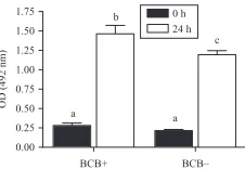
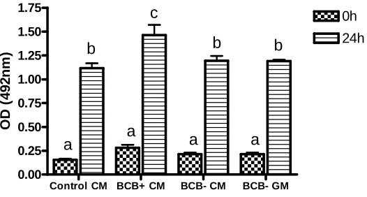
(39) Lamb oocyte competence selected by BCB Table 1 Immature prepubertal sheep oocytes exposed at different concentrations of Brilliant Cresyl Blue (BCB). Oocyte classification BCB concentration (mM). Total COC. BCBC, n (%). BCBK, n (%). 226 225 234 283. 44 (19)a,A 64 (28)b,A 85 (36)b,A 132 (47)c. 182 (81)a,B 161 (72)b,B 149 (64)b,B 151 (53)c. 13 26 39 52. Values in the same column (a,b,c) or road (A, B) with different letters differ significantly (Fisher test; P!0.05).. Although staining with 13 mM BCB showed a low percentage of stained oocytes (BCBC), the number of blastocysts obtained in this group (21%) was significant higher (P!0.05) than with 39 mM (10%) and 52 mM BCB (8%; Table 2). Of 174 inseminated oocytes from the control group (not exposed to BCB), 116 (67%) were cleavage oocytes and 14 (8%) reached the blastocyst stage. This percentage of blastocysts was significantly different from BCBC but not from BCBK oocytes. After 24 h of IVM there were no significant differences in the percentage of oocytes (stained with 13 mM BCB) reaching the MII stage in BCBC, BCBK and the control group (86, 72.5 and 80% respectively). After 17 h of IVF, the percentage of normal fertilisation (2PN) was significantly different (P!0.05) between the BCBC(40%) and BCBK groups (22%), and between BCBC and controls (23%) selected with 13 mM BCB (Table 3). The analysis of the cell number counting at day 8 postinsemination of all blastocysts produced in vitro from prepubertal sheep oocytes selected with 13 mM BCB is summarised in Table 4. BCBC oocytes produced blastocysts with a significantly (P!0.001) higher number of cells than BCBK oocytes, 69.71G6.19 and 45.91 G3.35 respectively. The ICM and TE cell number were higher in BCBC (18.82G1.77 and 50.88G5.06) than BCBK (12.55G1.12 and 33.36G3.16 respectively). The ICM:TE ratio was not significant between BCB selected groups (1:2.70 and 1:2.65 respectively). Before maturation, the mean diameter of BCBC oocytes was 123.66G2.72 (GS.E.M.), significantly higher (P!0.0001) than BCBK (106.82G2.88). After 24 h of IVM, the BCBC group maintained their diameter while. 519. BCBK showed a significant increase of 12 mm of the internal zona diameter (from 106.82G2.88 to 118.86 G3.26 mm; PZ0.006). Mitochondrial activity in prepubertal sheep oocytes selected by BCB Figure 1 shows representative images for the different mitochondrial distribution parameters. At the GV stage, 43.9% of oocytes presented homogeneous (Fig. 1B) and 56.1% showed peripheral (Fig. 1C) distribution. After maturation, 53.2% showed a homogeneous distribution while the peripheral distribution decreased up to 6.4%, the rest of the oocytes exhibited a polarised distribution (40.4%) marked by the position of active mitochondria around the metaphase spindle and polar body (Fig. 1D; P!0.001). No differences were found in mitochondrial distribution between BCBC and BCBK oocytes. Mitochondrial activity is represented in Fig. 2 by the analysis of the fluorescence intensity in oocytes prelabelled with the mitochondrial-specific probe. Our results indicate a relationship between mitochondrial activity, BCB oocyte status and maturation stage. Before IVM, BCBC and BCBK oocytes showed no significant differences in mitochondrial activity between groups (2834G223.42 and 3519G288.48 AU respectively). After IVM, BCBK oocytes mitochondrial activity descended abruptly (from 3519G288.48 to 1565G113.8 AU; P!0.0001) while activity in the BCBC group did not show any changes. Between matured BCBC and BCBK oocytes, mitochondrial activity differed significantly (P!0.0001). Considering the overall oocytes and comparing mitochondrial activity at the GV and MII stages, we observed a decreasing activity during meiosis (3175G253.9 to 2385G233 AUGS.E.M. respectively P!0.05). MPF activity in prepubertal sheep oocytes selected by BCB Results in MPF activity of oocytes with different cytoplasmic quality and stage of maturation assessed by CDC2 kinase activity are presented in Fig. 3. No differences were observed in MPF activity at collection. Table 2 Embryo development of prepubertal sheep oocytes selected with different Brilliant Cresyl Blue (BCB) concentrations. Oocyte classification BCBC, n (%) BCB concentration (mM). Inseminated oocytes. 13 26 39 52. 107 114 136 123. Cleavage a. 85 (79) 77 (68)b 90 (66)b 86 (70)a,b. BCBK, n (%) Blastocyst a,A. 22 (21) 19 (17)a,c,A 14 (10)b,c 10 (8)b. Inseminated oocytes 204 192 202 207. Cleavage 128 122 145 120. a,b. (63) (64)a,b (72)a (58)b. Blastocyst 18 (9)a,B 9 (5)a,B 13 (6)a 9 (4)a. Values in the same column (a,b,c) or row (A, B) with different letters differ significantly (Fisher test; P!0.05). www.reproduction-online.org. Reproduction (2011) 142 517–527.
(40) 520. M G Catalá and others. Table 3 Nuclear stage of prepubertal sheep Brilliant Cresyl Blue (BCB) selected oocytes at 17 h post-insemination. Oocyte classification (13 mM BCB). Fertilised oocytes, n (%) Oocytes at MII (%). Control BCBC BCBK. Inseminated oocytes. 80 86 72.5. 2PN. PS b. 68 60 64. 16 (23) 24 (40)a 14 (22)b. AS 2 (3). 3 (5) 4 (6). 3 (5). Different letters (a,b) within a column are significantly different (Fisher test; P!0.05). MII, metaphase II; 2PN, one sperm tail and 2 pronuclei; PS (polyspermic), two or more sperm tails or more than 2 pronuclei; AS (asynchronous), only one pronucleus.. time in BCBC and BCBK oocytes (0.285G0.03 and 0.212G0.01 optical density (OD) respectively). After IVM, MPF showed a significantly (P!0.05) increased activity in BCBC with respect to BCBK oocytes (1.479G0.09 and 1.184G0.05 OD respectively). In both cases BCBC and BCBK oocytes showed a significantly (P!0.001) increasing MPF activity from the GV to the MII stage of maturation. Relative mRNA expression of ATP1A1, COX1, CPEB and S100A10 of BCB selected oocytes Figure 4 shows the relative expression (RE) of the four maturation gene candidates analysed by real-time PCR in relation to BCB status and IVM. No differences in gene expression were observed in relation to BCB classification. The only difference we found was in BCBC oocytes that showed a significantly lower amount of S100A10 transcript after IVM (P!0.05).. Discussion This study was carried out to determine the best concentration of BCB stain to select competent prepubertal sheep oocytes for in vitro embryo production and to analyse mitochondrial distribution and activity, MPF activity and changes in the RE of ATP1A1, COX1, CPEB and S100A10 as maturation gene candidates in those BCB selected oocytes. In cows (Pujol et al. 2004, Alm et al. 2005), goats (Rodriguez-Gonzalez et al. 2002) and mice (Wu et al. 2007) the concentration of BCB stain with the best results was 26 mM BCB for 90 min, while in pigs 13 mM BCB was sufficient to increase sperm penetration of BCBC. oocytes (Roca et al. 1998, Egerszegi et al. 2010). In our work, we decided to work with 13 mM BCB for 60 min, as it was the most specific concentration, increasing blastocyst rate from 9% (BCBK) to 21% (BCBC). We considered BCB stain not to be detrimental for oocytes because no differences were found between treatments and the control group as described by Wongsrikeao et al. (2006) in pigs. Oocyte diameter is a determinant factor for completion of meiosis and acquisition of full competence for embryo development (Lonergan et al. 1994, Crozet et al. 2000). In our work, we found that the BCB test was helpful in selecting larger oocytes with 123.66G2.72 mm diameter (BCBC) compared with those of 106.82G2.88 mm (BCBK). This confirmed previous reports in cattle (152.6 vs 147 mm; Pujol et al. 2004), goats (136.6 vs 125.5 mm; Rodriguez-Gonzalez et al. 2002) and pigs (113.08 vs 100.29 mm; Roca et al. 1998) for BCBC and BCBK respectively. After IVM, we found a significant increase of 12 mm of the internal zona diameter in BCBK oocytes, while BCBC oocytes maintained the same diameter after 24 h of in vitro culture. Oocyte quality is assessed by male pronuclear formation (2PN zygotes), blastocyst yield and blastocyst cell number. In this study, we have observed significantly higher 2PN zygote and blastocyst production in BCBC than BCBK oocytes. This increase in 2PN zygote and blastocyst development has been shown in BCBC oocytes of buffalo (Manjunatha et al. 2007), cattle (Pujol et al. 2004, Alm et al. 2005, Bhojwani et al. 2007, Torner et al. 2008), goats (Rodriguez-Gonzalez et al. 2002), mice (Wu et al. 2007) and pigs (Egerszegi et al. 2010). Assessment of blastocyst cell number indicates that BCBC oocytes produce blastocysts with a higher cell number than BCBK. In prepubertal goat oocytes, Romaguera et al. (2010, 2011) did not find differences in blastocyst quality, assessed by blastomere ploidy and cryotolerance, between oocytes coming from different diameter follicles and between oocytes from adult and prepubertal goats, concluding that oocyte quality was positively related to blastocyst production but not to blastocyst quality. In cattle, Majerus et al. (2000) observed that the percentage of blastocysts obtained from calf oocytes was reduced in comparison with those from adult animals (26 vs 46%); however, the number of blastocyst cells was not different (89 and 100 respectively) and the ICM:TE ratio was similar (1:2.70 and. Table 4 Total cell numbers of in vitro produced blastocyst from prepubertal sheep Brilliant Cresyl Blue (BCB) selected oocytes at day 8 postinsemination. Cell number (meanGS.E.M.) N BCBC BCBK. 24 22. Total. TE a. 69.71G6.19 45.91G3.35b. Percentage/total cells (n) ICM. a. 50.88G5.06 33.36G3.16b. a. 18.83G1.77 12.55G1.12b. ICM. TE. 27.01 27.34. 72.99 72.66. ICM:TE ratio 1:2.70 1:2.65. ICM, inner cell mass; TE, trophectoderm. Different letters (a,b) within are significantly different (Student’s t-test; P!0.05). Reproduction (2011) 142 517–527. www.reproduction-online.org.
(41) Lamb oocyte competence selected by BCB B. A. C. 521. D. High. Low. Figure 1 Representative images of active mitochondrial distribution of prepubertal sheep oocytes taken by a confocal microscope. Images were spectrally (A) coded to represent staining intensity (red is the highest intensity). Representative images of (B) homogeneous, (C) peripheral and (D) polarised mitochondrial activity distribution in lamb oocytes.. www.reproduction-online.org. and BCBK oocytes separately, we found that at the GV stage there were no differences in mitochondrial activity between groups. However, after IVM, BCBK oocytes showed a significant reduction in mitochondrial activity while BCBC mitochondrial activity remained constant. This would indicate a positive relationship between mitochondrial activity at MII stage and embryo development. Stojkovic et al. (2001) showed that mitochondrial reorganisation was different between morphologically good and poor quality oocytes. In our study, mitochondria migrated throughout the IVM process. Oocytes at the GV stage presented a homogeneous (43.9%) or peripheral (56.1%) mitochondrial distribution. After 24 h of IVM, MII oocytes presented a homogeneous (53.2%) distribution or mitochondria polarised around the metaphase spindle and inside the polar body (PB; 40.4%). We have previously shown (Velilla et al. 2006), in prepubertal goat IVM oocytes, that total mitochondria migrate from a cortical and perinuclear distribution in GV oocytes to a polarised distribution opposite the metaphase spindle and inside the PB (86%) after IVM, whereas ovulated adult goat oocytes presented a mitochondrial distribution inside the PB and aggregated to the metaphase spindle (Velilla et al. 2006) as we have found here in lamb oocytes. In pigs, Torner et al. (2004) # 5000.00 Mean fluorescence activity (arbitrary units). 1:2.85 respectively). Selecting lamb oocytes according to the number of cumulus layers, Kelly et al. (2007) concluded that the percentage of day 8 blastocysts was affected by COC grade but the number of blastocyst cells was not significantly different (range 49.2–54.6 cells per blastocyst). To our knowledge, no studies on oocytes selected by BCB and embryo quality have been done. In this study, we have shown a positive relationship between BCBC oocytes and the number of blastomeres per blastocyst. Mitochondrial distribution and activity inside the oocyte could be a good marker of oocyte competence to develop to the blastocyst stage. The primary function of mitochondria is to generate ATP. Van Blerkom et al. (1995) described in human oocytes the relationship between ATP content and embryo developmental capacity where a transient decrease in ATP content can lead to embryo arrest. Therefore, these data suggest that mitochondrial activity is a determinant factor of quality and changes in mitochondrial activity can alter oocyte quality in a remarkable way. In cattle (Tarazona et al. 2006), horses (Torner et al. 2007), humans (Van Blerkom 2004) and pigs (Torner et al. 2004) an increase in mitochondrial activity after IVM was described. In cattle, Torner et al. (2008) observed higher mitochondrial activity in BCBK oocytes than in BCBC. These authors speculated that the reason for the increasing respiratory activity in low-quality oocytes was to provide ATP for still unfinished processes for cytoplasmic maturation. In pig oocytes, Egerszegi et al. (2010) found a higher mitochondrial activity in BCBC compared with BCBK oocytes before IVM, but after IVM, no differences were found between either kind of oocyte. They attribute this to BCBC oocytes increasing their respiratory activity to provide ATP for the energy-demanding processes of GVBD and the subsequent condensation of chromatin up to MII, while BCBK oocytes showed no changes in mitochondrial activity during meiosis and only a few of them reached MII stage. In our study with prepubertal sheep oocytes, we found a decrease in mitochondrial activity from the GV to the MII stage (3175G253.9–2385G233 AUGS.E.M.). Analysing BCBC. 4500.00 4000.00 3500.00. * *. 0h 24 h Total. 3000.00 2500.00 2000.00 1500.00 1000.00 500.00 0.00 BCB +. BCB – Oocyte classification. Figure 2 Mitochondrial activity of BCB selected oocytes assessed by fluorescence intensity before (0 h) and after IVM (24 h). Different symbols (#, *) indicate significant differences (*P!0.0001; #P!0.05). Reproduction (2011) 142 517–527.
(42) M G Catalá and others 1.75. b. OD (492 nm). 1.50. 0h 24 h c. 1.25 1.00 0.75 0.50. a. a. 0.25 0.00 BCB+. BCB–. Figure 3 MPF activity in BCB-exposed prepubertal sheep oocytes before (0 h) and after IVM (24 h). MPF activity is expressed as OD at 492 nm. a,b,cIndicates a significant difference in kinase activity among oocyte groups.. found that mitochondria moved from a homogeneous to a peripheral distribution; however, Brevini et al. (2005) showed a peripheral distribution in immature pig oocytes that became diffused after IVM. In our study, we did not find differences in mitochondrial distribution between BCB oocyte groups, while Egerszegi et al. (2010) concluded that, in pigs, BCBK oocytes showed more heterogeneous and non-aggregated mitochondrial distribution than BCBC oocytes. Different authors have reported differences in mitochondrial distribution patterns among species, during IVM and in vivo maturation (Stojkovic et al. 2001, Sun et al. 2001, Torner et al. 2004, Velilla et al. 2006, Torner et al. 2007, Dell’Aquila et al. 2009, Egerszegi et al. 2010). Previous studies showed that GV-oocytes do not present MPF activity (Dedieu et al. 1996) but, in our study, we detected MPF activity before IVM; this may be due to the BCB staining time. We presume that oocytes could restart meiosis and so they were in GVBD instead of GV when MPF analysis took place. After IVM, we observed significantly higher MPF activity in BCBC oocytes than in BCBK oocytes. Salamone et al. (2001) reported that calf oocytes undergo cleavage and blastocyst production at significantly lower rates than cow oocytes and this was correlated to a lower activity of MPF in these oocytes. Closer to our study, comparing prepubertal and adult sheep oocytes, Ledda et al. (2001) showed that the low competence in prepubertal oocytes could be due to morphological anomalies and alterations in physiological activity due to the evidence of low MPF activity after IVM. Bogliolo et al. (2004) showed a higher MPF activity in in vivo matured oocytes than in in vitro matured cat oocytes, suggesting a possibly incomplete cytoplasmic maturation after culture. High MPF activity was observed by Anguita et al. (2007) in oocytes with larger diameter and better competence to develop to the blastocyst stage. In addition, MPF activity has also been related to an increase in developmental competence of oocytes treated with caffeine during nuclear transfer (Kawahara et al. 2005). In relation to mitochondrial and MPF activities analysed in this work, we could speculate that there is a positive relationship between the ATP produced by the active mitochondria and the ATP production needed to phosphorylate p34cdc2 and activate the MPF complex. Reproduction (2011) 142 517–527. The genes analysed in this work were related to metabolism (ATP1A1 and COX1) and constitutive function of the cell (CPEB and S100A10). Oocytes selected by BCB did not show any differences in RE in any of the studied genes, in spite of the higher embryo development observed in BCBC oocytes. However, in bovine oocytes, Opiela et al. (2010) found higher RE of the COX1 gene in immature BCBC with respect to BCBK and Torner et al. (2008) showed a higher RE of S100A10 in matured BCBK than BCBC oocytes. During IVM in prepubertal sheep oocytes, we found a significant decreasing mRNA RE in S100A10 in BCBC oocytes. In conclusion, exposing prepubertal sheep oocytes to 13 mM BCB for 60 min stains the largest and most competent oocytes to develop to the blastocyst stage. After IVM, the more competent oocytes (BCBC) presented higher mitochondrial and MPF activity with respect to BCBK oocytes. BCBC oocytes produced blastocysts with higher numbers of cells than BCBK oocytes. Mitochondrial distribution and mRNA expression of ATP1A1, COX1, CPEB and S100A10 were not affected by oocyte quality.. Materials and Methods Chemical All chemicals were purchased from Sigma–Aldrich Chemical Co. unless otherwise specified.. Oocyte collection Ovaries from prepubertal ewes (3–6 months old), were obtained from a local abattoir and transported to laboratory in sterile dulbecco’s (PBS) held at 34–37 8C and were washed. Relative mRNA expression. 522. 7 6 5 4 3 2 1 0. S100A10 a,b. CPEB 20 15 10. a a,b. 5. b 0 0. 24. 0. BCB+. 24. 0. 24. 0. BCB+. BCB– CYTO C. 24 BCB–. ATP1A1 15. 20 15. 10. 10 5. 5. 0. 0 0. 24 BCB+. 0. 24 BCB–. 0. 24 BCB+. 0. 24 BCB–. Figure 4 Relative mRNA expression of S100A10, CPEB, COX1 and ATP1A1 transcripts before (0 h) and after IVM (24 h) of prepubertal sheep BCB selected oocytes. Different letters (a, b) indicate significant differences (P!0.05: one-way ANOVA with Tukey’s multiple comparison post-test to compare the four groups of each gene). www.reproduction-online.org.
Figure


+7

Documento similar
In JIVET the high percentage of small antral follicles makes oocyte collection by follicular aspiration difficult (reviewed by Paramio & Izquierdo 50 ).
Thus, it could be concluded that, although some mushroom extracts showed PL inhibitory activity in an in vitro test, under the in vitro digestion model
• The sequences at sites bound by Meis, Prep and Pbx1 suggest Prep tends to bind as a heterodimer with Pbx, without participation of Hox proteins.. In contrast, Meis tends to bind
In vitro and in vivo antitumor activity of platinum(II) complexes with thiosemi- carbazones derived from 2-formyl and 2-acetyl pyridine and con- taining
1.6 Recombinant production in E.coli and characterization of the proteolytic activity of different constructs of the Catalytic Domain of USP28 and USP25 with
The expansionary monetary policy measures have had a negative impact on net interest margins both via the reduction in interest rates and –less powerfully- the flattening of the
Jointly estimate this entry game with several outcome equations (fees/rates, credit limits) for bank accounts, credit cards and lines of credit. Use simulation methods to
In our sample, 2890 deals were issued by less reputable underwriters (i.e. a weighted syndication underwriting reputation share below the share of the 7 th largest underwriter
