F18-FDG-PET/CT in the evaluation
of patients with suspected recurrent or persistent
locally advanced cervical carcinoma
Lucely Cetina,* Alberto Serrano,* David Cantú-de-León,
†Delia Pérez-Montiel,
‡Enrique Estrada,
§Jaime Coronel,* Martha Hernández-Lucio,
||Alfonso Dueñas-González
||* Oncología Médica y División de Investigación Clínica, † Departamento de Oncología Ginecológica, ‡ Departamento de Patología, § Departamento de Medicina Nuclear, Instituto Nacional de Cancerología. || División de Investigación Básica, Instituto Nacional de Cancerología y Unidad de Investigación Biomédica en Cáncer,
Instituto de Investigaciones Biomédicas, Universidad Nacional Autónoma de México. ARTÍCULO ORIGINAL
C
1 a e
1 C a e
F18-FDG-PET/CT en la evaluación de pacientes F18-FDG-PET/CT en la evaluación de pacientes
s s e a e con sospecha de recurrencia o persistencia de
u
á c o
cáncer cervicouterino localmente avanzado
E N R E S U M E N
e e e s
Antecedentes. El cáncer cervical (CC) representa la segunda neoplasia más frecuente y la tercera causa de muerte por cáncer entre las mujeres. La enfermedad recurrente o persistente se encuentra en relación con la etapa clínica, pero puede ser alta hasta en 70%. La tomografía computada con emisión de positrones (PET/CT) es un estudio de imagen que puede detectar la absorción incrementada de glucosa en los tejidos neoplásicos. a Material y métodos. Se realizó PET/CTr l y é dt s en pacientes con CC confirmado, que habían sido previamente tratadas y que desarrollaron síntomas sugestivos de recurrencia o enfermedad persistente con o sin evidencia de la enfermedad en una tomografía computarizada. Se evaluaron la sensibilidad, especificidad, valores predictivos de PET/CT y tomografía computarizada. Resultados. Dieciséis pacientess l a con una edad promedio de 47.2 años se incluyeron en el estudio entre abril 2007 y junio 2008. Trece pacientes (81.2%) presentaron síntomas. PET/CT fue positivo en 14/16 (85.7%). De éstos, 12 catalogados como Verdaderos Positivos (TP) y dos, Falsos Positivos (FP); otros dos casos fueron catalogados como Verdaderos Negativos (TN) (12.5%). Los sitios anatómicos más frecuentes de captación detectados por el PET/CT fueron el cuello uterino, los ganglios linfáticos retroperitoneales, iliacos, obturadores y mediastinales. El número de sitios anatómicos promedio con alta captación de Fluoro-desoxi-D-glucosa (FDG) fueron dos (rango, 1-7 sitios). PET/CT y TC presentaron sensibilidad de 100 y 91.7%, respectivamente. Especificidad para ambos 50%; valor A TC
A C T A B S T R A C T A B S T R A C T
n ro
Background. Cervical cancer (CC) represents the second most common neoplasm and the third cause of death by cancer among women. Recurrent or persistent disease depends on the clinical stage, but can be as high as 70%. Positron emission tomography/computed tomography (PET/CT) is an image study that can detect increased glucose uptake in tumor tissues. Material and methods.t m t PET/CT was performed in patients with confirmed CC, who had been previously treated, who developed suspected symptoms of recurrence or persistent disease with or without evidence of disease on a CT scan. Sensitivity, specificity, predictive values from PET/CT, and CT scan were evaluated. Results. Sixteen patients with a mean age of 47.2 years were included in the study from April 2007 to June 2008. Thirteen patients (81.2%) were symptomatic. PET/CT was positive in 14/16 (85.7%), of these, 12 True positive (TP) and two, False positive (FP); meanwhile another two cases were True negative (TN) (12.5%). Cervix, retroperitoneal, iliac, obturator, and mediastinal lymph nodes were the most common anatomic sites detected by PET/CT. Mean number of anatomic sites with high Fluoro-deoxy-D-glucose (FDG) uptake was two sites (range 1-7 sites). PET/CT and CT scan had 100 and 91.7% sensitivity, respectively. Specificity for both was 50%. Positive predictive value (PPV) was 85.4 and 84.6%, respectively. Negative predictive value (NPV) was 100 and 66%, respectively, and accuracy was 88 vs. 81%, respectively.
n
INTRODUCTION
Cervical cancer (CC) accounted for the second most frequent tumor worldwide; during the year 2007, 555,100 new cases were reported, which repre-sented the third cause of death by cancer among women (309,800 deaths).1,2 Eighty percent of new
ca-ses were registered in countries with fewer financial resources. In México 9,927 new cases were registe-red during the year 2003 (12.8% of tumors in female population), and 4,326 deaths (rate 8.7 x 100,000)3
due to CC.
Recurrence of the disease is defined as tumoral growth or distant metastasis development at least 6 months after initial treatment; anatomical sites that exhibit higher frequency of recurrence are pelvis (central or lateral), pelvic lymph nodes (first or se-cond relay), extra-pelvic nodes, and distant metasta-sis localized in peritoneum, liver, lungs, and/or bones as well.4 The incidence of recurrent disease
fundamentally depends on the initial stage; 10-20% is registered for stages IB-IIA, while incidence beco-mes as high as 50-70% for stages IIB–IVA.5 The
2006 annual report of the International Federation of Gynecology (FIGO)6 confirmed that the most
important prognostic variable is the initial clinical stage: the 5-year survival rate is > 90% for stage IA; 89% for IB1; 75% for IB2; 73% for IIA; 65% for IIB; 40% for stages IIIA and -B, 22% for stage IVA, and only 9% for stage IVB.
Recurrent disease is suspected, clinically, by the presence of lumbar pain, sciatica pain, or limb ede-ma; however, in some cases, patients remain asymptomatic. Radiologic studies have been used for detecting recurrent disease; among these, computed tomography (CT) and nuclear magnetic resonance imaging (NMRI)7 are the most popular. However,
they have limitations; for example, CT is not able to differentiate tumor activity from post-radiotherapy fibrosis, and magnetic resonance imaging (MRI) is not able to differentiate among necrosis, edema,
he-morrhage, or inflammation.4,8 On the other hand,
positron emission tomography (PET) is a molecular imaging technique that employs radioactive molecu-les to detect images of molecular interactions of bio-logical processes in vivo, thus being able to establish the functional metabolic behavior of tis-sues, which allows distinguishing recurrent disease with higher accuracy.10
A glucose analog, 2-18F-Fluoro-deoxy-D-glucose (FDG), is utilized to evaluate the metabolic activity in tumors. This particular glucose analog is not me-tabolized and by intracellular trapping, it avidly accu-mulates. making it possible to be measured by PET.11
Progress in the knowledge of metabolic behavior of tumor cells establishes that these show increased glycolytic activity favored by several mechanisms, such as the increase of glucose transporting proteins at the cell surface [as example Glucose transporter 1 (GLUT 1) receptors] and the increase of several key enzymes such as hexokinase.12 In squamous CC, it
has been reported that 94% of primary lesions, whe-ther recurrent or persistent, show Glut 1 over-expre-ssion, while normal tissue, or in cases with cervical intraepithelial neoplasia (CIN), over-expression is mi-nimal, being no > 15% of basal layer cells –either normal or dysplastic tissues–.13
PET efficacy for detecting the recurrence of CC has been reported in some studies.14-18 In order to
reduce limitations of anatomical location of hyper-uptake sites detected with PET, their fusion and in-tegration with morphological information obtained by CT has been structured through a metabolic test, positron emission tomography/computed tomogra-phy (PET/CT), which is considered an effective ima-ging technique even for initial staima-ging of cervical cancer.19,20
OBJECTIVE
The purpose of this study was to analyze the ca-pability of PET/CT in detecting recurrence and/or
impact of this study on the early detection of CC recurrence.
o s
Key words. Positron emission tomography/computed tomography (PET/CT). Cervical cancer. Recurrence.
predictivo positivo (PPV) 85.4 y 84.6%, respectivamente. Valor predictivo negativo (NPV) 100 y 66%, respectivamente. Precisión 88 vs. 81%, respectivamente. CConclusiones. Elus ne . PET/CT tiene la capacidad para detectar el cáncer de cérvix recurrente o persistente; detecta el aumento de la actividad metabólica, principalmente, en el sitio primario o los ganglios linfáticos. Es necesario realizar estudios adicionales sobre PET/CT y esta entidad para confirmar el impacto real de este estudio sobre la detección temprana de la recurrencia de CC.
b s a e
persistence of locally advanced CC treated with stan-dard procedures.
MATERIAL AND METHODS
We conducted a retrospective study from April 2007 to June 2008 in women with confir-med locally advanced CC treated at the Instituto Nacional de Cancerología de México (INCan) and who underwent PET/CT due to suspicious persistent or recurrent disease. Patients had been treated according to international guideli-nes for their corresponding FIGO clinical stage. Criteria for requesting the PET/CT study were the following:
• Symptoms suggesting recurrence.
• Radiologic findings or clinical data suggesting re-currence.
• Positive Papanicolaou smear.
Abdominal and pelvic CT was also requested and findings were compared in all cases. Re-current or persistent disease was confirmed by means of biopsy or documentation of progressive disease through serial imaging studies or clini-cal disease course.
We retrieved information from the clinical chart such as age, initial FIGO stage, histopathological type, treatment, overall survival (OS), symptoms re-lated with the tumor activity, CT and PET/CT fin-dings, number and size of positive sites, biopsy results, and the Maximum standardized uptake va-lue (max SUV).
c n e PET/CT technique c n e PET/CT technique
PET/CT with FDG was conducted in the INCan Nuclear Medicine Department using a Siemens BIO-GRAPH 16 system. After Intravenous (IV) adminis-tration of a 370 MBq (10 mCi) dose of the F-18-labeled FDG, patient was placed to rest in an isolated room during 60 min in order to obtain the uptake phase.
CT images were obtained in simple phase, through 16 detectors, making axial cuts and coronal and sagittal reconstructions from cranium base to upper third of thighs. High-resolution bidimensional PET phase was conducted for 7 min, and finally, image reconstruction was carried out employing fil-tered back-projection. PET/CT studies were evalua-ted by two physicians, a Nuclear Medicine Specialist and a Radiologist.
E CT n l PET/CT analysisE C n lT PET/CT analysis
Max SUV indicates the activity found in a lesion and is expressed as related with injected dosage and the patient’s weight. Values > 3.0 were considered positive for malignancy.
A study was considered as True positive (TP) in PET/CT when it showed at least one lesion with a positive biopsy. A False positive (FP) study was defi-ned when at least one lesion considered as malig-nant in PET/CT was determined to be without malignancy by histopathologic study or clinical course. A True negative (TN) study showed no ab-normal findings and the patient was followed-up without clinical evidence of malignant disease. False negative (FN) was considered when the study was reported as negative but the biopsy was positive or when the clinical evidence of malignant disease was unquestionable. These definitions are considered re-liable and reported by other authors as valid.8
a s i a s s Statistical analysisa s i a s s Statistical analysis
Statistical analysis was performed by means of descriptive methods. Sensitivity, specificity, positive and negative predictive values, and accuracy for de-tecting recurrent disease by PET/CT and computeri-zed tomography (CT) were determined according to the following formulas:
• Sensitivity = (TP/TP + FN) x 100. • Specificity = (TN/TN + FP) x 100.
• Positive predictive value (PPV) = (TP/TP + FP) x 100.
• Negative predictive value (NPV) = (TN/TN + FN) x 100.
Where:
• TP = True positive. • TN = True negative. • FP = False positive. • FN = False negative.
Histopathologic findings and clinical course were considered as the gold standard for comparison.
RESULTS
planning for the radiation treatment, three additio-nal cases because they lacked CT –although they had pelvic magnetic resonance imaging (MRI) or ul-trasound (US)– and another three because they had no confirmatory biopsy.
The remaining 16 patients were included in this study; median age was 47.2 years (range 31-66 years); general characteristics are shown in table 1. All cases were staged as locally advanced tumors mainly stage IIB in eight cases (50%); predominant histology was squamous cell carcinoma (15/16 cases); initial treatment was concurrent chemoradiation in 13 patients (81.2%), chemoradiotherapy (Ch) followed by radical surgery in one patient, radiotherapy (RT) alone in one patient, and radical surgery and adju-vant RT in another patient. Complete clinical and cytological response was observed in six cases (37.5%), disease progression in one (6.2%), and per-sistence in the remaining nine patients (56.2%). The median recurrence-free interval was 13 months (ran-ge 6-36 months) among cases with complete response. Persistence or disease recurrence was suspected in 13/16 cases (81.2%) by the referred symptoms, the more frequent being pelvic pain in eight cases (61.5%), vaginal discharge in five (38.4%), and lo-wer limb edema and malaise in three (23.1%); fur-thermore, three patients had no symptoms. On the other hand, suspicion of recurrent or persistent di-sease was documented in 12/16 (75%) cases through the gynecological findings and in 13/16 (81.2%) by means of CT findings.
Suspicious anatomic sites detected by CT inclu-ded the following: Uterine cervix in nine (56.2%); enlargement of retroperitoneal lymph nodes in nine (56.3%); RT-related changes in 5 (31.2%); loss of the inteface in contiguous tissues in four (25%); enlar-gement of obturator lymph nodes in four (25%); pa-rametrial involvement in three (18.7%), and iliac lymph node enlargement in three cases (18.7%).
PET/CT was considered positive in 14 of the 16 patients (87.5%) (12 TP and 2 FP) and negative in 2/16 (12.5%) (2 TN). Table 2 depicts the anatomic sites detected by the metabolic study; the median number of hypermetabolic anatomical sites was 2.2
e l .
Table 1. Patients’ characteristics.
Age (years) Histology FI-GO stage Treatment Indication PET/CT DFI Symptoms CT PET/CT
60 Squamous IIB Ch-RT Recurrence 15 Present FP TN
49 Squamous IIIB Ch-RT Persistence Present TP TP
52 Squamous IIIB RT Persistence Present TP TP
42 Squamous IB2 Ch-RT Recurrence 6 Present FN TP
31 Squamous IIB Ch-RT-Sx Recurrence 14 Absent TP TP
47 Squamous IVB Ch-RT Persistence Present TP TP
47 Squamous IB2 Ch-RT Persistence Present TP TP
35 Squamous IVA Ch-RT Persistence Present TP TP
46 Squamous IIB Ch-RT Recurrence 12 Present TP TP
52 Adeno IIIB Ch-RT Persistence Present TP TP
33 Squamous IIB Sx-RT Persistence Present TP TP
66 Squamous IVB Ch-RT Progression Present TN FP
57 Squamous IIB Ch-RT Recurrence 10 Present TP TP
47 Squamous IIB Ch-RT Persistence Present FP FP
51 Squamous IIB Ch-RT Recurrence 36 Absent TP TP
41 Squamous IIB Ch-RT Persistence Absent TN TN
:: CT:
CT: Computed tomography. DFI:DFI: Disease-free interval (months). PPPET/CT:PET/CT: Positron emission tomography/computer tomography. Ch:: ChCh:Ch: Chemotherapy. RT:RT: Radiotherapy. Sx: Surgery. TTP: True positive. N:: TN: True negative. FFP: False positive. FN: False negative. d: Adeno: Adenocarcinoma.
2
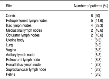
Table 2. Sites with increased metabolic activity detected by PET/CT.
Site Number of patients (%)
Cervix 8 (50)
Retroperitoneal lymph nodes 5 (41.6) Iliac lymph nodes 4 (33.3) Mediastinal lymph nodes 2 (16.6) Obturator lymph nodes 2 (16.6)
Uterine body 1 (8.3)
Lung 1 (8.3)
Vagina 1 (8.3)
Axillary lymph node 1 (8.3) Retrocrural lymph node 1 (8.3) Renal hilius lymph node 1 (8.3) Supraclavicular lymph node 1 (8.3)
Pelvis 1 (8.3)
/
(range 1-7 sites). The most frequent anatomic sites identified with hypermetabolic activity were uterine cervix in eight cases (50%), retroperitoneal lymph nodes in five (41.6%), iliac in four (33.3%), mediasti-nal in two (16.6%), and obturator lymph nodes in two cases (16.6%). Median max SUV was 6.9 (range 2.2-14). Positive PET/CT detected lesions size accor-ding to anatomic site:
• Lymph nodes, mean 22 mm (range 10-31 mm). • Uterine cervix, mean 53 mm (range 50-66 mm).
In addition, a 31-mm pulmonary nodule and a 54-mm lesion in the vagina were identified.
Regarding the two patients with FP PET/CTs, one was a 47-year-old woman with stage IIB squa-mous carcinoma treated with concurrent Ch and va-ginal brachytherapy; she referred mild pelvic pain 1 month after her treatment, and CT reported enlar-gement of obturator chains from 5-7 mm, slight en-largement of the cervix 3.8 x 3.5 cm, and this was associated with alterations in the adjacent fatty tis-sue, as well as thickening of the vaginal walls. PET/CT detected 5 x 2.5 cm uterine cervix with max SUV 2.2, posterior bladder wall with loss of soft-tissue interface and left parametrial thicke-ning; biopsy reported only necrosis. The patient has been followed up for 18 months with no eviden-ce of tumor activity.
The other FP case was a 66-year-old woman with IVB stage squamous carcinoma and left supracla-vicular adenopathy, treated with concurrent chemo-radiotherapy (Ch-RT) and vaginal brachytherapy; she developed left pelvic limb edema 2 months after treatment completion; CT identified thickened recto-sigmoid walls, alteration in adjacent fat, tissue density, and bladder-wall thickening; PET/CT de-monstrated uterine cervical-wall thickening and
thickening of the vaginal canal, max SUV 4.5. In the physical examination, a recto-vaginal fistula was do-cumented that led to a colostomy; during this proce-dure, biopsies were taken and reported as negative for neoplasm. The patient is alive and has been di-sease-free during the 8 months during which she has been followed up.
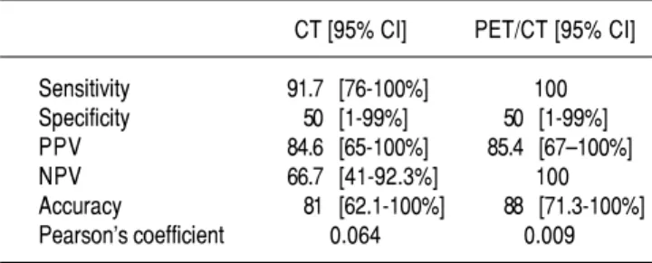
In patients with TP PET/CT, 11 had positive fin-dings in the CT and only five had an association with the biopsy report. Quantitative PET/CT analy-sis, CT, and biopsies are shown in table 3. Metabo-lic test sensitivity was superior to that detected for CT [100 vs. 91.7% (95% Confidence interval [CI], 76-100%), respectively], while specificity was similar in both studies [50% (95% CI, 1-99)]; the PPV was 85.4% (95% CI, 67-100) and for CT, 84.6% (95% CI, 65-100), while the NPV was 100 and 66.7% (95% CI, 41–92.3), respectively; Pearson’s correlation test showed statistical significance for PET/CT (p = 0.009).
Among the 12 TP PET/CTs, three of these patients were lost with no further treatment and nine patients were treated with Ch within a clinical trial (five cases) or standard therapy for recurrent cervical cancer (four cases). Both cases considered TN and one FP PET/CT were maintained under observation and are disease-free; the remaining FP case was subjected to colostomy for chronic RT effects. At present, only one patient has died as a di-rect consequence of disease, nine are lost to follow-up, and six are alive (37.5%); survival for the group with the positive PET/CT was 11.5 months (range 1-23 months), and in the group with negative or FP PET/CT, this was 14.5 months (range 9-19 months).
DISCUSSION
Recurrent or persistent cervical cancer is usually evaluated by means of CT and/or MRI, which can identify the presence of new abnormal masses or changes caused by cancer growth in an already known lesion;21 but this type of imaging studies has
limitations in differentiating tumor infiltration from reactive changes, fibrosis, and scar tissue;8,14,22,23
additionally, both studies exhibit a reduced capabili-ty for discriminating lymph-node status, especially when the latter are relatively small.14
In our study, PET/CT showed high sensitivity and PNV for identifying recurrent or persistent di-sease, which allowed to locate hyperuptake areas in uterine cervix, retroperitoneal, iliac, mediastinal and/or obturator lymph nodes with greater frequen-cy than the CT scan; also, the PET/CT showed a
e l
Table 3. Statistical analysis of CT and PET/CT in recurrent or persis-tent locally advanced cervical cancer.
CT [95% CI] PET/CT [95% CI]
Sensitivity 91.7 [76-100%] 100 Specificity 50 [1-99%] 50 [1-99%] PPV 84.6 [65-100%] 85.4 [67–100%] NPV 66.7 [41-92.3%] 100 Accuracy 81 [62.1-100%] 88 [71.3-100%] Pearson’s coefficient 0.064 0.009
:
specificity value identical to that of the CT, while its PPV was slightly superior to that of the CT.
The first report of PET in CC was informed by Sugawara, et al., in 1999.24 More than a dozen
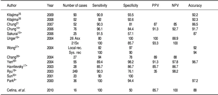
stu-dies have reported the usefulness of PET for detec-ting CC recurrence or persistence, as shown in table 4;8,14-18,21,24-30 reported sensitivity is highly variable
and ranges from 80-100%; specificity from 57.1-100%, and accuracy, from 86.5-97.2%, respectively. Yen, et al.,31 in a study of 150 cases, showed that
PET reduces the CT or MRI downstage proportion from 38.4-15.2% and that it added a higher capacity for identifying pelvic recurrence.
In this study, PET/CT was able to identify, with higher frequency, hyperuptake areas from the pri-mary site, and from retroperitoneal, iliac, mediasti-nal, and obturator nodes. In the literature, PET has informed to have a higher capability to detect lymph-node compromise8,15,17,30 with 100% sensitivity
for the increased metabolic-activity detection in hilar, scalene, mediastinal, and iliac lymph nodes, thoracic wall, liver, and vertebral spine, while its sensitivity is less (75-85%) in pulmonary lesions, retrovesicular region, and para-aortic nodes.18,31
Lai, et al.,32 found higher sensitivity and specificity
as compared with CT or MRI for detecting any re-current lesion (91 vs. 67%; p = 0.001), mainly from a metastatic type (92 vs. 60%; p = 0.0003), while all three imaging methods exhibited a similar capacity
for identifying local lesions (90 vs. 84%; p = 0.631). PET maintains its potential to detect lymph nodes (77.3 vs. 45.5%; p = 0.043), distant metastasis (66.7 vs. 26.7%; p = 0.028), and local tumors (100 vs. 74%; p = 0.462), even in second or third recurrent episodes.33
Sakurai, et al.,15 informed that metabolic
activi-ty and standardized uptake value (SUV) depend on tumor-lesion size (> 1 cm); SUV was reported to have an average of 3.90 and 2.31 in tumor and non-tumor lesions, respectively (p > 0.05). In our study, SUV in recurrent or persistent lesions was 6.4 in average.
PET is based on the increased glucose uptake by malignant cells, but lack of anatomical information and accurate location of suspicious lesions have comprised PET deterrents.21 To date, only two
stu-dies have reported the PET/CT diagnostic value for detecting CC recurrence.21,26 Chung, et al.,21 in 52
cases with evident recurrence –by means of clinical, cytological, serological, or imaging studies– repor-ted sensitivity of 90.3%, specificity of 81%, and accu-racy of 86.5%. Kitajima, et al., in his study with 52 patients compared the diagnostic capability of PET and PET/CT; the authors informed sensitivity, spe-cificity, and accuracy for PET of 80, 77.8, and 78.8%, respectively, while the figures for PET/CT were 92, 92.6, and 92.3%, respectively, with no sta-tistical difference with respect to sensitivity, but
Table 4. Sensitivity, specificity, predictive values, and accuracy based on FDG-PET or PET/CT in suspected cervical cancer recurrence in the literature.
Author Year Number of cases Sensitivity Specificity PPV NPV Accuracy
Kitajima25 2009 90 90.9 93.5 92.2
Kitajima26 2008 52 92 92.6 92.3
Chung21 2007 52 90.3 81 87 85 86.5
Chung14* 2006 76 96.1 84.4 91.3 92.7 91.7
Sakurai15* 2006 25 91.5 57.1 87
Unger16* 2004 26 Asx 80 100 100 88.9
21Sx 100 85.7 93.3 100
Wong27* 2004 Local rec. 82 97 92
Sys. rec 100 90 94
Chang28* 2004 27 94 78 89 88
Yen29* 2004 55 89.4 98.2 91.3 97.8 96.7
Havrilevsky17* 2003 28 85.7 86.7 85.7 86.7
Ryu18* 2003 249 90.3 76.1 35 98.2
Sun30* 2001 20 90 100
Park8* 2000 36 100 94.4 97.2
Cetina, et al. 2010 16 100 50 85.7 100 88
with a statistical difference in specificity and accura-cy (p = 0.00077 and 0.00051, respectively).26 An
up-dated version of this study has been published recently; the study included 90 patients with results that continued to favor PET/CT.25
Our study has the limitations inherent in the number of analyzed cases and its retrospective natu-re, which could have exerted an influence on the ob-served results; in addition, we did not register the clinical impact that PET/CT can have on the thera-peutic decisions made or on the role of PET/CT as a prognostic factor for survival. Observations found in this work are similar to those reported in the world literature, and among these, the following: PET/CT has the capability to detect recurrent tumor activity in asymptomatic patients.
The advantages of adequate recognition and loca-lization of recurrent lesions by PET or PET/CT are reflected in the therapeutic approach and the patient’s survival. In our study, we did not record the therapeutic impact related with the metabolic study, as previously mentioned; however, other au-thors have reported that PET or PET/CT can chan-ge the therapeutic approach in up to 65.5% of cases21,26,29,32 because it reduces the need of
fur-ther diagnostic tests and unnecessary treatments. Chung, et al.,14 reported a study that included 76
cases with recurrence CC; among these, 20 patients were asymptomatic with a positive PET, of them, eight underwent pelvic exenteration, considering five cases successful; the remaining patients were treated with Ch, RT, or palliative surgery, and sur-vival for these patients was 85.6% at 3 years. Huss-ain, et al.,34 sustain that PET could be considered as
the most reliable study for determining the best can-didates for pelvic exenteration.
Early detection for recurrent or persistent disease evidently has an impact on the treatment options offered to patients, and could be considered a prog-nostic factor as well, because survival expectations in symptomatic patients are very limited when com-pared with those for asymptomatic patients, that is, 11 vs. 42 months (p = 0.001), respectively.35 A
mul-ticenter study observed that 50% of 327 patients with recurrent disease were asymptomatic.36
The presence of symptoms suggesting recurrent disease has a sensitivity of 71% and specificity of 95%, while these diagnostic parameters are lower for pelvic examination and vaginal cytology37 because
they depend on the physicians’ medical experience and also on RT-induced tissue changes. These limi-tations in diagnostic capability are present even for CT and MRI, especially in asymptomatic
pa-tients;16,18 this has encouraged different groups to
evaluate PET as a follow-up method for early identi-fication of the persistence and/or recurrence of CC.
Brooks, et al.,38 detected 9 of 78 asymptomatic
patients with recurrent disease (12%) and 21 of 25 symptomatic patients (84%) by means of PET; the 3-year survival estimation was 100% among asymptomatic patients and 35% for those with symptoms (p < 0.05). Grigsby, et al.,39 among
152 symptom-free cases followed by PET/CT, iden-tified 20 patients with persistent tumor with a 5-year survival that was less in those with persis-tence (92 vs. 43%), and this was 0% for patients with new areas of abnormal uptake; in this study, FDG persistent uptake at 3 months after local treatment showed to be an important predictive factor for death due to CC death in the multivaria-te analysis. With the inmultivaria-tention of optimizing PET use, it has been suggested that it be conducted in groups of patients with a high risk of recurrence.29
At present, the main recommendation is to perform PET/CT in cases with suspicion of recurrence, and in some instances as a follow-up study.9
CONCLUSIONS
PET/CT has higher sensitivity and NPV compa-red with CT for detecting CC recurrence or persistence and permits differentiating CC and diffe-rentiation at the lymph-node level with higher accu-racy than other clinical, serological, or imaging methods. We consider that PET/CT must be perfor-med in all symptomatic patients with any evidence or suspicious manifestations of disease, and that the usefulness of PET/CT as a follow-up method in pa-tients with cervical cancer must be evaluated in comparative and prospective clinical trials.
REFERENCES
1 . Jemal A, Siegel R, Ward E, Murray T, Xu J, Thun MJ. Cancer statistics, 2007. CA Cancer J Clin 2007; 57(1): 43-66. 2 . International Agency for Research on Cancer. Globocan 2002.
Available from: www-dep iarc.fr/
3. Registro Histopatológico de Neoplasias Malignas. Compendio de mortalidad/morbilidad 2003. Secretaría de Salud (SSa). México. Disponible en: www.dgepi.salud.gob.mx/diveent/DI-VEENT-INDEX.htm
4. Jeong YY, Kang HK, Chung TW, Seo JJ, Park JG. Uterine cer-vical carcinoma after therapy: CT and MR imaging findings.
Radiographics 2003; 23: 969-80.
5 . Kesic V. Management of cervical cancer. Eur J Surg Oncol 2006; 32: 832-7.
6th Annual Report on the Results of Treatment in Gynecolo-gical Cancer. Int J Gynaecol Obstet 2006; 95 (Suppl. 1): S43-S103.
7 . National Comprehensive Cancer Network. Cervical Can-cer. Practice Guidelines in Oncology v.1.2009. Available from: www.nccn.org/professionals/physician_gls/PDF/ cervical/pdf
8. Park DH, Kim KH, Park SY, Lee BH, Choi CW, Chin SY. Diagnosis of recurrent uterine cervical cancer: computed to-mography versus positron emission toto-mography. Korean J
Ra-diol 2000; 1: 51-5.
9 . Magné N, Chargari C, Vicenzi L, Gillon N, Messai T, Magné J, et al. New trends in the evaluation and treatment of cervical cancer: the role of FDG-PET. Cancer Treat Rev 2008; 34: 671-81.
10. Lai CH, Yen TC, Chang TC. Positron emission tomography imaging for gynecologic malignancy. Curr Opin Obstet
Gyne-col 2007; 19: 37-41.
1 1 . Bomanji JB, Costa DC, Eli PJ. Clinical role of positron emission tomography in oncology. Lancet Oncol 2001; 3: 1 5 7 - 6 4 .
12. Jerusalem G, Hustinx R, Beguin Y, Fillet G. PET scan imaging in oncology. Eur J Cancer 2003; 39: 1525-34.
13. Yen TC, See LC, Lai CH, Yah-Huei CW, Ng KK, Ma SY, et al. 18F-FDG uptake in the squamous cell carcinoma of the cervix is correlated with glucose transporter 1 expression. J Nucl Med 2004; 45: 22-9.
14. Chung HH, Kim SK, Kim TH, Lee S, Kang KW, Kim JY, Park SY. Clinical impact of FDG-PET imaging in post-therapy sur-veillance of uterine-cervical cancer: from diagnosis to progno-sis. Gynecol Oncol 2006; 103: 165-70.
15. Sakurai H, Suzuki Y, Nonaka T, Ishikawa H, Shioya M, Kiyo-hara H, et al. FDG-PET in the detection of recurrence of uteri-ne cervical carcinoma following radiation therapy-tumor volume and FDG uptake value. Gynecol Oncol 2006; 100: 601-7.
16. Unger JB, Ivy JJ, Connor P, Charrier A, Ramaswamy RR, Am-pil FL, Monsour RP. Detection of recurrent cervical cancer by whole-body FDG-PET scan in asymptomatic and symptomatic women. Gynecol Oncol 2004; 94: 212-6.
17. Havrilesky LJ, Wong TZ, Álvarez-Secord A, Berchuk A, Clar-ke-Pearson DL, Jones EL. The role of PET scanning in the de-tection of recurrent cervical cancer. Gynecol Oncol 2003; 90: 186-90.
18. Ryu SY, Kim MH, Choi SC, Choi CW, Lee KH. Detection of early recurrence with 18F-FDG PET in patients with cervical cancer. J Nucl Med 2003; 44: 347-52.
19. Yildirim Y, Sehirali S, Avci ME, Yilmaz C, Ertopcu K, Tinar S, et al. Integrated PET/CT for the evaluation of para-aortic nodal metastasis in locally advanced cervical cancer patients with negative conventional CT findings. Gynecol Oncol 2008; 108: 154-9.
20. Sironi S, Buda A, Picchio M, Perego P, Moreni R, Pellegrino A, et al. Lymph-node metastasis in patients with clinical early-stage cervical cancer: detection with integrated FDG-PET/CT.
Radiology 2006; 238: 272-9.
21. Chung HH, Jo H, Kang WJ, Kim JW, Park NH, Song YS, et al. Clinical impact of integrated PET/CT on the management of suspected cervical cancer recurrence. Gynecol Oncol 2007; 104: 529-34.
22. Hricak H, Yu KK. Radiology in invasive cervical cancer. Am J
Roentgenol 1996; 167: 1101-8.
2 3 . Amit A, Beck D, Lowenstein L, Lavie O, Bar Shalom R, Ke-dar Z, Israel O. The role of hybrid PET/CT in the evalua-tion of patients with cervical cancer. Gynecol Oncol 2006; 100: 65-9.
24. Sugawara Y, Eisbruch A, Kosuda S, Recker BE, Kison PV, Wahl RL. Evaluation of FDG PET in patients with cervical can-cer. J Nucl Med 1999; 40: 1125-31.
25. Kitajima K, Murakami K, Yamasaki E, Domeki Y, Kaji Y, Su-gimura K. Performance of FDG-PET/CT for diagnosis of recu-rrent uterine cervical cancer. Eur Radiol 2008; 18: 2040-7. 26. Kitajima K, Murakami K, Yamasaki E, Domeki Y, Kaji Y,
Mo-rita S, et al. Performance of integrated FDG-PET/contrast-en-hanced CT in the diagnosis in recurrent uterine cancer: comparison with PET and enhanced CT. Eur J Nucl Med Mol
Imag 2009; 36: 362-72.
27. Wong TZ, Jones EL, Coleman RE. Positron emission tomogra-phy with 2-deoxy-2[(18)F]fluoro-D-glucose for evaluating lo-cal and distant disease in patients with cervilo-cal cancer. Mol
Imag Biol 2004; 6: 55-62.
28. Chang TC, Law KS, Hong JH, Lai CH, Ng KK, Hsueh S, et al. Positron emission tomography for unexplained elevation of serum squamous cell carcinoma antigen levels during follow-up for patients with cervical malignancies: a phase II study.
Cancer 2004; 101: 164-71.
29. Sun SS, Chen TC, Yen RF, Shen YY, Changlai SP, Kao A. Va-lue of whole body 18F-fluoro-2-deoxyglucose positron emis-sion tomography in the evaluation of recurrent cervical cancer.
Anticancer Res 2001; 21: 2957-61.
30. Yen TC, See LC, Chang TC, Huang KG, Ng KK, Tang SG, et al. Defining the priority of using 18F-FDG PET for recurrent cervical cancer. J Nucl Med 2004; 45: 1632-9.
31. Yen TC, Lai CH, Ma SY, Huang KG, Huang HJ, Hong JH, et al. Comparative benefits and limitations of 18F-FDG-PET and CT-MRI in documented or suspected recurrent cervical cancer.
Eur J Nucl Med Mol Imaging 2006; 33: 1399-407.
32. Lai CH, Huang KG, See LC, Yen TZ, Tsai CS, Chang TC, et al. Restaging recurrent cervical carcinoma with dual-phase [18F]-Fluoro-2-deoxy-D-glucose positron emission tomography.
Cancer 2004; 100: 544-52.
33. Lin CT, Yen TC, Chang TC, Ng KK, Tsai CS, Ho KC, Lai CH. Role of [18F]fluoro-2-deoxy-D-glucose positron emission to-mography in re-recurrent cervical cancer. Int J Gynecol
Can-cer 2006; 16: 1994-2003.
34. Husain A, Akhurst T, Larson S, Alektiar K, Barakat RR, Chi DS. A prospective study of the accuracy of 18-F-fluoro-deoxyglucose positron emission tomography (18FDG PET) in identifying sites of metastasis prior to pelvic exenteration.
Gy-necol Oncol 2007; 106: 177-80.
35. Bodurka-Bavers D, Morris M, Eifel PJ, Levenback C, Bevers MW, Lucas KR, Wharton JT. Post-therapy surveillance of wo-men with cervical cancer: an outcome analysis. Gynecol Oncol 2000; 78: 187-93.
36. Zola P, Fuso L, Mazzola S, Piovano E, Perotto S, Gadducci A, et al. Could follow-up different modalities play a role in asymptomatic cervical cancer relapses diagnosis? An Italian multicenter retrospective analysis. Gynecol Oncol 2007; 107(Suppl.): S150-S154.
3 7 . Soisson AP, Geszler G, Soper JT, Berchuk A, Clarke-Pear-son DL. A compariClarke-Pear-son of symptomatology, physical exa-mination, and vaginal cytology in the detection of recurrent carcinoma after hysterectomy. Obstet Gynecol 1990; 76: 106-9.
38. Brooks RA, Rader JS, Dehdashti F, Mutch DG, Powell MA, Thaker PH, et al. Surveillance FDG-PET detection of asympto-matic recurrences in patients with cervical cancer. Gynecol
On-col 2009; 112: 104-9.
39. Grigsby PW, Siegel BA, Dehdashti F, Rader J, Zoberi I. Posttherapy [18F] flourodeoxyglucose positron emission to-mography in carcinoma of the cervix: response and outcome.
Correspondence and reprint request:
David Cantú-de-León, MD
Departmento de Oncología Ginecológica Instituto Nacional de Cancerología Av. San Fernando No. 22 Col. Sección XVI, Tlalpan
14080 México, D.F.
Phone: (+52) (55) 5628-0400 Ext.: 277 Fax: (+52) (55) 5573-7037
E-mail: [email protected]