P
Rev Invest Clin. 2019;71:70-8
Small Airway Disease in COPD
Associated to Biomass Exposure
Alejandra Ramírez-Venegas
1*, Carlos Arturo Torres-Duque
2,
Nicolás Eduardo Guzmán-Bouilloud
1,
Mauricio González-García
3and Raúl H. Sansores
41Department of Research in Tobacco and COPD, Instituto Nacional de Enfermedades Respiratorias Ismael Cosío
Villegas, Mexico City, Mexico; 2Research Department, Fundación Neumológica Colombiana, La Sabana University,
Bogota, Colombia; 3Research Department, La Sabana University, Bogota, Colombia; 4Department of Respiratory
Medicine, Fundación Médica Sur, Mexico City, Mexico
Received for publication: 28-06-2018 Approved for publication: 27-08-2018 doi: 10.24875/RIC.18002652
ABSTRACT
Chronic obstructive pulmonary disease (COPD) is a complex and heterogeneous entity that may result from different causative agents and risk factors and may follow diverse clinical courses, including COPD secondary to biomass smoke exposure. At pres-ent, this phenotype is becoming more important for two reasons: first, because at least almost half of the world’s population is exposed to biomass smoke, and second, because the possibility of it being diagnosed is increasing. Biomass smoke exposure COPD affects primarily women and is related with insults to the airways occurred during early life. Although constituents of biomass smoke and tobacco smoke are similar, the physiopathological changes they induce differ depending not only on the chemical composition (related with the type of fuel used) but also on the particle size and the inhalation pattern. Evidence has shown that biomass smoke exposure affects the airway, predominantly the small airways causing anthracofibrosis and peri-bronchiolar fibrosis changes that will clinically translate into chronic bronchitis symptoms, with a high impact on the quality of life. In this review, we focus especially on the main epidemiological and clinical differences between COPD secondary to biomass exposure and COPD caused by tobacco exposure. (REV INVEST CLIN. 2019;71:70-8)
Key words: Chronic obstructive pulmonary disease. Biomass. Small airway disease phenotype.
Corresponding author:
*Alejandra Ramírez-Venegas
Department of Research in Tobacco and COPD Instituto Nacional de Enfermedades Respiratorias Ismael Cosío Villegas (INER)
Calzada de Tlalpan, 4502 Col. Sección XVI, Del. Tlalpan C.P. 14080, Mexico City, Mexico E-mail: [email protected]
INTRODUCTION
Chronic obstructive pulmonary disease (COPD) is a heterogeneous disease in terms of clinical presenta-tion, lung funcpresenta-tion, comorbidities, exacerbations, and quality of life, among other features1. Conventionally,
two major phenotypes of COPD were considered, i.e., emphysema and bronchiolitis also referred to as “small airways disease.” However, many of the old concepts
have changed. Today, it is generally accepted that COPD is not one single entity, but rather a complex and heterogeneous group of disorders, which may re-sult from different causative agents and risk factors and have diverse clinical courses2-5; such is the case
from developing countries6. Research data from
BE-COPD have developed slowly in comparison with data from tobacco-exposed COPD (TE-COPD); neverthe-less, there is sufficient information to describe the clinical, functional, histological, and imaging features of the BE-COPD phenotype7,8.
In this review, we analyze the epidemiology of the disease and the damage caused mainly to the small airway. We also describe differences between the BE-COPD and the TE-BE-COPD phenotypes.
DIFFERENCES IN THE EPIDEMIOLOGY
OF BIOMASS AND TOBACCO
EXPOSURES
The origins of biomass exposure
Biomass is defined as an organic matter that can be used as a fuel including wood, animal waste (manure), crop, and forestry residues, among others9. Biomass,
either wood or its derivatives, shares many properties and substances with tobacco. However, regarding their epidemiology, the main difference between bio-mass exposure and tobacco exposure and, therefore, with COPD expression, is related with the socioeco-nomic context in which these exposures occur. The tobacco epidemic developed as a result of innovations in the tobacco industry between 1913 and 1920; later, during the 60s, the cigarette industry experi-enced almost uninterrupted growth in the United States and over the world10, killing thousands of
mil-lions of people. In contrast, biomass exposure has existed since humans began using organic fuel mate-rials for cooking and heating, affecting billions of women and children11; unfortunately, for centuries,
there has been no awareness of the impact on the health of exposed individuals12. These individuals,
es-pecially women, have gone unnoticed by governments and also by the research community.
While tobacco smoking was promoted by the tobacco industry for recreational purposes, developing a need to smoke tobacco13, the use of biomass has been a
necessity for women in rural areas for subsistence, cooking, and household heating since poverty has pre-vented them from having access to electricity and safer fuels14.
The World Health Organization has estimated that there are around 1100 billion persons exposed to tobacco globally, while for biomass exposure, it is half of the world’s population, or about 3 billion people, who rely on solid fuels for cooking and heating. This proportion is higher in developing countries, especial-ly in rural areas14.
Biomass as an indoor air contaminant:
a cause of COPD in women
As women tend to stay indoors most of the time, exposure to contaminants inside the homes domi-nates the list of exposure to various pollutants world-wide, especially in developing countries. In most cul-tures, women have a leading role in domestic cooking, while men are at work or away from home. Globally, almost 50% of deaths from COPD in developing coun-tries could be attributed to biomass exposure, and approximately 75% of these are in women16.
COPD is the disease with the highest evidence, re-ported in different studies that women exposed to indoor smoke are 3 times more likely to suffer from COPD in the form of chronic bronchitis than women who cook with electricity, gas, or other cleaner fu-els17. For example, in Colombia, it was found that the
use of a biomass stove for 10 or more years was associated with a greater risk of COPD (GOLD Stage 1 or greater; OR, 1.5; 95% CI 1.22-1.86)18. At
pres-ent, COPD associated with indoor air pollution from biofuels is considered a public health issue with a double significance: it is a gender-specific disease, oc-curring almost exclusively in women, and, on the other hand, an increasing number of women are being affected worldwide17.
Early COPD: factors that contribute
to airway damage since childhood
It is very common for women to have several peri-ods of exposure to intense cooking smoke each day, occurring indoors in unvented open fires that oper-ate at low temperatures. This produces a great va-riety of air contaminants. The levels of pollutants inside homes burning biomass in unvented open fires are incredibly high, in the milligram per cubic meter range19-22. Women are exposed to biomass
they begin to cook. Therefore, women and girls re-ceive the largest cumulative exposures throughout their lives, since they spend an average of 4-8 h daily in the kitchen, usually in an enclosed space with poor ventilation. Consequently, during their lifetime, women are exposed to biomass smoke for 30-40 years, the equivalent of 60,000 h of expo-sure, or inhaling a total volume of 25 million L of polluted indoor air23.
Celli et al. recently wrote that the beginning of the disease is not a firmly defined concept3. The natural
history of a disease is sometimes said to start at the moment of exposure to a causal agent in an individ-ual susceptible to it3,24. The age of onset of biomass
and tobacco exposures is different. Biomass exposure begins much earlier in life (in utero and from the neo-natal period) than does active smoking (usually in the teenage years), thereby increasing the risk for COPD in relative terms25,26. In addition, young children and
infants, who are typically carried on the back or placed near their mother to sleep, are also exposed to bio-mass smoke14. There is a particular concern when
young children are exposed to smoke because data suggest that smoke exposure during the window of developmental susceptibility in early life is particu-larly detrimental26. Chronic insult beginning in
child-hood may act as an early stimulus that affects airway structure and function27. The early years are crucial
because diseases associated to exposure can affect lung function throughout life. Children exposed to bio-mass smoke have a larger number of acute respira-tory infections, including pneumonia, and asthma compared to unexposed children28,29. The
conse-quence is a lower growth in lung function, leading to COPD25.
Conversely, exposure to tobacco usually begins in adolescence, is intermittent, for very short periods, and most of the time occurs in open spaces. Assum-ing that smokAssum-ing exposure was the “only” causal agent of COPD, the absence of an early exposure in the first year of life would prevent the detrimental factors from damaging the airway in these children, i.e., lung growth in the first year of life is not affected in those future smokers30.
In other words, TE-COPD begins when a young indi-vidual starts to smoke, most often in adolescence, while BE-COPD starts in early childhood31.
Variations in biomass and tobacco
smokes inhalation lead to differences
in damage location
The differences between biomass and tobacco COPD phenotypes begin with the smoke source. Although bio-mass smoke has many of the same constituents as tobacco smoke, the exact composition differs depend-ing on the source of the fuel, combustion efficiency, and relative humidity. Although the particle size may be similar in both tobacco and biomass smoke21,32,
differ-ences in chemical composition could lead to variation in the pathophysiological processes. Another difference between BE-COPD and TE-COPD is the pattern of smoke inhalation. Individuals inhaling biomass smoke use a consistent tidal breathing pattern. This type of inhalation pattern probably prevents the damage from spreading beyond the small airway, leading to an airway-predominant COPD phenotype. The greatest damage from inhaling biomass components is located in the small airway that functions as the final part of the fun-nel, where an important inflammatory reaction takes place followed by remodeling of the small airway8.
Conversely, cigarette smokers usually smoke in a two-phase pattern: first, the smoke is drawn into the mouth without direct inhalation into the lungs, then there is a pause, and finally, the smoke is inhaled into the lungs with an additional volume of air32. The average
inhala-tion volumes have been measured at nearly 25% of vital capacity; this corresponds to close to twice the average tidal volume32. The larger inhalation volume in
cigarette smokers compared to those exposed to bio-mass smoke may allow the smoke to reach more deep-ly into the lungs and may increase the deposition of tobacco smoke in the lung parenchyma, leading to an emphysema-predominant COPD phenotype.
This contrasting epidemiological context between women using biomass fuel and smokers allows for a better understanding of the differences in the clinical picture and the functional, histologic, and tomograph-ic findings of COPD between these two phenotypes33.
AIRWAY DAMAGE IN COPD FROM
EXPOSURE TO BIOMASS SMOKE
in contrast to COPD due to cigarette smoke, is pre-dominantly a disease of the airways with mild or minimum emphysema6-8,34-38. Although recent
stud-ies have focused on the small airways’ damage in BE-COPD8,38, bronchial anthracofibrosis affecting also
the central airways seems a trait more frequent and severe in BE-COPD than in TE-COPD, which could cause bronchial stenosis8,39-41. In the following
para-graphs, we present some of the evidence and charac-teristics of the airways’ damage in BE-COPD.
Histological and tomographic findings
Pathological studies of samples obtained from bron-chial and lung biopsies and from autopsies in persons chronically exposed to biomass smoke, with or with-out a diagnosis of COPD, revealed an important thickening of the bronchial wall, mainly of its basal membrane, squamous-cell metaplasia, goblet cell hy-perplasia, peribronchiolar fibrosis, and bronchiectasis with a remarkable anthracotic pigment deposition in the bronchi and pulmonary interstitium7,8,33,42-44.
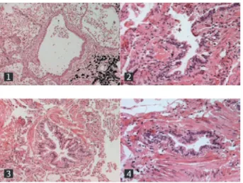
Among these findings, in the study by Rivera et al.,33
the autopsies of 10 women with BE-COPD and 10 women with TE-COPD showed greater remodeling and more fibrosis in the small airway in BE-COPD com-pared to TE-COPD. This is a clear evidence that the damage to the small airway is the main pathological feature in BE-COPD. Figure 1 shows an example of the severity of damage to the small airway in BE-COPD.
Importantly, these pathological changes are well cor-related with the radiographic findings, whether in the chest X-ray or the computed tomography36,37,42,45,46.
High-resolution computed tomography (HRCT) scans show peribronchial thickening, bronchial dilation, lam-inar subsegmental atelectasis, mosaic perfusion pat-tern, parenchymal bands, and no significant emphy-sema36-38,46. Using parametric response mapping, an
imaging tool that allows the quantification of small airway disease and emphysema in COPD, Fernandes et al. confirmed the absence of important emphyse-ma in patients with BE-COPD but, interestingly, sug-gested that these patients had a distinct pattern of small airway disease38.
Clinical findings
The vast majority of persons with BE-COPD are wom-en47, who strikingly and consistently have a body
mass index (BMI) higher than that of people with TE-COPD6,8,34,48. Individuals exposed to biomass
smoke have a high risk of chronic bronchitis (cough and phlegm for ≥ 3 months per year for at least 2 consecutive years)49,50 and patients with BE-COPD
frequently have respiratory symptoms: cough, expec-toration, and dyspnea36,42,45,51. Some studies show
that these symptoms are more frequent or have more impact in BE-COPD than in TE-COPD36,45,52,53, but
other studies do not48,51,54,55. With regard to the
physical examination, rhonchus and wheezing are relatively frequent in BE-COPD45. The high frequency
of cough, expectoration, rhonchus, and wheezing is clearly indicating the predominant damage to the air-ways in BE-COPD.
Quality of life
It is well known that COPD negatively affects the quality of life. Some studies have shown that this negative impact is higher in BE-COPD. Camp et al., using the St. George’s Respiratory Questionnaire, found worse symptoms and more compromised activ-ity indexes in women with BE-COPD compared to those with TE-COPD36. Another study including 138
women with COPD showed that among women with the same degree of obstruction, those with BE-COPD had worse health status (poorer quality of life and worse dyspnea) than those with TE-COPD, with no differences in comorbidities52.
Figure 1. Membranous bronchioles with structural remodel-ing from mild (1) to severe (4) in chronic obstructive
Functional findings
By definition, patients with BE-COPD have post-bron-chodilator airflow obstruction: FEV1/FVC < 0.7 or < lower limit of normal. Compared with TE-COPD, ob-struction in BE-COPD is milder, both overall and after adjusting for age36,42,45,48,54,56-58. Regarding the
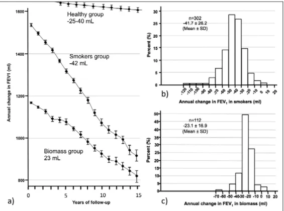
an-nual decline in lung function, the time-course behavior of FEV1 in a Mexican cohort of patients with COPD associated with biomass or tobacco during a 15-year follow-up period showed that the rate of FEV1 decline was significantly slower in patients whose COPD was caused by biomass smoke than in those caused by tobacco exposure. The annual rate of decline was significantly higher in the TE-COPD group than in the BE-COPD group (42 mL vs. 23 mL, respectively, p < 0.001). The proportion of rapid decliners in patients with COPD caused by biomass fuel was very low (1%) compared with the one seen in patients whose COPD was caused by tobacco exposure (11%) (Fig. 2)57.
With respect to gas exchange, some studies have shown that carbon dioxide arterial pressure (PaCO2) is higher (lower ventilation) and oxygen arterial pres-sure (PaO2) and oxygen arterial saturation (SaO2) are lower in BE-COPD than in TE-COPD36,45,54,57. The
low-er oxygenation rates obslow-erved in BE-COPD may be explained in part by hypoventilation. It remains to be determined whether this behavior is related with a higher BMI in these patients, most of whom are wom-en over 50 years of age. Recwom-ently, Olloquequi et al. found that patients with COPD exposed to both bio-mass and cigarette smoke exhibit significantly lower oxygen saturation than those exposed to only bio-mass or only cigarette smoke55.
Normal or mildly altered diffusing capacity (DLCO) and DLCO/alveolar volume (DLCO/AV) ratio are consistent-ly observed in BE-COPD when they are compared to TE-COPD, in which these parameters are significantly reduced37,45,55. This finding correlates with the lower
grade of emphysema found in HRCT in patients with BE-COPD8,36-38,46 at all levels of COPD severity. This
functional picture of decreased DLCO with normal DLCO/AV has been described in cases with significant-ly compromised small airways with little emphysema (pseudophysiological emphysema)59. Compromised
diffusion correlates better with decreased FEV1 in women with TE-COPD than in those with BE-COPD, posing the greater contribution of emphysema to air-flow obstruction in TE-COPD45.
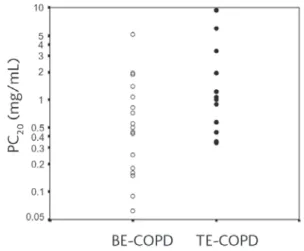
Women with BE-COPD have greater bronchial hyper-reactivity than women with TE-COPD (Fig. 3)60.
Fur-ther, research is needed to determine if this correlates with the higher frequency of the asthma–COPD over-lap phenotype observed in BE-COPD48. There is
evi-dence also that small airway resistance measured by oscillometry is considerably more affected in BE-COPD than in TE-BE-COPD61.
Exercise tolerance in BE-COPD
The walked distance in the 6 min walking test in BE-COPD does not seem significantly different compared to TE-COPD36,52,54,55, although Zhao et al. found a
lower distance in BE-COPD8. Camp et al. reported
lower SaO2 measured by pulse oximetry at the end of the test in women with BE-COPD36.
Clinical phenotypes in BE-COPD
Golpe et al.48 evaluated that the frequency of clinical
phenotypes defined by the Spanish COPD guidelines62
in patients with COPD caused by biomass or tobacco smoke. The asthma–COPD overlap phenotype was more common in BE-COPD, but the difference disap-peared after adjusting for sex. Similar to the findings discussed in the previous sections, they found a great-er frequency of emphysema phenotype in TE-COPD. No difference was found in the frequencies of chron-ic bronchitis or exacerbator phenotypes48.
Pulmonary hypertension in BE-COPD
A recent study found that pulmonary hypertension on echocardiography was more common in patients with BE-COPD than in those with TE-COPD63. In previous
studies, González et al. based on radiographic evalu-ation, and Sandoval et al. showed a higher rate of pulmonary hypertension among individuals with COPD related to wood smoke exposure compared to those exposed to tobacco smoke45,64. The origin of
pulmonary hypertension in BE-COPD patients does not appear to be related only to hypoxic pulmonary vasoconstriction but also to direct effects caused by the inhaled substances or indirect inflammatory-me-diated effects65.
Exacerbations and survival in BE-COPD
Golpe et al. did not find differences in the annual rate of exacerbations comparing BE-COPD and TE-COPD48,
but it should be noted that no prospective data are available on this aspect. After adjusting for age, sex, and disease severity, Ramírez-Venegas et al. and Gol-pe et al. did not find differences in survival between BE-COPD and TE-COPD54,58.
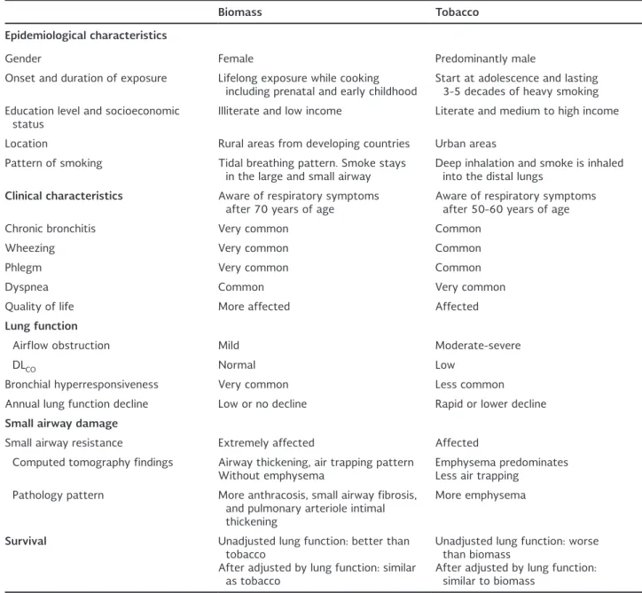
Table 1 summarizes the most important epidemio-logical, clinical, functional, histoepidemio-logical, and imaging differences between BE-COPD and TE-COPD.
Summary
No doubt BE-COPD is a disorder clearly associated with poverty and sociocultural issues. There is a direct impact on lung growth in these women that damage mainly the small airway.
However, in spite of these women being exposed to very high levels of pollutants while cooking, the con-sequent decline in FEV1 seems not as detrimental as that induced by tobacco. These findings could indicate that BE-COPD may be classified as the benign version of COPD. Even so, despite having a higher baseline FEV1, the histological and tomographic findings in BE-COPD show a more intense small airway damage in comparison with TE-COPD. Furthermore, in BE-COPD gas exchange abnormalities occur earlier, and pa-tients have a worse quality of life than smokers.
Moreover, mortality is similar to the one observed in smokers.
CONCLUSION
The effect on the small airway of biomass is as harm-ful as that caused by smoke in the lung parenquima. Therefore, BE-COPD seems equally damaging than TE-COPD.
Table 1. COPD characteristics of subjects exposed to biomass in comparison with those of smokers.
Biomass Tobacco
Epidemiological characteristics
Gender Female Predominantly male Onset and duration of exposure Lifelong exposure while cooking
including prenatal and early childhood Start at adolescence and lasting 3-5 decades of heavy smoking Education level and socioeconomic
status Illiterate and low income Literate and medium to high income Location Rural areas from developing countries Urban areas
Pattern of smoking Tidal breathing pattern. Smoke stays
in the large and small airway Deep inhalation and smoke is inhaled into the distal lungs
Clinical characteristics Aware of respiratory symptoms
after 70 years of age Aware of respiratory symptoms after 50-60 years of age Chronic bronchitis Very common Common
Wheezing Very common Common Phlegm Very common Common Dyspnea Common Very common Quality of life More affected Affected
Lung function
Airflow obstruction Mild Moderate-severe
DLCO Normal Low
Bronchial hyperresponsiveness Very common Less common Annual lung function decline Low or no decline Rapid or lower decline
Small airway damage
Small airway resistance Extremely affected Affected Computed tomography findings Airway thickening, air trapping pattern
Without emphysema Emphysema predominatesLess air trapping Pathology pattern More anthracosis, small airway fibrosis,
and pulmonary arteriole intimal thickening
More emphysema
Survival Unadjusted lung function: better than
tobacco
After adjusted by lung function: similar as tobacco
Unadjusted lung function: worse than biomass
After adjusted by lung function: similar to biomass
REFERENCES
1. Agusti A, Calverley PM, Celli B, et al. Characterisation of COPD heterogeneity in the ECLIPSE cohort. Respir Res. 2010;11:122. 2. Han MK, Agustí A, Calverley PM, et al. Chronic obstructive pul-monary disease phenotypes. Am J Respir Crit Care Med. 2010; 182:598-604.
3. Celli BR, Agustí A. COPD: time to improve its taxonomy? ERJ Open Res. 2018;4:00132-2017.
4. Agustí A, Celli B. Natural history of COPD: gaps and opportuni-ties. ERJ Open Res. 2017;3:00117-2017.
5. Agustí A, Sobradillo P, Celli B. Addressing the complexity of chronic obstructive pulmonary disease. Am J Respir Crit Care Med. 2011;183:1129-37.
6. Pérez-Padilla R, Ramírez-Venegas A, Sansores-Martínez R. Clin-ical characteristics of patients with biomass smoke-associated COPD and chronic bronchitis, 2004-2014. Chronic Obstr Pulm Dis. 2014;1:23-32.
7. Assad N, Balmes J, Mehta S, Cheema U, Sood A. Chronic ob-structive pulmonary disease secondary to household air pollu-tion. Semin Respir Crit Care Med. 2015;36:408-21.
8. Zhao D, Zhou Y, Jiang C, et al. Small airway disease: a different phenotype of early stage COPD associated with biomass smoke exposure. Respirology. 2018;23:198-05.
9. Salvi SS, Barnes PJ. Chronic obstructive pulmonary disease in non-smokers. Lancet. 2009;374:733-43.
10. Slade J. The tobacco epidemic: lessons from history. J Psychoac-tive Drugs. 1989;21:281-91.
11. Kodgule R, Salvi S. Exposure to biomass smoke as a cause for airway disease in women and children. Curr Opin Allergy Clin Immunol. 2012;12:82-90.
12. Biomass.net. Following the Energy Trail With Biomass History. Available from: http://www.biomass.net/Biomass-History.html. [Last accessed on 2018 Apr 10th].
13. Hurt RD, Murphy JG, Dunn WF. Did we finally slay the evil drag-on of cigarette smoking in the late 20th century? Unfortunately,
the answer is no-the dragon is still alive and well in the 21st
century and living in the third world. Shame on us! Chest. 2014; 146:1438-43.
14. Gordon SB, Bruce NG, Grigg J, et al. Respiratory risks from household air pollution in low and middle income countries. Lan-cet Respir Med. 2014;2:823-60.
15. Global Health Observatory. World Health Organization. Avail-able from: http://www.who.int/gho/phe/en/. [Last accessed on 2018 Apr 10th].
16. Eisner MD, Anthonisen N, Coultas D, et al. An official American thoracic society public policy statement: novel risk factors and the global burden of chronic obstructive pulmonary disease. Am J Respir Crit Care Med. 2010;182:693-18.
17. Ramírez-Venegas A, Sansores RH, Velázquez-Uncal M, Pérez-Bautista O. Nonsmokers and biomass exposure. ERS Monogr. 2015;69:35-46.
18. Caballero A, Torres-Duque CA, Jaramillo C, et al. Prevalence of COPD in five Colombian cities situated at low, medium, and high altitude (PREPOCOL study). Chest. 2008;133:343-9.
19. Balakrishnan K, Sambandam S, Ramaswamy P, Mehta S, Smith KR. Exposure assessment for respirable particulates associated with household fuel use in rural districts of Andhra Pradesh, India. J Expo Sci Environ Epidemiol. 2004; 14:S14-25.
20. Balakrishnan K, Sankar S, Parikh J, et al. Daily average exposures to respirable particulate matter from combustion of biomass fuels in rural households of southern India. Environ Health Per-spect. 2002;110:1069-75.
21. Naeher LP, Brauer M, Lipsett M, et al. Woodsmoke health ef-fects: a review. Inhal Toxicol. 2007;19:67-6.
22. Brauer M, Hirtle RD, Hall AC, Yip TR. Monitoring personal fine particle exposure with a particle counter. J Expo Anal Environ Epidemiol. 1999;9:228-36.
23. Salvi S, Barnes PJ. Is exposure to biomass smoke the biggest risk factor for COPD globally? Chest. 2010;138:3-6.
24. Postma DS, Bush A, van den Berge M. Risk factors and early origins of chronic obstructive pulmonary disease. Lancet. 2015; 385:899-909.
25. Heinzerling AP, Guarnieri MJ, Mann JK, et al. Lung function in woodsmoke-exposed Guatemalan children following a chimney stove intervention. Thorax. 2016;71:421-8.
26. Hayatbakhsh MR, Sadasivam S, Mamun AA, et al. Maternal smoking during and after pregnancy and lung function in early adulthood: a prospective study. Thorax. 2009;64:810-4.
27. Kulkarni N, Pierse N, Rushton L, Grigg J. Carbon in airway mac-rophages and lung function in children. N Engl J Med. 2006; 355:21-30.
28. Mishra V, Retherford RD. Cooking smoke increases the risk of acute respiratory infection in children. Natl Fam Health Surv Bull. 1997;8:1-4.
29. Po JYT, FitzGerald JM, Carlsten C. Respiratory disease associated with solid biomass fuel exposure in rural women and children: systematic review and meta-analysis. Thorax. 2011;66:232-9. 30. Martinez FD. Early-life origins of chronic obstructive pulmonary
disease. N Engl J Med. 2016;375:871-8.
31. Stocks J, Hislop A, Sonnappa S. Early lung development: lifelong effect on respiratory health and disease. Lancet Respir Med. 2013;1:728-42.
32. Bernstein DM. A review of the influence of particle size, puff volume, and inhalation pattern on the deposition of cigarette smoke particles in the respiratory tract. Inhal Toxico. 2004; 16:675-89.
33. Rivera RM, Cosío MG, Ghezzo H, Salazar M, Pérez-Padilla R. Comparison of lung morphology in COPD secondary to cigarette and biomass smoke. Int J Tuberc Lung Dis. 2008;12:972-7. 34. Torres-Duque CA, García-Rodriguez MC, González-García M. Is
chronic obstructive pulmonary disease caused by wood smoke a different phenotype or a different entity? Arch Bronconeumol. 2016;52:425-31.
35. Perret JL, Abramson MJ. Biomass smoke COPD: a phenotype or a different disease? Respirology. 2018;23:124-5.
36. Camp PG, Ramírez-Venegas A, Sansores RH, et al. COPD phe-notypes in biomass smoke-versus tobacco smoke-exposed Mexican women. Eur Respir J. 2014;43:725-34.
37. González-García M, Maldonado Gómez D, Torres-Duque CA, et al. Tomographic and functional findings in severe COPD: com-parison between the wood smoke-related and smoking-related disease. J Bras Pneumol. 2013;39:147-54.
38. Fernandes L, Gulati N, Fernandes Y, et al. Small airway imaging phenotypes in biomass- and tobacco smoke-exposed patients with COPD. ERJ Open Res 2017; 3: 00124-2016.
39. Kim YJ, Jung CY, Shin HW, Lee BK. Biomass smoke induced bron-chial anthracofibrosis: presenting features and clinical course. Respir Med. 2009;103:757-65.
40. Gupta A, Shah A. Bronchial anthracofibrosis: an emerging pul-monary disease due to biomass fuel exposure. Int J Tuberc Lung Dis. 2011;15:602-12.
41. Kim H, Cha SI, Shin KM, et al. Clinical relevance of bronchial anthracofibrosis in patients with chronic obstructive pulmonary disease exacerbation. Tuberc Respir Dis. 2014;77:124-31. 42. Morán-Mendoza O, Pérez-Padilla JR, Salazar-Flores M,
Vázquez-Alfaro F. Wood smoke-associated lung disease: a clinical, func-tional, radiological and pathological description. Int J Tuberc Lung Dis. 2008;12:1092-8.
43. Palacios DM, Méndez O. NeumopatÍa por humo de leña. Un estudio en autopsias. Biomédica. 1998;18:153.
44. Restrepo J, Reyes P, De Ochoa P, Patiño E. Neumoconiosis por inhalación del humo de leña. Acta Med Colomb. 1983;8:191-4. 45. González M, Páez S, Jaramillo C, Barrero M, Maldonado D. En-fermedad pulmonar obstructiva crónica (EPOC) por humo de leña en mujeres: comparación con la EPOC por tabaquismo. Acta Med Colomb. 2004;29:17-25.
46. Moreira MA, Barbosa MA, Queiroz MC, et al. Pulmonary chang-es on HRCT scans in nonsmoking femalchang-es with COPD due to wood smoke exposure. J Bras Pneumol. 2013;39:155-63. 47. Sana A, Somda SM, Meda N, Bouland C. Chronic obstructive
pulmonary disease associated with biomass fuel use in women: a systematic review and meta-analysis. BMJ Open Respir Res. 2018;5:e000246.
48. Golpe R, Sanjuán López P, Cano Jiménez E, Castro Añón O, Pérez de Llano LA. Distribución de fenotipos clínicos en pacientes con enfermedad pulmonar obstructiva crónica por humo de bio-masa y por tabaco. Arch Bronconeumol. 2014;50:318-24. 49. González-Garcia M, Caballero A, Jaramillo C, Torres-Duque CA.
Chronic bronchitis: high prevalence in never smokers and under-diagnosis-a population-based study in Colombia. Chron Respir Dis. 2018,1:1-8.
50. Kurmi OP, Semple S, Simkhada P, Smith WC, Ayres JG. COPD and chronic bronchitis risk of indoor air pollution from solid fuel: a systematic review and meta-analysis. Thorax. 2010;65:221-8. 51. Moreira MA, Moraes MR, Silva DG, et al. Comparative study of
52. González-García M, Gómez V, Perlaza I, Casas A. Diferencias en el impacto sobre el estado de salud entre la EPOC por cigarrillo y por humo de leña. Arch Bronconeumol. 2014;50:59. 53. Ramírez-Venegas A, Velázquez-Uncal M, Pérez-Hernández R, et
al. Prevalence of COPD and respiratory symptoms associated with biomass smoke exposure in a suburban area. Int J Chron Obs Pulmon Dis. 2018;13:1727-34.
54. Ramírez-Venegas A, Sansores RH, Pérez-Padilla R, et al. Sur-vival of patients with chronic obstructive pulmonary disease due to biomass smoke and tobacco. Am J Respir Crit Care Med. 2006;173:393-7.
55. Olloquequi J, Jaime S, Parra V, et al. Comparative analysis of COPD associated with tobacco smoking, biomass smoke expo-sure or both. Respir Res. 2018;19:1-8.
56. Torres-Duque CA, Caballero A, González-García M, Jaramillo C. Chronic obstructive pulmonary disease in people exposed to wood smoke. PREPOCOL: a population based study. Am J Respir Crit Care Med. 2013;187:A3644.
57. Ramírez-Venegas A, Sansores RH, Quintana-Carrillo RH, et al. FEV1 decline in patients with chronic obstructive pulmonary dis-ease associated with biomass exposure. Am J Respir Crit Care Med. 2014;190:996-2.
58. Golpe R, Mengual-Macenlle N, Sanjuan-López P, et al. Prognostic indices and mortality prediction in COPD caused by biomass smoke exposure. Lung. 2015;193:497-3.
59. Gelb AF, Zamel N, Hogg JC, Müller NL, Schein MJ. Pseudophys-iologic emphysema resulting from severe small-airways disease. Am J Respir Crit Care Med. 1998;158:815-9.
60. González-García M, Torres C, Jaramillo C, Maldonado D, Bustos D. Bronchial hyperresponsiveness in women with chronic ob-structive pulmonary disease related to wood smoke. Int J Chron Obstruct Pulmon Dis. 2012;7:367-73.
61. Guzmán-Bouilloud NE, Velázquez-Uncal M, Aranda-Chávez A, et al. Roll of small airways in patients with chronic obstructive pulmonary disease associated to biomass smoke. Am J Respir Crit Care Med. 2017;195:A5009.
62. Miravitlles M, Soler-Cataluña JJ, Calle M, et al. Guía española de la EPOC (GesEPOC). Actualización 2014. Arch Bronconeumol. 2014;50:1-16.
63. Sertogullarindan B, Gumrukcuoglu HA, Sezgi C, Akil MA. Fre-quency of pulmonary hypertension in patients with COPD due to biomass smoke and tobacco smoke. Int J Med Sci. 2012; 9:406-12.
64. Sandoval J, Salas J, Martínez-Guerra ML, et al. Pulmonary arte-rial hypertension and cor pulmonale associated with chronic domestic woodsmoke inhalation. Chest. 1993;103:12-20. 65. Mehra D, Geraghty PM, Hardigan AA, Foronjy R. A Comparison