P
www.permanyer.com
Rev Inves Clin. 2016;68:75-83 IN DEPTH REVIEWS
Aging and Pulmonary Fibrosis
Moisés Selman
1*, Ivette Buendía-Roldán
1and Annie Pardo
21Instituto Nacional de Enfermedades Respiratorias Ismael Cosío Villegas, SSA, Mexico City, Mexico; 2Facultad de Ciencias, Universidad Nacional Autónoma de México, Mexico City, Mexico
Corresponding author:
*Moisés Selman
Instituto Nacional de Enfermedades Respiratorias Calzada de Tlalpan, 4502
Col. Sección XVI, Del. Tlalpan C.P. 14080, Ciudad de México, México E-mail: mselmanl@yahoo.com.mx
moises.selma@salud.gob.mx Received for publication: 22-10-2015 Accepted for publication: 04-11-2015
ABSTRACT
Idiopathic pulmonary fibrosis is a chronic, progressive, and usually fatal lung disorder of unknown etiology. The disease likely results from the interaction of genetic susceptibility architecture, environmental factors such as smoking, and an abnormal epigenetic reprogramming that leads to a complex pathogenesis. Idiopathic pulmonary fibrosis occurs in middle-aged and mainly elderly adults, and in this context age has emerged as its strongest risk factor. However, the mechanisms linking it to aging are uncertain. Recently, nine molecular and cellular hallmarks of aging have been proposed: genomic instability, telomere attrition, epigenetic alterations, loss of proteostasis, deregulated nutrient sensing, mitochondrial dysfunction, cellular senescence, stem cell exhaustion, and altered intercellular communication. In this review, we provide an overview of these molecular mechanisms and their involvement in the pathogenesis of idiopathic pulmonary fibrosis, while emphasizing that the studies on this disease are few and the findings are not definitive. (REV INVES CLIN. 2016;68:75-83)
Key words: Aging. Lung fibrosis. Senescence. Telomeres.
INTRODUCTION
Biological lung aging is characterized by structural changes and progressive loss of physiological integrity, leading to impaired function1. Although the mechanisms
that contribute to the aging process are uncertain, nine putative hallmarks associated with the aging phenotype have recently been proposed2. However, in
what way and magnitude they participate in the aging lung is unknown.
FUNCTIONAL AND STRUCTURAL
MODIFICATIONS OF THE LUNGS
AND THORAX DURING AGING
In general, “normal aging” is characterized by narrowing of the intervertebral disk spaces and increased preva-lence of hyperkyphosis. In fact, 20-40% of older adults present an excessive curvature of the thoracic spine3. In
addition, there are changes in the intrinsic function of the muscles, which are associated with reduced inspiratory
Sin contar con el consentimiento previo por escrito del editor
, no podrá reproducirse ni fotocopiarse ninguna parte de esta publicación.
and expiratory respiratory muscle strength. This pro-cess, together with a decrease in the mitochondrial adenosine triphosphate (ATP) reserves, contribute in older individuals to having difficulties in sustaining a sudden rise in metabolic demand, increasing the risk of respiratory failure in acute lung diseases.
A common finding with aging is a decrease of lung elas-ticity4. This affects the small airways and alveolar septa,
and may explain at least two frequent observations in the elderly. The first is a premature collapse of the pe-ripheral airways, which provokes the so-called “air-trap-ping” that is more evident during expiration (Fig. 1). The other is the increase in size of the alveolar ducts and alveoli, which was previously called “senile emphyse-ma”, although this is not an appropriate term since it lacks the characteristic destruction of the alveolar walls seen in emphysema (Fig. 2). Nevertheless, the alveolar over-distention results in an increase of the residual volume of about 5-10% per decade5.
Other studies performed in older individuals (> 75 years old) without known respiratory disease have also re-ported the presence of reticular opacities (suggestive of fibrosis), as well as airway dilation, bronchial thick-ening, and bronchiectasis when compared with younger (< 55 years old) subjects6. Changes in the airways are
associated with dysfunction of the mucociliary escala-tor, decreased capacity to clear mucus and particles from the lungs, and a reduction in cough strength7. We
have found similar alterations in an ongoing study on aging lung in asymptomatic individuals (Selman, et al., unpublished results) (Fig. 3).
Physiological age-related pulmonary changes are charac-terized by a decrease of approximately 30 ml each year in forced expiratory volume in one second (FEV1) and forced vital capacity (FVC)8,9. Likewise, the mentioned
increase in the closing volume by the premature collapse of the small airways, combined with diverse age-related changes in the pulmonary circulation, result in a hetero-geneous distribution of the ventilation/perfusion ratio. This, together with a decrease in the diffusing lung capac-ity for carbon monoxide (DLCO), causes an age-related decline in the arterial tension for oxygen (PaO2)10-12.
LUNG DISEASES ASSOCIATED
WITH AGING
There are two types of lung disorders associated with aging: those that may occur at any period of life but whose severity is affected by aging, and those that oc-cur virtually only in old people. Among the first type, asthma, obstructive sleep apnea, and pulmonary ede-ma in the setting of congestive heart failure are some of the most common13,14. Likewise, decreased
respira-tory muscle strength, attenuated cough, dysfunction of mucociliary clearance, and altered immune response increase the risk for lung infections in elderly patients13.
By contrast, chronic obstructive pulmonary disease (COPD) and idiopathic pulmonary fibrosis (IPF) are two diseases usually diagnosed in individuals over 50 years old and whose incidence and prevalence in-crease remarkably with age. Thus, the prevalence of COPD in persons aged 65 years and older in the
Figure 1. Expiratory high-resolution computed tomography scan revealing inhomogeneous lung attenuation due to air trapping identified by the presence of areas of low attenuation next to regions with normal attenuation (arrows).
Figure 2. Early stage of centriacinar emphysema in a 73-year-old asymptomatic individual. High-resolution computed to-mography demonstrates numerous tiny low attenuation areas throughout the lung field (arrow).
Sin contar con el consentimiento previo por escrito del editor
, no podrá reproducirse ni fotocopiarse ninguna parte de esta publicación.
general population is at least 10-15%15. The real
in-cidence and prevalence of IPF are uncertain, but in the USA it has been reported that the incidence is about 10 per 100, 000 persons per year, which increases to approximately 90 per 100, 000 per year in people aged 65 years and older16,17.
IDIOPATHIC PULMONARY FIBROSIS:
THE INFLUENCE OF AGING
Idiopathic pulmonary fibrosis is a progressive, irrevers-ible, and usual fatal lung disorder of unknown etiolo-gy18. It has been proposed that the disease is triggered
by an aberrant activation of alveolar epithelial cells (AEC), which in turn induces the migration, proliferation, and activation of fibroblasts/myofibroblasts, leading to the exaggerated accumulation of extracellular matrix and the subsequent destruction of the lung architec-ture19. As mentioned before, IPF occurs in middle-aged
and mainly elderly adults, suggesting a mechanistic link between chronological age and this disease. However, the biopathological mechanisms that link aging with the pathogenesis of IPF have not been elucidated.
Recently, nine putative cellular and molecular hallmarks were proposed to contribute to the aging processes
and aging phenotype2. Although studies in IPF are few,
almost all of these hallmarks have been examined and results suggest that an accelerated aging process occurs in this disease.
GENOMIC INSTABILITY
Age-dependent accumulation of DNA damage is a well-recognized component of the aging phenotype2. Several
studies have reported the presence of genomic instabil-ity in IPF patients20-22. The incidence of microsatellite
instability (MSI) and loss of heterozygocity (LOH) were determined in cytological sputum specimens from 26 IPF patients and 26 matched controls using 10 highly poly-morphic microsatellite markers20. Fifty percent of the
patients displayed genetic alterations, either MSI or LOH. The most commonly affected microsatellite markers were THRA1 and D8S133. Subsequently, a one-base-pair de-letion was detected in the polyadenine tract in exon 3 of the transforming growth factor (TGF)-beta RII recep-tor gene in AECs isolated by microdissection from IPF lungs. Furthermore, in these areas, low expression of the receptor was confirmed21. Finally, 40 microsatellite
mark-ers were evaluated in 52 sputum/venous blood DNA pairs from IPF patients22. Twenty specimens (38.5%)
exhibited LOH in at least one of the examined loci; LOH
Figure 3. High-resolution computed tomography showing several abnormalities detected in elderly asymptomatic subjects.
A: Central airway dilation. B: Bronchiectasis. C: and D: Peripheral, subpleural septal thickening (arrows).
Sin contar con el consentimiento previo por escrito del editor
, no podrá reproducirse ni fotocopiarse ninguna parte de esta publicación.
was observed in microsatellite DNA markers located in MYCL1, FHIT, SPARC, p16Ink4, and TP53 genes. Taken together, these findings indicate that genetic instability likely affecting genes involved in critical cellular pathways is a relatively frequent phenomenon that could account for the pathogenesis of IPF.
TELOMERE ATTRITION
Telomere shortening is considered one of the most influential mechanisms of cellular aging. When telo-meres become critically short, they activate a DNA damage response that provokes cellular senescence or apoptosis23. Abnormal telomere shortening has been
associated with several progressive disease pheno-types that share the short telomere defect as a driving mechanism23. Telomerase mutations cause
approxi-mately 20% of the cases of familial IPF (identified by the presence of two or more individuals in a family having pulmonary fibrosis), and all of these patients character-istically have very short telomeres24-26. Furthermore,
20-30% of patients with sporadic IPF that do not have mu-tations in telomerase components displayed telomere lengths less than the 10th percentile when compared with control subjects27. The mechanisms by which
telo-mere defects contribute to IPF are uncertain. It has been proposed that telomerase mutations (familial IPF) or exaggerated proliferative response (sporadic IPF) lead to telomere shortening in the alveolar epithelium and that this is critical for the development of the disease. A recent study supports this notion28. In this work,
late-generation telomerase-null mice induced by delet-ing telomeric repeat-binddelet-ing factor 2 (Trf2) was gener-ated, and in this conditional mutant model, where telo-mere dysfunction was restricted to type 2 AECs (AEC2), the stem cell function of this subpopulation was im-paired, leading to senescence. Moreover, when telo-mere dysfunction was induced in purified adult AEC2s, ex vivo cells survived but remained senescent28. These
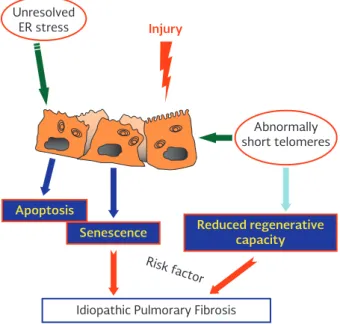
results indicate that AEC2-dependent telomere dys-function and senescence limit alveolar repair and can signal mesenchymal abnormalities (Fig. 4).
CELLULAR SENESCENCE
Cellular senescence has been considered a critical event in biological aging. It refers to a permanently arrested state of cell growth together with the achievement of
the senescence-associated secretory phenotype, char-acterized by the release of a variety of inflammatory, growth-regulating, and tissue-remodeling factors2,29.
Recently, AEC senescence was revealed in IPF lungs30.
In this study, strong staining of β-galactosidase, a marker of senescence, and p21/waf-1, a senescence-associated cyclin-dependent kinase inhibitor, was ob-served in the lung epithelium. These results were con-firmed in a second study where nuclear staining of p21 was clearly demonstrated only in epithelial cells covering actively fibrosing lesions, while β-Gal-positive staining was observed in epithelial cells covering fibro-blastic foci31. Alveolar epithelial senescence, likely related
to shortening of telomeres, may contribute to the high secretory profile exhibited by these cells in IPF.
On the other hand, studies on fibroblasts have given elusive results. Recently, a study demonstrated that fibroblasts within fibroblastic foci of IPF lungs show features of senescence. Expression of p16 and p21 was seen in fibroblasts within the foci and in the overly-ing epithelial cells32. Moreover, fibroblast expression of
NADPH oxidase-4 (Nox4) was increased in IPF lung
Unresolved ER stress
Apoptosis
Senescence Reduced regenerativecapacity
Idiopathic Pulmorary Fibrosis
Risk f actor
Abnormally short telomeres
Injury
Figure 4. Alveolar epithelial cells play a critical role in the pathogenesis of idiopathic pulmonary fibrosis. Unsolved en-doplasmic reticulum stress and extreme shortening of telo-meres may lead to epithelial cell death or senescence, and the senescence-associated secretory phenotype character-ized by the upregulation of genes encoding a complex pro-inflammatory and profibrotic transcriptional response. ER: endoplasmic reticulum.
Sin contar con el consentimiento previo por escrito del editor
, no podrá reproducirse ni fotocopiarse ninguna parte de esta publicación.
increased production of reactive oxygen species (ROS)37.
Mitochondrial DNA (mtDNA) is damaged by ROS gen-erated during oxidative metabolism, and the accumu-lation of damaged mtDNA and decreased mitophagy result in loss of fidelity in the synthesis of new mito-chondria proteins, leading to senescence and aging2.
Excessive production of ROS and disruption of the oxi-dant/antioxidant balance in the lung have been found in IPF38. In the expired breath condensate, the
concen-trations of H2O2 and 8-isoprostane, which are mark-ers of oxidative stress, are usually increased in IPF patients compared with normal controls, indicating high levels of oxidative stress39. Likewise, a marked
reduction of levels of glutathione, a major antioxidant molecule, has been observed in bronchoalveolar la-vage, sputum, and plasma of patients with IPF40,41.
Recently, a study demonstrated that AEC2 of IPF lungs exhibit an age-related mitochondrial dysfunction with altered structure and impaired mitophagy42.
Deficien-cy of PTEN-induced putative kinase 1 (PINK1) was identified as a fundamental mechanism leading to ac-cumulation of dysfunctional mitochondria and, more-over, the mitochondrial phenotype observed in IPF lungs and susceptibility to lung fibrosis was recapitulated in an animal model of aging and PINK1 deficiency. Importantly, several chronic degenerative diseases associated with aging, such as Parkinson’s disease and neuropsychiatric disorders, present mutations or deficit of PINK1 and show swollen and dysfunc-tional mitochondria and poor mitophagy, indicating that this may be a common phenomenon in aging-associated diseases.
LOSS OF PROTEOSTASIS
Aging and some aging-related diseases are associ-ated with impaired proteostasis. Protein homeostasis involves mechanisms for the stabilization of correct-ly folded proteins and mechanisms for the degrada-tion of proteins by two principal proteolytic systems implicated in protein quality control: the autophagy-lysosomal system and the ubiquitin-proteasome sys-tem2. There is a strong body of evidence indicating
that aging is associated with disturbed proteostasis, which may contribute to age-associated disorders. Furthermore, maintenance of appropriate autophagic activity prevents or slows down the functional failure fibroblasts, and the use of a specific inhibitor
attenu-ated βgal activity, suggesting that Nox4 contributes to cellular senescence of IPF fibroblasts. More recent-ly, a study showed that IPF fibroblasts displayed an accelerated entry to replicative senescence, accompa-nied by an accumulation of senescent cells with fea-tures of myofibroblasts characterized by high expres-sion of alpha smooth muscle actin (α-SMA)33.
There is also some “systems senescence” in IPF patients, e.g., immune senescence or endocrine senescence, which may contribute to the development or progression of IPF. For example, a marked downregulation of CD28 on circulating CD4 T-cells has been found in IPF patients compared with age-matched controls34. CD28 is a
ma-jor co-stimulatory molecule responsible for the optimal activation of naive T-cells. It is also involved in prolif-eration, survival, and glucose metabolism. The T-cells lose CD28 expression with age, often taken as a hall-mark of aging human T-cells35.
Deterioration of the endocrine system also occurs during aging and is thought to contribute to increased suscep-tibility to aging-associated diseases. In this context, we evaluated the blood levels of dehydroepiandrosterone (DHEA) and its sulfate ester (DHEA-S), the most abundant adrenal steroids in humans. Under physio-logical conditions, DHEA/DHEA-S reach a peak be-tween the ages of 25 and 30 years and thereafter gradually decline so that, by the age of 60, the con-centrations are only 10-20% of corresponding values in young adults36. We found that IPF patients had a
disproportionate decrease in the circulating levels of DHEA-S compared with age-matched controls. More-over, DHEA displayed a strong antifibrotic effect on fibroblasts, affecting migration, proliferation, differ-entiation to myofibroblasts, collagen synthesis, and survival, indicating that its exaggerated decline may participate in the pathogenesis of the disease36.
MITOCHONDRIAL DYSFUNCTION
Mitochondria play a key role in cellular homeostasis, bioenergetic capacity, and longevity since they are the highest producers of ATP and regulate programmed cell death. Aging is associated with the expansion of dys-functional mitochondria, with alterations in mitochon-drial dynamics and quality control processes resulting from an imbalance of fission and fusion events and
Sin contar con el consentimiento previo por escrito del editor
, no podrá reproducirse ni fotocopiarse ninguna parte de esta publicación.
associated with cellular proteotoxicity and accumula-tion of intracellular damage in aging43.
Recent work has approached the putative role of au-tophagy in IPF, while studies on the ubiquitin system are scant.
Autophagy is a complex process involving multiple pro-teins and steps, including the formation of an initia-tion complex and development of a double-membrane phagophore; elongation of the membrane and com-pletion of an autophagosome vesicle around cargo; lysosomal fusion; dissolution of the inner membrane allowing hydrolases to degrade the cargo; and recy-cling of the components44.
In the first approach in IPF, it was reported that LC3-II levels (commonly used as a marker of autophagy) were significantly lower in whole tissue homogenate of lungs from patients with IPF compared with control lungs. In experimentally induced lung fibrosis, it was shown that the inhibition of mTORC1, a primary modulator of autophagy, with rapamycin attenuated the fibrotic response45. In this study, they also found that
inhibi-tion of autophagy potentiated fibroblast to myofi-broblast differentiation and activation. A subsequent study, using biochemical evaluation of in vitro models, demonstrated that autophagy inhibition is sufficient to induce acceleration of epithelial cell senescence and myofibroblast differentiation in lung fibroblasts31.
More recently it was shown that an aberrant PTEN/ Akt/mTOR axis desensitizes IPF fibroblasts from po-lymerized collagen-driven stress by suppressing au-tophagic activity, which produces an IPF fibroblast phenotype resistant to apoptosis in collagen46.
Most studies regarding the role of autophagy in lung fibrosis have focused on fibroblasts. A more recent work suggests that epithelial cells may also be affected. In an experimental model induced by bleomycin, it was shown that Atg4b-deficient mice exhibited reduced au-tophagy and a significantly higher inflammatory and fibrotic response compared with the wild-type litter-mate. Importantly, the study found that Atg4b dis-ruption resulted in increased apoptosis, affecting pre-dominantly alveolar and bronchiolar epithelial cells47.
These findings indicate that autophagy protects epi-thelial cells against bleomycin-induced stress and apop-tosis, and participates in the attenuation of the in-flammatory and fibrotic responses.
Importantly, evidence suggests that there is an age-related decline in autophagy and selective targeting of mitochondria for autophagic degradation that enhanc-es the lung fibrotic renhanc-esponse in experimental models48.
This reduction seems to be exaggerated or accelerated in IPF, a natural aging-associated human fibrosis.
Oxidative stress, endoplasmic reticulum (ER) stress, and hypoxia, all mechanisms that participate in the patho-genesis of IPF, are well-known inducers of autophagy. However, this protective mechanism is dysfunctional, likely contributing to the pathobiology of the disease.
The ubiquitin-proteasome system is the major degra-dation pathway for short-lived proteins in eukaryotic cells. Its relevance for preservation of protein homeo-stasis in the lung is emerging for chronic lung dis-eases49. In this context, inhibition of this system by
specific proteasome inhibitors has been shown to provide antifibrotic effects in the mouse model of bleomycin-induced lung damage50.
However, the regulation of proteasome function in IPF has not been explored in detail. Recently, a study showed that the proteasome is activated in the process of TGF-β-induced human myofibroblast differentiation51. The activation resulted from
in-creased formation of 26S proteasomes. In IPF lungs, the expression of the subunit Rpn6 was upregu-lated specifically in myofibroblasts and hyperplastic AECs overlying fibroblast foci. Elevated levels of K48-polyubiquitin protein conjugates in these cells and the positive correlation of whole lung Rpn6 protein levels with K48-polyubiquitinated proteins suggest that activation of ubiquitin-dependent protein deg-radation by the 26S proteasome may be a patho-logic feature of fibrotic remodeling occurring specifi-cally in IPF51.
STEM CELL EXHAUSTION
The balance between stem cell self-renewal and dif-ferentiation is critical to orchestrate tissue homeo-stasis and the response for repair/replacement of damaged tissues. In this context, a major hallmark of aging is a reduced ability to regenerate, which has been associated with a decline in proliferative activ-ity, impaired function, and exhaustion of tissue-spe-cific stem and progenitor cells2. There is an emerging
Sin contar con el consentimiento previo por escrito del editor
, no podrá reproducirse ni fotocopiarse ninguna parte de esta publicación.
body of evidence indicating that reduced function of adult stem cells plays an important role in the develop-ment of age-related diseases52. So far, no studies in
IPF have been published. In a recent report, bone mar-row-derived mesenchymal stem cells (B-MSC) derived from old animals were found to display a remarkable downregulation of multiple chemokine receptors such as CCR7, CX3CR1, and CXCR5 as well as other genes involved in migration53. When lungs were injured with Escherichia coli lipopolysaccharide, aged endogenous B-MSCs not only failed to migrate appropriately to the injury site, but once there they also failed to pro-duce enough of the anti-inflammatory agents that characterize their younger forms. Interestingly, there were similar differences between B-MSCs obtained from young and aged human individuals; that is, old cells showed a downregulation of cytokine receptors, decrease in activation, and migration.
DEREGULATED NUTRIENT-SENSING
Insulin-like growth factor (IGF-1) and insulin signaling are known as the “insulin and IGF-1 signaling” and represent the most conserved aging-controlling path-way in evolution2. Among its multiple targets are the
mammalian target of rapamycin (mTOR) complexes, which are also involved in aging and recently have been implicated in lung fibrosis. For example, in a recent work, aberrant mTOR signaling activation was provoked in AECs using conditional Tsc1 knock-down mice that were then injured with bleomycin54. Mice with increased
mTOR activation exhibited high mortality and exagger-ated lung fibrosis compared with control mice. Moreover, mTOR inhibition with rapamycin rescued bleomycin-mediated lung injury and fibrosis. These findings were associated to decreased autophagy that, as mentioned, seems to contribute to abnormal repair and fibrosis.
Supporting the role of mTOR complexes in the fibrot-ic response, a recent study in IPF lung fibroblasts dem-onstrated that TGF-β, a major profibrotic mediator, induced the Rictor component of mTORC2, which led to Akt activation55. Moreover, the use of a specific
inhibitor of the active site mTOR attenuated the ex-pression of profibrotic matrix-regulatory proteins in TGF-β-stimulated IPF fibroblasts and inhibited the fi-brotic response in a murine bleomycin lung model55.
Overactivation of mTOR has been found in fibroblast foci and alveolar epithelial cells of IPF lungs54,56.
EPIGENETIC ALTERATIONS
Epigenetic mechanisms are heritable changes in gene activity that are independent of alterations in the underlying DNA sequence. In a more extensive defini-tion, epigenetic includes the set of covalent modifica-tions to DNA, posttranslational modificamodifica-tions to his-tones, and the regulatory effect of non-coding RNAs that influence the expression of genes and the struc-ture of chromatin. All these epigenetic processes do not act independently, but strongly interact to form a com-plex regulatory system that can dynamically adjust the gene expression. Epigenetic marks are remodeled and may actively modulate the processes of aging.
DNA METHYLATION
AND IDIOPATHIC PULMONARY FIBROSIS
DNA methylation is a covalent modification that occurs on cytosine, mostly located in CG dinucleotides (CpG). Cytosine methylation primarily happens in CpG-rich se-quences, dubbed as CpG islands, resulting in the con-stitutive silencing of chromatin regions.
Aging is characterized by hypomethylation of sites outside promoter CpG islands, while CpG islands near promoters are typically hypermethylated, and there is some evidence indicating that these modifications in DNA methylation may be a sensor for both chron-ological and bichron-ological age57.
Studies in IPF are scant and initially focused on putative meaningful candidate genes. Thus for example, Thy-1 (CD90), an important regulator of fibroblast behavior, is absent in myofibroblasts within fibroblastic foci in IPF, and its downregulation is mediated at least partially by the hypermethylation of the promoter58. Likewise,
dif-ferent levels of methylation of three CpG islands in the promoter of α-SMA in fibroblasts and myofibroblasts correlate with the levels of expression of this gene59.
On the other hand, we have demonstrated that IPF fi-broblasts have reduced expression of the proapoptot-ic p14ARF attributable to promoter hypermethylation, suggesting that epigenetic mechanisms may underlie their resistance to apoptosis60.
Global methylation and gene expression patterns have been recently examined in IPF lungs61. By
comprehen-sive high-throughput arrays, 4.6 million CpG sites
Sin contar con el consentimiento previo por escrito del editor
, no podrá reproducirse ni fotocopiarse ninguna parte de esta publicación.
distributed across the human genome as well as the gene expression changes were examined in 94 IPF lungs and 67 controls. Over 2,000 differentially methylated regions associated with 1,514 unique genes were iden-tified, with the majority of the methylation changes located outside of promoter CpG islands. Functional analyses identified several enriched canonical pathways that have been implicated in the pathogenesis of IPF, including CXCR4 signaling, thrombin signaling, Wnt/β -catenin signaling, and epithelial adherens junction sig-naling. Analysis of binding motifs in promoters revealed overrepresentation of regulators of lung development, specifically, β-catenin, GLI1, and FOXC2; this is impor-tant since the upregulation of developmental pathways is involved in the aberrant activation of epithelial cells62.
These findings support the notion that several biologi-cally relevant methylation-expression changes may con-tribute to the development of IPF.
NON-CODING RNA AND IDIOPATHIC
PULMONARY FIBROSIS
Two main sub-groups of regulatory-type non-coding RNA (ncRNA) have been described: the short ncRNAs (< 30 nucleotides long), that include microRNAs (miRNA), short interfering RNAs (siRNA), and piwi-interacting RNAs (piRNA); and the long ncRNAs that contain over 200 nucleotides and seem to control genome activity at the chromatin level.
Epigenetic deregulation of ncRNAs, primarily miRNAs, has been observed in IPF. In fact, different studies have shown that approximately 10% of miRNAs are deregu-lated and an imbalance between profibrotic and antifi-brotic miRNAs are thought to be linked to the develop-ment or progression of IPF63,64. The downregulated
miRNAs include 326, let 7d, 26a, 29, miR-200, and miR-17~92, while miR-21, miR-154, 199a-5p, and 145 are upregulated. In general, all these miR-NAs play roles in the TGF-β1 signaling pathway, fibrop-roliferation, lung epithelial cell development, and epithe-lial to mesenchymal transition, and their deregulation results in the facilitation of many profibrotic processes.
It is important to emphasize that all these epigenetic mechanisms are integrated through complex crosstalk pathways and feedback loops. For example, an asso-ciation between aberrant DNA methylation and miR-NA expression has been recently identified in IPF65.
Thus, increased DNA methylation in the promoter of the miR-17~92 clusters silence its expression, which in turn results in the upregulation of genes strongly related to the fibroproliferative response and the fi-broblast phenotype in IPF.
Finally, whether some of the mentioned epigenetic changes observed in IPF are related to aging is uncertain. It has been proposed that there is a stochastic age-re-lated DNA methylation drift, which is bidirectional (both hyper- and hypomethylation), is not uniform across the genome, and is quite variable between individuals of the same age66. It is tempting to think that in few of them,
the drift particularly affects genes whose up- or down-regulation results in a profibrotic reprogramming.
CONCLUSIONS
Aging is a multifaceted process that results in pro-gressive decline in homeostasis and increased risk of disease or death. Incidence and prevalence of IPF in-crease remarkably with aging. Before 50 years of age, IPF is rare, but over 60 years old, the prevalence may be as high as 300/100,000, indicating a strong link between aging and IPF. Most of the hallmarks of aging seem to be involved in the development or progres-sion of IPF. However, studies to date were performed in small cohorts and have produced heterogeneous results. In the future it will be necessary to integrate the genetic and epigenetic data to identify regulatory pathways associated with aging and identify which of them may be implicated in the pathogenesis of IPF.
REFERENCES
1. Thannickal VJ, Murthy M, Balch WE, et al. Blue journal confer-ence. Aging and susceptibility to lung disease. Am J Respir Crit Care Med. 2015;191:261-9.
2. López-Otín C, Blasco MA, Partridge L, Serrano M, Kroemer G. The hallmarks of aging. Cell. 2013;153:1194-217.
3. Lowery EM, Brubaker AL, Kuhlmann E, Kovacs EJ. The aging lung. Clin Interv Aging. 2013;8:1489-96.
4. Fukuchi Y. The aging lung and chronic obstructive pulmonary dis-ease: similarity and difference. Proc Am Thorac Soc. 2009; 6:570-2. 5. Zaugg M, Lucchinetti E. Respiratory function in the elderly.
An-esthesiol Clin North America. 2000;18:47-58.
6. Copley SJ, Wells AU, Hawtin KE, et al. Lung morphology in the elderly: comparative CT study of subjects over 75 years old versus those under 55 years old. Radiology. 2009;251:566-73. 7. Svartengren M, Falk R, Philipson K. Long-term clearance from small airways decreases with age. Eur Respir J. 2005;26:609-15. 8. Knudson RJ, Slatin RC, Lebowitz MD, Burrows B. The maximal
expiratory flow-volume curve. Normal standards, variability, and effects of age. Am Rev Respir Dis. 1976;113:587-600. 9. Griffith KA, Sherrill DL, Siegel EM, Manolio TA, Bonekat HW,
Enright PL. Predictors of loss of lung function in the elderly: the
Sin contar con el consentimiento previo por escrito del editor
, no podrá reproducirse ni fotocopiarse ninguna parte de esta publicación.
Cardiovascular Health Study. Am J Respir Crit Care Med. 2001; 163:61-8.
10. Cardús J, Burgos F, Diaz O, et al. Increase in pulmonary ventila-tion-perfusion inequality with age in healthy individuals. Am J Respir Crit Care Med. 1997;156:648-53.
11. Guenard H, Marthan R. Pulmonary gas exchange in elderly sub-jects. Eur Respir J. 1996;9:2573-7.
12. American Thoracic Society and American College of Chest Phy-sicians. ATS/ACCP Statement on cardiopulmonary exercise test-ing. Am J Respir Crit Care Med. 2003;167:211-77.
13. Akgün KM, Crothers K, Pisani M. Epidemiology and management of common pulmonary diseases in older persons. J Gerontol A Biol Sci Med Sci. 2012;67:276-91.
14. Peppard PE, Young T, Barnet JH, Palta M, Hagen EW, Hla KM. Increased prevalence of sleep-disordered breathing in adults. Am J Epidemiol. 2013;177:1006-14.
15. Mannino DM, Homa DM, Akinbami LJ, Ford ES, Redd SC. Chron-ic obstructive pulmonary disease surveillance-United States, 1971-2000. MMWR Surveill Summ. 2002;51:1-16.
16. Raghu G, Weycker D, Edelsberg J, Bradford WZ, Oster G. Inci-dence and prevalence of idiopathic pulmonary fibrosis. Am J Respir Crit Care Med. 2006;174:810-16.
17. Raghu G, Chen SY, Yeh WS, et al. Idiopathic pulmonary fibrosis in US Medicare beneficiaries aged 65 years and older: incidence, preva-lence, and survival, 2001-11. Lancet Respir Med. 2014; 2:566-72. 18. King TE, Pardo A, Selman M. Idiopathic pulmonary fibrosis.
Lan-cet. 2011;378:1949-61.
19. Selman M, Pardo A. Revealing the pathogenic and aging-related mechanisms of the enigmatic idiopathic pulmonary fibrosis. An integral model. Am J Respir Crit Care Med. 2014;189:1161-72. 20. Vassilakis DA, Sourvinos G, Spandidos DA, Siafakas NM, Bouros D. Frequent genetic alterations at the microsatellite level in cytologic sputum samples of patients with idiopathic pulmonary fibrosis. Am J Respir Crit Care Med. 2000;162:1115-19. 21. Uematsu K, Yoshimura A, Gemma A, et al. Aberrations in the
fragile histidine triad (FHIT) gene in idiopathic pulmonary fibro-sis. Cancer Res. 2001;61:8527-33.
22. Demopoulos K, Arvanitis DA, Vassilakis DA, Siafakas NM, Span-didos DA. MYCL1, FHIT, SPARC, p16(INK4) and TP53 genes associated to lung cancer in idiopathic pulmonary fibrosis. J Cell Mol Med. 2002;6:215-22.
23. Stanley SE, Armanios M. The short and long telomere syn-dromes: paired paradigms for molecular medicine. Curr Opin Genet Dev. 2015;33:1-9.
24. Armanios MY, Chen JJ, Cogan JD, et al. Telomerase mutations in families with idiopathic pulmonary fibrosis. N Engl J Med. 2007;356:1317-26.
25. Tsakiri KD, Cronkhite JT, Kuan PJ, et al. Adult-onset pulmonary fibrosis caused by mutations in telomerase. Proc Natl Acad Sci U S A. 2007;104:7552-7.
26. Lawson WE, Loyd JE, Degryse AL. Genetics in pulmonary fibro-sis--familial cases provide clues to the pathogenesis of idio-pathic pulmonary fibrosis. Am J Med Sci. 2011;341:439-43. 27. Cronkhite JT, Xing C, Raghu G, et al. Telomere shortening in
familial and sporadic pulmonary fibrosis. Am J Respir Crit Care Med. 2008;178:729-37.
28. Alder JK, Barkauskas CE, Limjunyawong N, et al. Telomere dys-function causes alveolar stem cell failure. Proc Natl Acad Sci U S A. 2015;112:5099-104.
29. Byun HO, Lee YK, Kim JM, Yoon G. From cell senescence to age-related diseases: differential mechanisms of action of senescence-associated secretory phenotypes. BMB Rep. 2015; 48;549-58. 30. Minagawa S, Araya J, Numata T, et al. Accelerated epithelial cell
senescence in IPF and the inhibitory role of SIRT6 in TGF-β -induced senescence of human bronchial epithelial cells. Am J Physiol Lung Cell Mol Physiol. 2011;300:L391-401.
31. Araya J1, Kojima J, Takasaka N, et al. Insufficient autophagy in idiopathic pulmonary fibrosis. Am J Physiol Lung Cell Mol Physi-ol. 2013;304:L56-69.
32. Hecker L, Logsdon NJ, Kurundkar D, et al. Reversal of persistent fibrosis in aging by targeting Nox4-Nrf2 redox imbalance. Sci Transl Med. 2014;6:231ra47.
33. Yanai H, Shteinberg A, Porat Z, et al. Cellular senescence-like features of lung fibroblasts derived from idiopathic pulmonary fibrosis patients. Aging (Albany NY) 2015;7:664-72.
34. Gilani SR, Vuga LJ, Lindell KO, et al. CD28 down-regulation on circulating CD4 T-cells is associated with poor prognoses of pa-tients with idiopathic pulmonary fibrosis. PLoS One. 2010;5:e8959. 35. Larbi A, Pawelec G, Wong SC, Goldeck D, Tai JJ, Fulop T. Impact of age on T cell signaling: a general defect or specific altera-tions? Ageing Res Rev. 2011;10:370-8.
36. Mendoza-Milla C, Valero Jiménez A, Rangel C, et al. Dehydroepi-androsterone has strong antifibrotic effects and is decreased in idiopathic pulmonary fibrosis. Eur Respir J. 2013;42:1309-21.
37. López-Lluch G, Santos-Ocaña C, Sánchez-Alcázar JA, et al. Mi-tochondrial responsibility in ageing process: innocent, suspect or guilty. Biogerontology. 2015;16:599-620.
38. Lenz AG, Costabel U, Maier KL. Oxidized BAL fluid proteins in pa-tients with interstitial lung diseases. Eur Respir J. 1996;9:307-12. 39. Psathakis K, Mermigkis D, Papatheodorou G, et al. Exhaled
markers of oxidative stress in idiopathic pulmonary fibrosis. Eur J Clin Invest. 2006;36:362-7.
40. Cantin AM, Hubbard RC, Crystal RG. Glutathione deficiency in the epithelial lining fluid of the lower respiratory tract in idio-pathic pulmonary fibrosis. Am Rev Respir Dis. 1989;139:370-2. 41. Beeh KM, Beier J, Haas IC, Kornmann O, Micke P, Buhl R. Gluta-thione deficiency of the lower respiratory tract in patients with idiopathic pulmonary fibrosis. Eur Respir J. 2002;19:1119-23. 42. Bueno M, Lai YC, Romero Y, et al. PINK1 deficiency impairs
mi-tochondrial homeostasis and promotes lung fibrosis. J Clin In-vest. 2015;125:521-38.
43. Zhang C, Cuervo AM. Restoration of chaperone-mediated au-tophagy in aging liver improves cellular maintenance and he-patic function. Nat Med. 2008;14:959-65.
44. Patel AS, Morse D, Choi AM. Regulation and functional signifi-cance of autophagy in respiratory cell biology and disease. Am J Respir Cell Mol Biol. 2013;48:1-9.
45. Patel AS, Lin L, Geyer A, et al. Autophagy in idiopathic pulmo-nary fibrosis. PLoS One. 2012;7:e41394.
46. Nho RS, Hergert P. IPF fibroblasts are desensitized to type I col-lagen matrix-induced cell death by suppressing low autophagy via aberrant Akt/mTOR kinases. PLoS One. 2014;9:e94616. 47. Cabrera S, Maciel M, Herrera I, et al. Essential role for the ATG4B
protease and autophagy in bleomycin-induced pulmonary fibro-sis. Autophagy. 2015;11:670-84.
48. Sosulski ML, Gongora R, Danchuk S, Dong C, Luo F, Sanchez CG. Deregulation of selective autophagy during aging and pulmo-nary fibrosis: the role of TGFβ1. Aging Cell. 2015;14:774-83. 49. Balch WE, Sznajder JI, Budinger S, et al. Malfolded protein
struc-ture and proteostasis in lung diseases. Am J Respir Crit Care Med. 2014;189:96-103.
50. Mutlu GM, Budinger GRS, Wu M, et al. Proteasomal inhibition after injury prevents fibrosis by modulating TGF-β(1) signalling. Thorax. 2012;67:139-46.
51. Semren N, Welk V, Korfei M, et al. Regulation of 26S protea-some activity in pulmonary fibrosis. Am J Respir Crit Care Med. 2015;192:1089-101.
52. Boyette LB, Tuan RS. Adult stem cells and diseases of aging. J Clin Med. 2014;3:88-134.
53. Bustos ML, Huleihel L, Kapetanaki MG, et al. Aging mesenchymal stem cells fail to protect because of impaired migration and antiinflammatory response. Am J Respir Crit Care Med. 2014; 189:787-98.
54. Gui YS, Wang L, Tian X, et al. mTOR Overactivation and com-promised autophagy in the pathogenesis of pulmonary fibrosis. PLoS One. 2015;10:e0138625.
55. Chang W, Wei K, Ho L, et al. A critical role for the mTORC2 pathway in lung fibrosis. PLoS One. 2014;9:e106155. 56. Park JS, Park HJ, Park YS, et al. Clinical significance of mTOR,
ZEB1, ROCK1 expression in lung tissues of pulmonary fibrosis patients. BMC Pulm Med. 2014;14:168.
57. Benayoun BA, Pollina EA, Brunet A. Epigenetic regulation of age-ing: linking environmental inputs to genomic stability. Nat Rev Mol Cell Biol. 2015;16:593-610.
58. Sanders YY, Pardo A, Selman M, et al. Thy-1 promoter hyper-methylation: a novel epigenetic pathogenic mechanism in pul-monary fibrosis. Am J Respir Cell Mol Biol. 2008;39:610-18. 59. Hu B, Gharaee-Kermani M, Wu Z, Phan SH. Epigenetic regulation
of myofibroblast differentiation by DNA methylation. Am J Pathol. 2010;177:21-8.
60. Cisneros J, Hagood J, Checa M, et al. Hypermethylation-mediated silencing of p14(ARF) in fibroblasts from idiopathic pulmonary fibrosis. Am J Physiol Lung Cell Mol Physiol. 2012;303: L295-303. 61. Yang IV, Pedersen BS, Rabinovich E, et al. Relationship of DNA
methylation and gene expression in idiopathic pulmonary fibro-sis. Am J Respir Crit Care Med. 2014;190:1263-72.
62. Selman M, Pardo A, Kaminski N. Idiopathic pulmonary fibrosis: aberrant recapitulation of developmental programs? PLoS Med. 2008;5:e62.
63. Pandit KV, Milosevic J. MicroRNA regulatory networks in idio-pathic pulmonary fibrosis. Biochem Cell Biol. 2015;93: 129-37. 64. Nho RS. Alteration of Aging-Dependent MicroRNAs in
Idiopath-ic Pulmonary Fibrosis. Drug Dev Res. 2015;76:343-53. 65. Dakhlallah D, Batte K, Wang Y, et al. Epigenetic regulation of
miR-17~92 contributes to the pathogenesis of pulmonary fibro-sis. Am J Respir Crit Care Med. 2013;187:397-405.
66. Issa JP. Aging and epigenetic drift: a vicious cycle. J Clin Invest. 2014;124:24-9.
Sin contar con el consentimiento previo por escrito del editor
, no podrá reproducirse ni fotocopiarse ninguna parte de esta publicación.