The impact of bacterial infections on
survival of patients with decompensated cirrhosis
Edna StraussHospital do Coração-São Paulo, Brazil.
ABSTRACT
Introduction. Bacterial infection in cirrhotic patients is a severe complication that requires early recogni-tion and specific therapeutic care. Material and methods. In this review the various aspects of diagnosis and management of infections that may impact survival in cirrhosis are analyzed. Results. Active search for infections allows early detection and its treatment with suitable antibiotics has reduced mortality rates in spontaneous bacterial peritonitis, the main infection in patients with decompensated cirrhosis. Other common infections, such as bacteremia and septicemia or urinary tract, lung, skin and soft tissue infections must be thoroughly investigated so that antibiotic treatment can be started early. As intestinal bacterial translocation is one of the most important mechanisms for development of bacterial infections, selective intestinal decontamination is able to prevent these infections in populations at risk. After the first episode of spontaneous bacterial peritonitis, poorly absorbed oral antibiotics, such as quinolones, must be started and continued. Moreover, when there is upper gastrointestinal bleeding, infection prevention should be based on oral administration of quinolones or intravenous administration of cephalosporins, both for seven days, to avoid morbidity and early lethality. With the advent of resistance to commonly used anti-biotics and recent reports of multiresistant bacteria, there is a need for stricter control when administe-ring antibiotics to cirrhotic patients. Conclusion. Existing knowledge of therapy and prophylaxis for bacterial infections in cirrhotic patients, which undoubtedly improve survival, should be disseminated and applied in clinical practice for the benefit of the population at large.
Key words. Treatment of bacterial infections. Prophylaxis of bacterial infections. Mortality in cirrhosis. Spontaneous bacterial peritonitis. Urinary tract infections.
Correspondence and reprint request: Edna Strauss. Department of Internal Medicine. Universidade de São Paulo, Av. Brigadeiro Luiz Antonio 2589, São Paulo, Brazil. E-mail: [email protected]
Manuscript received: June 30, 2013. Manuscript accepted: August 31, 2013.
INTRODUCTION
Advances in the diagnosis and treatment of chro-nic liver diseases have significantly changed the na-tural history of cirrhosis. Although it is a morphological term that describes architectural dis-rupture of the liver, pathologists nowadays prefer to avoid this term emphasizing etiology of disease, grade of activity and features suggestive of progression or regression.1,2 Clinicians, on the other hand,
con-sider cirrhosis as a dynamic and potentially reversi-ble disease, clearly separated in two stages: compensated and decompensated.3 The compensated
phase, divided in two stages without or with ga-troesophageal varices, has a good prognosis with a survival probability of more than 10 years. Decom-pensation, related to portal hypertension, mainly upper gastrointestinal bleeding and ascites or the association of both, result in much lower (around two years) survival probabilities.4
THE PREVALENCE AND SIGNIFICANCE OF INFECTIONS IN CIRRHOSIS
The global prevalence of bacterial infections in hospitalized cirrhotic patients varies from 33 to 47%.8,9 These rates are very high compared with the
overall prevalence of < 10% for infections in pa-tients hospitalized due to other conditions. Mo-reover, the lethality rates in patients with infections are significantly higher than those observed in hos-pitalized cirrhotic patients with severe liver decom-pensation who do not develop infections.7 Bacterial
infections are currently considered the leading cause of death in decompensated cirrhosis.10
The presence of infections in cirrhotic patients is a factor that triggers other complications, such as hepatic encephalopathy, the persistence of gastroin-testinal bleeding and the development of hepatorenal syndrome. However, as cirrhotic patients are fre-quently in an immunocompromised state, clinical and/or laboratory data that can facilitate the diag-nosis of infections is not always available. Hyper-thermia or leukocytoses with a left shift occur in less than half of patients with any type of bacterial infection. Even so, early diagnosis and rapid thera-peutic intervention are fundamental in preventing morbidity and lethality. It is mandatory to search for infections in any patient that presents with asci-tes, episodes of upper gastrointestinal bleeding, he-patic encephalopathy or clinically suspected sepsis or septic shock.11
According to a recent investigation of the CLIF consortium, the CANONIC study, bacterial infection is the most common precipitating event of acute-on-chronic liver failure (ACLF), corresponding to 33%. ACLF is more frequent in patients with spontaneous bacterial peritonitis or pneumonia than in those with infections in other sites. Risk of death varied from 22 to 77% according to the grade of ACLF. Pa-tients with one organ failure are in grade 1 (with three sub-groups), patients in grade 2 have two or-gan failures and grade 3 patients have three or more organ feilures.12
The most common bacterial infection in cirrhosis are spontaneous bacterial peritonitis (SBP) and uri-nary tract infections, followed by respiratory tract infections and soft tissue infections, as well as bacte-remias and septicemias.13 Early detection and
treat-ment with suitable antibiotics have reduced mortality rates in SBP from 80 to 20% in the last three decades.14 However, mortality rates for
respi-ratory tract infections (around 50%) and septice-mias (70%) remain extremely high.7
While community-acquired infections are easier to control, nosocomial infections have much higher risks of morbidity and lethality. SBP, besides being the most frequent infection in cirrhosis, is exclusive to this disease and, like urinary tract infection, can occur repeatedly. Among community-acquired infec-tions, the most common are those due to Gram-negative bacilli (GNB), especially Escherichia coli, while Gram-positive bacilli (GPB), such as strepto-cocci and staphylococci, predominate in pneumonias and bacteremias associated with procedures.13-15
Those health-care infections are more prevalent than community-acquired or nosocomial infection. This prospective cohort study has shown that potentially preventable second infections are pre-dictors of mortality independent of liver disease severity.16
THE PATHOGENESIS OF BACTERIAL INFECTIONS
The intrinsic mechanisms that make cirrhotic pa-tients more likely to develop bacterial infections have not yet been fully elucidated. The most widely accepted hypothesis is that cirrhosis impairs the body’s immune defenses and is thus a disease that leads to immunodeficiency. Various factors have been proposed to explain the immunological altera-tions observed. Portal hypertension, by producing portal-systemic anastomoses, diverts blood that would go to the liver, thus preventing detoxification. In cirrhosis there is also a qualitative dysfunction of the reticuloendothelial system, with monocyte dysfunction and a reduction in complement levels in both the blood and AF. Neutrophil phagocytosis is also adversely affected, particularly in alcoholic cirr-hosis.17,18
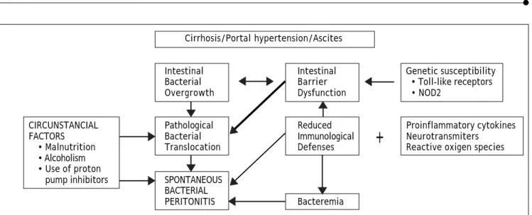
The human gut harbors ten times more microbial cells than eukaryotic cells of the host and bacterial translocation (BT) from the intestinal lumen to re-gional lymph nodes is a physiological process. Pa-thological BT that occurs in cirrhosis is related not only to bacterial overgrowth but also to intestinal barrier dysfunction, qualitative parameters of microbiota and immune dysfunction, with no clear evidence of superiority of one factor over the others. Indeed, recent studies have demonstrated in cirrho-tic patients with sepsis that BT was not caused by an abnormal small bowel gut microbiota.19 Portal
of cirrhosis can accelerate pathological BT mainly due to pro-inflammatory cytokines, as tumor-necro-sis factor, neurotransmitters, as norepinephrine and reactive oxygen species.20 Systemic cytokinemia might
affect the structural and functional integrity of intestinal tight junctions, increasing paracellular permeability. The state of immunodeficiency found in decompensated cirrhosis induces a persistent acti-vation of immune system cells with production of proinflammatory cytokines. More recently it has been shown that activated intestinal macrophages in patients with cirrhosis release IL6 and NO that may disrupt intestinal barrier function, enhancing per-meability to bacterial products.21 Other factors as
susceptibility genes may also be envolved. In fact, recent studies indicate that gene variants, as Toll-like receptors and NOD2 (nucleotide-binding oli-gomerization domain 2) linked to impaired mucosal barrier function represent gentic risk factor for SBP and other infections in cirrhosis.22,23 Although the
presence of microorganisms transported by bacterial translocation can be solved by opsonization of bacte-ria in the AF or by other forms of phagocytosis, when these defense mechanisms are exhausted the infectious process develops.24 Thus, viable and
non-viable bacteria and bacterial products such as endo-toxins/lipopolysaccharides and bacterial DNA that have crossed the intestinal barrier are found in me-senteric lymph nodes. These are what are known as the intrinsic factors, which are associated with cir-rhosis itself .
The circumstantial factors that facilitate the de-velopment of bacterial infections in cirrhotic pa-tients, especially those with some type of decompensation, include malnutrition, which is very
common in cirrhosis of any etiology, chronic alcoho-lism and more recently the use of acid suppressive therapy has benn associated with development of SBP and other infections. Proton pump inhibitors, widely used in cirrhosis, facilitates enteric bacterial overgrowth and translocation, as demonstrated in clinical studies25 (Figure 1). Medical procedures
with iatrogenic potential, such as those involving catheters or probes, as well as other invasive proce-dures13 are the main iatrogenic factors that can
trig-ger bacterial infections (Table 1).
SPONTANEOUS BACTERIAL PERITONITIS
SBP is the most common, severe infection that develops in cirrhotic patients with ascites. SBP is defined as infection of the AF in the absence of an intra-abdominal focus of infection. During a one-year follow-up, 10% of cirrhotic patients with asci-tes are likely to develop SBP. Furthermore, after a first episode, SBP can recur frequently if infection prophylaxis is not administered.15,26
Cirrhosis/Portal hypertension/Ascites
Intestinal Intestinal Genetic susceptibility
Bacterial Barrier • Toll-like receptors
Overgrowth Dysfunction • NOD2
CIRCUNSTANCIAL Pathological Reduced Proinflammatory cytokines
FACTORS Bacterial Immunological Neurotransmiters
• Malnutrition Translocation Defenses Reactive oxigen species • Alcoholism
• Use of proton
pump inhibitors SPONTANEOUS BACTERIAL
PERITONITIS Bacteremia
Figure 1. Pathogenesis of spontaneous bacterial peritonitis.
+
Table 1. Infections in liver cirrhosis.
Predisposing iatrogenic factors
• Intravenous catheters (Gram+, Gram-, Candida)
• Urinary catheters (Gram-; Enterococcus)
• Endoscopic sclerotherapy of varices → bacteremia (Gram+ ® S. viridans)→ 5-30%
• Diagnostic or therapeutic paracentesis (Gram -/+) • Ligation of esophageal varices – rare
Diagnosis
The signs and symptoms of SBP are sometimes unapparent, making it necessary to always bear in mind that this type of infection may be present so that an early diagnosis can be made. Late diag-nosis significantly worsens the patient’s progdiag-nosis as it delays the start of treatment. In fact, around 10 to 30% of cases of SBP are asymptomatic. It is for this reason that paracentesis of AF is perfor-med in all hospitalized cirrhotic patients with as-cites.9 In the community, cirrhotic patients with
ascites but without the signs and symptoms of any complications do not need to have this test. The commonest clinical manifestations of SBP in-clude enlargement of ascitis , failure of diuretic treatment, the onset of hepatic encephalopathy or, less frequently, abnormal laboratory findings indi-cating leukocytosis, metabolic acidosis or kidney dysfunction. Minor increases in temperature (37.8 °C) or abdominal pain, with or without a positive rebound test, may be present. When paralytic ileus, arterial hypotension or hypothermia are present, the infection is at a severe stage and the prognosis is poor.27
SBP is diagnosed by analyzing AF collected by paracentesis. The mandatory test is a poly-morphonuclear(PMN) count, for which values > 250 cell/mm3 indicate a positive diagnosis. If SBP is
suspected the flowchart in figure 2 is useful to con-firm the diagnosis and criteria for indications of the-rapy. AF culture is usually positive in 35 to 65% of patients. It should always be collected and put direc-tly into blood culture flasks at the bedside, increa-sing positivity to 70 or 90%.28 It can be useful to
collect peripheral blood for blood cultures at the same time, as the same microorganism is frequently found in AF and peripheral blood. Although various studies involving other laboratory tests have been carried out, these tests do not contribute to a diag-nosis of SBP. Hence, the usefulness of measuring AF pH and lactate levels, as well as, more recently, AF lactoferrin in the diagnosis of SBP has not been confirmed.29-31
Procalcitonin (PCT) and C reactive protein (CRP) are two acute-phase serum proteins commonly used as early markers of infection in general population. But as both are produced by hepatocytes, patients with cirrhosis may present reduced levels32
Never-theless, the predictive power of CRP and PCT for
de-Figure 2. Flowchart for the diagnosis and treatment of spontaneous bacterial peritoinitis.
Hospitalized cirrhosis with ascitis / Paracentesis for diagnosis Risk factors for SBP
Any complication of cirrhosis Child C / Upper gastrointestinal bleeding /
Previous history of SBP / Malnutrition A.F. with T. Prot ≤ 1.5 g/dL / Use of proton pump inhibitors
Normal PMN/Negative culture PMN < 250/mm3 PMN > 250/mm3 Very high PMN / Polymicrobial culture/
Glucose < 50 mg/dL / T.Protein > 1 g/dL / LDH ↑
Positive culture Negative culture Positive culture Secondary
Bacteria ascites CNNA SBP bacterial peritonitis
-repeat paracentesis in 48
h-PMN < 250/mm3 PMN > 250/mm3
No treatment Treatment CT localized CT no localized
Follow-up Cefotaxima 2 g-12/12 h - 5 to 10 days lesion lesion
Albumin - 1.5 mg/kg day 1 and 1 g/kg day 3 if creatinine > 1 mg/dL and/or
tecting infection has been similar in patients with and without cirrhosis33 Besides that, as a surrogate
marker of systemic inflammatory response syn-drome (SIRS) CRP was recently shown to predict short-term mortality in patients with cirrhosis, most of them with bacterial infections.34
Various studies suggest the use of urine dipsticks, which indicate leukocyte esterase activity of ac-tivated granulocytes by an immediate color change on the strip. Their use to determine the increase in neutrophils in AF has been studied and has proved to have good accuracy. There are various controlled studies showing a sensitivity of around 85%, but these findings have yet to be confirmed.35 Because
they are fast and low-cost, dipsticks can be used when the therapeutic decision is made, but neutro-phil count in the same fluid continues to be the gold standard for therapy. A new strip calibrated specifi-cally for ascitic fluid was recently tested in 1,304 experiments with a median PMN count of 492 cell/mm3 showing good sensibility and negative
predictive value (100%) but specificity of 58% and positive predictive value of 76%.36
With regard of the identification of the bacterial pathogen, real-time PCR assays may be of potential utility, but compared to standard culture techniques the information is not entirely interchangeable.37
More recently, the application of a direct susceptibi-lity testing based on a Matrix Assisted Laser Des-orption Ionization-Time of Flight (MALDI-TOF) from positive blood cultures has been proposed for early detection of resistant bacteria and their anti-biotic susceptibility.38
It is worth remembering that there are two va-riants of SBP: culture-negative neutrocytic ascites (CNNA) and bacterascites (BA). In the former, the PMN count is > 250 cell/mm3 but the AF culture is
negative. This group of patients presents with the same signs and symptoms and has the same progno-sis as those with a positive culture. Other clinical causes of increased AF PMN count, although less frequently associated with ascites and cirrhosis, need to be evaluated, namely, peritoneal carcinoma-tosis, peritoneal tuberculosis, hemorrhagic ascites and pancreatitis.39,40
In BA, conversely, bacterial growth is observed in AF culture, but PMN counts are below 250 cell/mm3. This group is heterogeneous and may not
be infected. If left untreated, spontaneous remis-sion occurs in 60% of cases, particularly asympto-matic ones. If there are symptoms or signs that are compatible with SBP, the patient must be treated, even if there is no increase in PMN count in AF.
As BA is diagnosed about two days after paracente-sis because of the waiting time for culture results, asymptomatic patients should undergo another paracentesis. If there is an elevated neutrophil count, treatment should be administered. When PMN count remains bellow 250 cell/mm3,frequently
the culture is no longer positive and no treatment should be given.41
When infection of AF in cirrhotic patients is se-condary to intestinal perforation or abscesses in ab-dominal organs, the process is known as secondary bacterial peritonitis (2BP), known to correspond to less than 10% of the cases of peritonitis with ascites. The clinical features of secondary bacterial peritoni-tis may be similar to those of SBP, but local inflam-matory response is more severe and mortality is higher.42 While the latter is treatable only with
anti-biotics, 2BP usually requires surgical intervention (Figure 2). An important factor in the differential diagnosis is that 2BP is usually polymicrobial whe-reas only one microorganism is isolated in SBP.43
Laboratory findings, such as AF low glucose levels, high LDH and total protein levels are known as the Runyon’s criteria for diagnosis of 2BP44 Elevated
alkaline phosphatase or carcinoembryonic antigen levels in the AF analysis, have also been used in this differential diagnosis with varying degrees of reliability.44,45 In cases where 2BP is suspected,
antibiotic therapy must also cover anaerobic micro-organisms and enterococci. Due to the high mortali-ty rate in less decompensated cirrhosis a more aggressive approach including imaging tests as abdominal computed tomography must be carried out for early surgical approach that can improve prognosis.42
Therapy
Treatment of SBP is empirical and should be started immediately following diagnosis. If required, treatment can be modified when the results of the culture and the antibiogram are available. As around 70% of cases of SBP are caused by Gram negative bacteria (GNB) and a minority by gram po-sitive bacteria (GPB), third-generation cephalospo-rins, which cover around 95% of the germs involved in this condition, are considered the treatment of choice46 (Figure 2). This form of therapy is effective
such as ceftriaxone and ceftazidine, as well as amoxicillin and clavulanic acid, are effective.49 The
quinolones have also been tested, and both cipro-floxacin and ocipro-floxacin yielded good results.50 In
un-complicated SBP, i.e., SBP without gastrointestinal bleeding, hepatic encephalopathy, ileus or renal fa-ilure, oral quinolone treatment (ofloxacin) can be considered.51 However, aminoglycosides should
be avoided as they are less effective in treating SBP and have high rates of nephrotoxicity.52
The widespread use of quinolones, including for SBP prophylaxis, has led to an increase in quinolone-resistant infections. As greater control was gained over Gram-negative germs, the frequency of SBP due to Gram-positive germs increased. Nevertheless, cephalosporins are still effective in the treatment of infectious episodes.13
Antibiotic resistance has become a problem with the decrease of therapeutic options and poor clinical outco-mes.53 Multi-drug resistance (MDR) has developed,
in-cluding an increased prevalence of methicillin resistant Staphylococcus aureus (MRSA) , MDR enterococci, ex-tended spectrum beta-lactamase (ESBL) producing Echerichia coli and Klebsiella penumoniae.54
A recent study found a significant percentage of multiresistant bacteria in cirrhotic patients and pro-posed the use of broad-spectrum antibiotics such as carbapenems and glycopeptides when treating noso-comial SBP. In this study of 92 episodes of infections, 18% were caused by multi-resistant germs.55 Risk
factors for this resistance include having used anti-biotics in the previous three months, the presence of diabetes and nosocomial SBP.56
Making an early diagnosis and starting appro-priate treatment as soon as possible is not suffi-cient; in addition, close attention must be paid to the patient’s condition in order to avoid other com-plications, which could worsen the prognosis. Asci-tes is the risk factor for the development of bacterial infection, which can lead to renal failure in around 30% of cases. As renal failure can greatly increase the likelihood of mortality in cirrhotic patients, some authors investigated the possibility of reducing this risk by expanding plasma volume with albumin (1.5 g/kg when SBP is diagnosed and a further 1 g/kg on the third day of treatment) in a controlled study with patients who were using cefo-taxime. There was a reduction in both renal failure and mortality (from 29 to 10%).57 A more recent
stu-dy established that the use of albumin as coadjuvant therapy for SBP to prevent renal failure should be limited to more serious cases of cirrhosis, with total bilirubin ≥ 4 mg% or BUN > 30 mg/dL or creatinine
> 1 mg/dL58 (Figure 2). In a recent study,
hospitali-zed cirrhotic patients with other infections than SBP were randomized to receive only antibiotics or antibiotics plus albumin in similar doses. Associa-tion of albumin has shown beneficial effects on the renal and circulatory function and a potential survival benefit.59
DIFFERENT INFECTIONS IN DECOMPENSATED CIRRHOSIS
• Urinary tract infections (UTI) rival SBP in terms of frequency and share most of the etiopa-thogenic mechanisms. Like SBP, they usually re-cur in the same patient and require a variety of different medical measures. Their incidence is higher in females than in males, and patients are usually asymptomatic, presenting only with bac-teriuria.9,60,61 Although prophylaxis for SBP is
also effective for urinary infections, there is usually cross-resistance to antibiotics because of the predominance of Gram-negative cocci in patients who have not received prophylaxis and the development of Gram-positive infections in patients being treated. The best approach when there is UTI, is to base the choice of therapy on culture results and susceptibility data and moni-tor its effectiveness in successive tests. Figure 3 shows a flowchart for diagnosis and treatment of both lower and upper urinary tract infections in cirrhosis, with the first choice antibiotics and other possibilities.
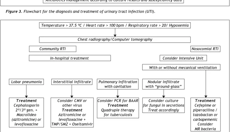
• Respiratory tract infections (RTI), although less frequent, are usually more serious, carrying the highest risk of mortality among the prevalent infections in cirrhosis.62 They are diagnosed
cli-nically or by radiological means, since blood cul-tures are seldom positive and culture of secretions is not easily obtained. A much broader spectrum of microorganisms cause these infections, the main causative organism being Streptococcus pneumoniae. Anaerobic germs or even GNB, such as K. pneumoniae, can also be found. Cirrhotic patients with hydrothorax can develop spontaneous bacterial empyema, which is consi-dered to have the same physiopathological me-chanism as SBP.63 Cephalosporins are usually
Figure 3. Flowchart for the diagnosis and treatment of urinary tract infection (UTI). Clinical symptoms, urinalysis and urine culture Leukocytes > 10/field – culture > 105 colonies/mm3
Assymptomatic bacteriuria Non-complicated Pyelonephritis and upper UTIs Cystitis and lower UTIs
Chronic bladder Treatment Treatment
colonization without infection Quinolones (1st choice) Cefotaxime/ceftriaxone (1st choice) Amoxaciline/clavulanic Piperacilina-tazobactan
Cephalosporines Imipenem-cilastatina Nitrofurantoine
No treatment
Antibiotics management according to culture results and susceptibility data
Figura 4. Flowchart for the diagnosis and treatment of respiratory tract infection (RTI).
Temperature > 37.5 ºC / Heart rate > 100 bpm / Respiratory rate > 20/ Hypoxemia
Chest radiography/Computer tomography
Community RTI Nosocomial RTI
In-hospital treatment Consider Intensive Unit
With or without mecanical ventilation
Lobar pneumonia Interstitial infiltrate Pulmonary infitration Nodular infiltrate with cavitation with “ground-glass”
Treatment Consider CMV or Consider PCR for BAAR Consider culture Treatment
Cephalosporin other virus Treatment for fungui in secretions Cefepime or
2º/3º gen. + Treatment Quadruple therapy Treat accordingly piperacilina /
Macrolideo Azitromicine or for tuberculosis tazobactan or
(azitromicine) or levofloxacine + carbapemenic
levofloxacine TMP/SMZ + Oseltamivir Consider
MR bacteria
• Skin and soft tissue infections, such as cellu-litis and lymphangitis, are relatively frequent, particularly in the lower extremities of cirrhotic patients with edemas. When detected and treated in time, the prognosis is usually good, particu-larly if they are community acquired. Although such infections are frequently resolved, in a re-cent retrospective case-control study there was a higher percentage of patients with renal failure (21.3 vs. 5.4%) and hyponatremia (40 vs. 20%),
and three-month mortality rates were also higher (23 vs. 4%).64 The most frequently found
• Infections related to catheters and other pro-cedures can also occur in decompensated cirrho-tic patients while they are hospitalized. Measures to avoid these iatrogenic infections must be follo-wed strictly; these include systematic hand hy-giene, the use of chlorhexidine for all skin preparations and full-barrier precautions during catheter insertion or other procedures (Table 2). A recent study has shown that second infections, largely preventable as RTI due to aspiration or UTI related to catheters were linked to higher mortality rates.16 Among them fatality rate was
higher for Clostridium difficile infections. Diffi-cult of treat infections as Clostridium difficillis have been treated with fecal microbiota trans-plantation.65 A meta analysis of eleven studies
with 273 patients has shown 89.7% of resolutions. So, fecal microbiota transplant holds conside-rable promise as therapy for these difficult to treat infections but follow-up registries are still needed.66
• Bacteremia and septicemia develop more fre-quently in decompensated cirrhosis.67 Outbreaks
of bacteremia manifest as a minor increase in body temperature and general malaise and are confirmed by blood culture. The clinical and la-boratory findings can be those of spontaneous or procedure-related transient bacteremia. However, whenever bacteria are in the general circulatory system, there is a risk of systemic inflammatory response syndrome (SIRS) developing, with a
cli-nical and laboratory picture of septicemia. Sepsis is particularly serious in cirrhotic patients and should always be suspected so that therapeutic measures can be taken immediately, as the con-dition can progress to multiple organ failure as a result of the immunological dysfunction inherent to cirrhosis.67
Diagnosis of SIRS and severe sepsis can be more difficult in decompensated cirrhotic patients because of their low baseline blood pressure secondary to hy-perdynamic circulation in the more advanced stages of cirrhosis. By promoting the release of cytokines into the circulatory system, the bacterial infection worsens splanchnic and systemic vasodilations, which are already present because of portal hypertension. This increased vasodilation leads to a reduction in effective arterial blood volume and activation of the neurohormonal (rennin-angiotensin-aldosterone) system, causing vasoconstriction and renal failure. Renal failure in turn results in elevated levels of inflammatory cytokines such as TNF-α and inter-leukin-6 and vasodilatory hormones such as nitric oxide (NO). A vicious circle of progressive altera-tions, including cirrhotic cardiomyopathy, hepatic encephalopathy, coagulopathy and other dysfunctions in organs or systems, is thus established, characte-rizing multiple organ failure.67 As in non-cirrhotic
patients, in cirrhosis there is a direct association between the number of failing organs or systems and lethality.68
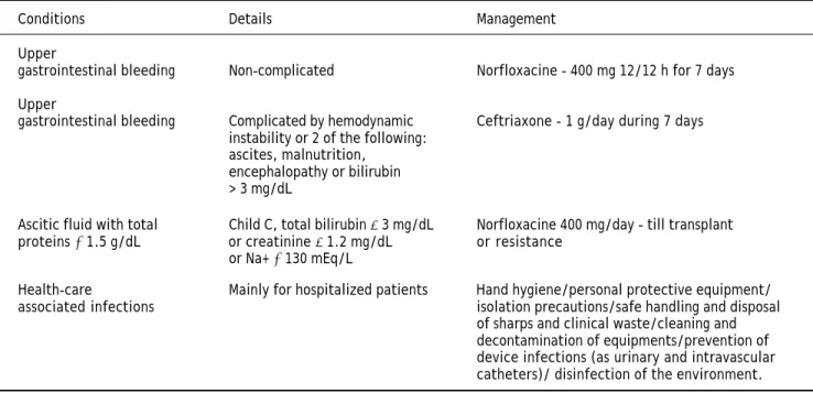
Table 2. Primary prophylaxis of bacterial infections in cirrhosis.
Conditions Details Management
Upper
gastrointestinal bleeding Non-complicated Norfloxacine - 400 mg 12/12 h for 7 days
Upper
gastrointestinal bleeding Complicated by hemodynamic Ceftriaxone - 1 g/day during 7 days instability or 2 of the following:
ascites, malnutrition, encephalopathy or bilirubin > 3 mg/dL
Ascitic fluid with total Child C, total bilirubin ≥ 3 mg/dL Norfloxacine 400 mg/day - till transplant proteins ≤ 1.5 g/dL or creatinine ≥ 1.2 mg/dL or resistance
or Na+ ≤ 130 mEq/L
Health-care Mainly for hospitalized patients Hand hygiene/personal protective equipment/ associated infections isolation precautions/safe handling and disposal
Some infected cirrhotic patients with sepsis deve-lop adrenal dysfunction. Its pathogenesis is complex and poorly understood. The majority develop reversi-ble dysfunction due to decreased production of corti-sol or ACTH. Although high doses of corticosteroids are detrimental to septic shock patients, low doses remain controversial.69-71
PROPHYLAXIS FOR BACTERIAL INFECTIONS IN CIRRHOSIS AND SBP
If one accepts the bacterial translocation theory, the intestines are the main source of bacteria which migrate to regional lymph nodes, ascites and gene-ral circulatory system causing SBP and other GNB infections in cirrhosis. Thus, to avoid these infections, the ideal agent should be safe, minimally absor-bed and effective. Additionally, it should eliminate GNB while preserving GPB and anaerobic flora, a process known as selective intestinal decontamina-tion. Oral administration of poorly absorbed anti-biotics has been shown to be effective in preventing a relapse of SBP(72) and has been used for around two decades. Nevertheless, such antibiotics should only be indicated for cirrhotic patients who really need them because of the risk of developing resis-tant bacteria. Below, we discuss three conditions for which the use of prophylactic antibiotics is recom-mended to prevent the onset of infections and thus improve the survival of patients with decompensated cirrhosis.
Antibiotic therapy in variceal upper gastrointestinal bleeding (UGIB)
It is estimated that 20% of cirrhotic patients with UGIB present with bacterial infections on admission to hospital and that around 50% develop them while they are hospitalized.73 Worsening portal
hyper-tension and a greater risk of the recurrence of hemorrhage were observed in patients with UGIB who presented with infectious complications. The development of infections in this group of patients is associated with a fivefold increase in the risk of a re-currence of bleeding, with an adverse impact on sur-vival.74,75
The main bacterial infections that must be sys-tematically investigated in cirrhotic patients with UGIB are urinary tract infection, SBP, respira-tory tract infection and spontaneous bacteremia. All cirrhotic patients hospitalized for UGIB must be screened for infections with blood cultures, pa-racentesis of AF for neutrophil count and culture
(for the latter AF is collected directly in blood culture flasks), urine sediment analysis and a chest X-ray.9,76
The use of antibiotic prophylaxis has been asso-ciated with a reduced frequency of infectious events and improved survival,76,77 resulting in a decreased
risk for infections and mortality in patients treated with this prophylaxis of 58% and 29%, respective-ly.77 Various prophylactic schemes have been
advo-cated, but the most frequently used are oral quinolones, particularly norfloxacin administered in a 400 mg dose twice a day for seven days.78 Another
controlled study was carried out comparing antibio-tic prophylaxis with ceftriaxone (1g IV/day) and nor-floxacin (400 mg twice daily) for seven days in cirrhotic patients with UGIB with at least two of the following criteria: ascites and/or malnutrition and/or encephalopathy and/or bilirubin > 3 mg/dL. The authors reported a significant reduction in the frequency of infections (11 vs. 26%) and SBP (2 vs. 12%) in the group treated with ceftriaxone.79 Table 2
summarizes the main indications and the drugs most commonly used in primary prophylaxis of bacterial infections in cirrhosis.
Prophylaxis for relapse of SBP
After a first episode SBP frequently recurs, with relapses in 43% of patients in the first 6 months and 68% in a year. Prophylaxis is therefore recommended for any patient recovering from an episode of SBP, as it has been shown that recurrence falls from 68 to 20% and that the probability of developing SBP due to GNB falls from 60 to 3%.72 Quinolones
(norfloxa-cin) are the drug of choice and are indicated for these cirrhotic patients in doses of 400 mg/day to be taken continuously until the ascites resolves or the patient has a transplant.80 As alternative prophylactic drugs,
ciprofloxacin or sulfamethoxazole and trimethoprim are also indicated.15,81 More recently the use of
rifaxi-min, a poorly absorbable antibiotic with low risk of bacterial resistance is been indicated,82 and a
retros-pective analysis comparing its use with norfloxacine has shown better results,83 that should be confirmed
in prospective studies.
Ascites with low protein levels
allowing infections to develop more easily. In cases with AF protein levels > 1 g/dL the prevalence of SBP in two years was zero, while the corresponding figure in patients with AF protein levels < 1g/dL reached 20%.84 Although some controlled studies
failed to provide convincing evidence to justify this approach in all cirrhotic patients with low AF pro-tein levels, recent investigations recommend prophylaxis with 400 mg/day of norfloxacin when patients with decompensated cirrhosis are Child C, with total bilirubin ≥ 3 mg/dL or creatinine ≥ 1.2 mg/dL or Na+ ≥ 130 mEq/L.85
Alternative non-antibiotic therapies, such as pro-biotics, prokinetic agents and supplementation with oral bile acids have been proposed. Probiotics would modulate gut microflora, stabilize mucosal barrier function and restor neutrophyl phagocytic capaci-ty.86 It has already been shown to be effective for
other clinical conditions,87 but must be evaluated
carefully before being adopted. A recent trial has shown that the addition of probiotics to norfloxacin does not improve its efficacy in primary or seconda-ry prophylaxis of SBP.88 As β-adrenergic blockers
shorten intestinal transit time with anti-bacterial effects some studies using propranolol have been carried out and a meta-analysis has shown a relative risk reduction of 12% in the development of SBP.89
Combination of norfloxacin with cisapride has also been evaluated.90
In conclusion, there has been great progress in the control of bacterial infections in decompensated cirrhosis in recent decades. Various controlled cli-nical studies reviewed according to the strict crite-ria of evidence-based medicine have proved that prophylactic and therapeutic measures have mana-ged to reduce morbidity and increase survival in cirrhotic patients even in the decompensated sta-ges.61,91 However, problems remain to be solved in
terms of both therapy and prophylaxis. With the advent of resistance to commonly used antibiotics92
and the recent description of multiresistant bacte-ria,93 there is a need for stricter control of the
administration of antibiotics to cirrhotic patients. Further research in this area is also required. Great care must be taken when using antibiotic prophylaxis and the agreed limits must not be exceeded. To sum up, this body of knowledge about therapy and prophylaxis for bacterial infections in cirrhotic patients, which undoubtedly helps to improve survival, cannot continue to be restricted to specialists but should be disseminated and applied in clinical practice for the benefit of the population at large.
REFERENCES
1. Hytiroglou P, Snover DC, Alves V, Balabaud C, Bhathal PS, Bioulac-Sage P, Crawford JM, et al. Beyond “cirrhosis”: a proposal from the International Liver Pathology Study Group. Am J Clin Pathol 2011; 137: 5-9.
2. Pinzani M. Exploring beyond cirrhosis. Hepatology 2012; 56: 778-80.
3. Albilllos A, Garcia-Tsao G. Classification of cirrhosis: the clinical use of HVPG measurements. Dis Markers 2011; 31: 121-8.
4. D’Amico G. Natural History of Cirrhosis. In: AASLD (ed.). Cirrhosis: current challenges and futures directions. San Francisco, California: AASLD; 2011, p. 26-9.
5. Wiest R, Garcia-Tsao G. Bacterial translocation (BT) in ci-rrhosis. Hepatology 2005; 41: 422-33.
6. Arvaniti V, D’Amico G, Fede G, Manousou P, Tsochatzis E, Pleguezuelo M, Burroughs AK. Infections in patients with cirrhosis increase mortality four-fold and should be used in determining prognosis. Gastroenterology 2010; 139: 1246-56, 1256 e1241-1245.
7. Strauss E. Bacterial infections associated with hepatic en-cephalopathy: prevalence and outcome. Ann Hepatol
2003; 2: 41-5.
8. Navasa M, Fernandez J, Rodes J. Bacterial infections in liver cirrhosis. Ital J Gastroenterol Hepatol 1999; 31: 616-25. 9. Caly WR, Strauss E. A prospective study of bacterial
in-fections in patients with cirrhosis. J Hepatol 1993; 18: 353-8.
10. Fernandez J, Gustot T. Management of bacterial infections in cirrhosis. J Hepatol 2012; 56(Suppl.): S1-S12.
11. Bittencourt PL, Farias AQ, Strauss E, Mattos AA. Variceal bleeding: consensus meeting report from the Brazilian So-ciety of Hepatology. Arq Gastroenterol 2010; 47: 202-16. 12. Moreau R, Jalan R, Gines P, Pavesi M, Angeli P, Cordoba J, Durand F, et al. Acute-on-chronic liver failure is a distinct syndrome that develops in patients with acute decompen-sation of cirrhosis. Gastroenterology 2013; 144: 1426-37. 13. Fernandez J, Navasa M, Gomez J, Colmenero J, Vila J, Arroyo V, Rodes J. Bacterial infections in cirrhosis: epide-miological changes with invasive procedures and norfloxa-cin prophylaxis. Hepatology 2002; 35: 140-8.
14. Garcia-Tsao G. Bacterial infections in cirrhosis: treat-ment and prophylaxis. J Hepatol 2005; 42(Suppl.): S85-92. 15. Strauss E, Caly WR. Spontaneous bacterial peritonitis: a
the-rapeutic update. Expert Rev Anti Infect Ther 2006; 4: 249-60. 16. Bajaj JS, O’Leary JG, Reddy KR, Wong F, Olson JC, Subra-manian RM, Brown G, et al. Second infections independen-tly increase mortality in hospitalized patients with cirrhosis: the North American consortium for the study of end-stage liver disease (NACSELD) experience. Hepatology
2012; 56: 2328-35.
17. Rimola A, Soto R, Bory F, Arroyo V, Piera C, Rodes J. Reti-culoendothelial system phagocytic activity in cirrhosis and its relation to bacterial infections and prognosis. He-patology 1984; 4: 53-8.
18. Rajkovic IA, Williams R. Abnormalities of neutrophil phago-cytosis, intracellular killing and metabolic activity in alco-holic cirrhosis and hepatitis. Hepatology 1986; 6: 252-62. 19. Steed H, Macfarlane GT, Blackett KL, Macfarlane S, Miller
MH, Bahrami B, Dillon JF. Bacterial translocation in cirrho-sis is not caused by an abnormal small bowel gut microbio-ta. FEMS Immunol Med Microbiol 2011; 63: 346-54. 20. Bellot P, Frances R, Such J. Pathological bacterial
21. Du Plessis J, Vanheel H, Janssen CE, Roos L, Slavik T, Sti-vaktas PI, Nieuwoudt M, et al. Activated intestinal macro-phages in patients with cirrhosis release NO and IL-6 that may disrupt intestinal barrier function. J Hepatol 2013; 58: 1125-32.
22. Appenrodt B, Grunhage F, Gentemann MG, Thyssen L, Sauerbruch T, Lammert F. Nucleotide-binding oligomeriza-tion domain containing 2 (NOD2) variants are genetic risk factors for death and spontaneous bacterial peritonitis in liver cirrhosis. Hepatology 2012; 51: 1327-33.
23. Bruns T, Peter J, Reuken PA, Grabe DH, Schuldes SR, Bren-moehl J, Scholmerich J, et al. NOD2 gene variants are a risk factor for culture-positive spontaneous bacterial pe-ritonitis and monomicrobial bacterascites in cirrhosis. Li-ver Int 2011; 32: 223-30.
24. Bernardi M. Spontaneous bacterial peritonitis: from pa-thophysiology to prevention. Intern Emerg Med 2013; 5(Suppl. 1): S37-44.
25. Trikudanathan G, Israel J, Cappa J, O’Sullivan DM. Associa-tion between proton pump inhibitors and spontaneous bac-terial peritonitis in cirrhotic patients - a systematic review and meta-analysis. Int J Clin Pract 2011; 65: 674-8. 26. Tito L, Rimola A, Gines P, Llach J, Arroyo V, Rodes J.
Recur-rence of spontaneous bacterial peritonitis in cirrhosis: frequency and predictive factors. Hepatology 1988; 8: 27-31.
27. Garcia-Tsao G. Spontaneous bacterial peritonitis: a histo-rical perspective. J Hepatol 2004; 41: 522-27.
28. Runyon BA. Spontaneous bacterial peritonitis: an explo-sion of information. Hepatology 1988; 8: 171-5.
29. Stassen WN, McCullough AJ, Bacon BR, Gutnik SH, Wadiwa-la IM, McLaren C, Kalhan SC, et al. Immediate diagnostic criteria for bacterial infection of ascitic fluid. Evaluation of ascitic fluid polymorphonuclear leukocyte count, pH, and lactate concentration, alone and in combination. Gas-troenterology 1986; 90: 1247-54.
30. Viallon A, Zeni F, Pouzet V, Lambert C, Quenet S, Aubert G, Guyomarch S, et al. Serum and ascitic procalcitonin levels in cirrhotic patients with spontaneous bacterial peritonitis: diagnostic value and relationship to pro-inflammatory cyto-kines. Intensive Care Med 2000; 26: 1082-8.
31. Barreales M, Fernandez I. Spontaneous bacterial peritoni-tis. Rev Esp Enferm Dig 2011; 103: 255-63.
32. Gabay C, Kushner I. Acute-phase proteins and other sys-temic responses to inflammation. N Engl J Med 1999; 340: 448-54.
33. Papp M, Vitalis Z, Altorjay I, Tornai I, Udvardy M, Harsfalvi J, Vida A, et al. Acute phase proteins in the diagnosis and prediction of cirrhosis associated bacterial infections. Li-ver Int 2011; 32: 603-11.
34. Cervoni JP, Thevenot T, Weil D, Muel E, Barbot O, She-ppard F, Monnet E, et al. C-reactive protein predicts short-term mortality in patients with cirrhosis. J Hepatol
2012; 56: 1299-304.
35. Koulaouzidis A, Leontiadis GI, Abdullah M, Moschos J, Ga-sem J, Tharakan J, Maltezos E, et al. Leucocyte esterase reagent strips for the diagnosis of spontaneous bacterial peritonitis: a systematic review. Eur J Gastroenterol He-patol 2008; 20: 1055-60.
36. Mendler MH, Agarwal A, Trimzi M, Madrigal E, Tsushima M, Joo E, Santiago M, et al. A new highly sensitive point of care screen for spontaneous bacterial peritonitis using the leukocyte esterase method. J Hepatol 2010; 53: 477-83.
37. Hardick J, Won H, Jeng K, Hsieh YH, Gaydos CA, Rothman RE, Yang S. Identification of bacterial pathogens in
asci-tic fluids from patients with suspected spontaneous bac-terial peritonitis by use of broad-range PCR (16S PCR) coupled with high-resolution melt analysis. J Clin Microbiol
2012; 50: 2428-32.
38. Bhattacharya S. Early diagnosis of resistant pathogens: how can it improve antimicrobial treatment? Virulence
2013; 4: 172-84.
39. Runyon BA, Hoefs JC. Culture-negative neutrocytic asci-tes: a variant of spontaneous bacterial peritonitis. Hepa-tology 1984; 4: 1209-11.
40. Runyon BA. Management of adult patients with ascites due to cirrhosis: an update. Hepatology 2009; 49: 2087-107.
41. EASL, Gines P, et al. EASL clinical practice guidelines on the management of ascites, spontaneous bacterial perito-nitis and hepatorenal syndrome in cirrhosis. J Hepatol
2010; 53: 397-417.
42. Soriano G, Castellote J, Alvarez C, Girbau A, Gordillo J, Ba-liellas C, Casas M, et al. Secondary bacterial peritonitis in cirrhosis: a retrospective study of clinical and analytical characteristics, diagnosis and management. J Hepatol
2009; 52: 39-44.
43. Runyon BA, Hoefs JC. Ascitic fluid analysis in the differen-tiation of spontaneous bacterial peritonitis from gastroin-testinal tract perforation into ascitic fluid. Hepatology
1984; 4: 447-50.
44. Akriviadis EA, Runyon BA. Utility of an algorithm in diffe-rentiating spontaneous from secondary bacterial perito-nitis. Gastroenterology 1990; 98: 127-33.
45. Evans LT, Kim WR, Poterucha JJ, Kamath PS. Spontaneous bacterial peritonitis in asymptomatic outpatients with cir-rhotic ascites. Hepatology 2003; 37: 897-901.
46. Felisart J, Rimola A, Arroyo V, Perez-Ayuso RM, Quintero E, Gines P, Rodes J. Cefotaxime is more effective than is ampicillin-tobramycin in cirrhotics with severe infections.
Hepatology 1985; 5: 457-62.
47. Rimola A, Salmeron JM, Clemente G, Rodrigo L, Obrador A, Miranda ML, Guarner C, et al. Two different dosages of cefotaxime in the treatment of spontaneous bacterial pe-ritonitis in cirrhosis: results of a prospective, randomi-zed, multicenter study. Hepatology 1995; 21: 674-79. 48. Runyon BA, McHutchison JG, Antillon MR, Akriviadis EA,
Montano AA. Short-course versus long-course antibiotic treatment of spontaneous bacterial peritonitis. A rando-mized controlled study of 100 patients. Gastroenterology
1991; 100: 1737-42.
49. Gomez-Jimenez J, Ribera E, Gasser I, Artaza MA, Del Valle O, Pahissa A, Martinez-Vazquez JM. Randomized trial com-paring ceftriaxone with cefonicid for treatment of spon-taneous bacterial peritonitis in cirrhotic patients.
Antimicrob Agents Chemother 1993; 37: 1587-92. 50. Terg R, Cobas S, Fassio E, Landeira G, Rios B, Vasen W,
Abecasis R, et al. Oral ciprofloxacin after a short course of intravenous ciprofloxacin in the treatment of sponta-neous bacterial peritonitis: results of a multicenter, ran-domized study. J Hepatol 2000; 33: 564-9.
51. Navasa M, Follo A, Llovet JM, Clemente G, Vargas V, Ri-mola A, Marco F, et al. Randomized, comparative study of oral ofloxacin versus intravenous cefotaxime in spon-taneous bacterial peritonitis. Gastroenterology 1996; 111: 1011-17.
53. Salerno F, Cazzaniga M. Therapeutic strategies and emer-gence of multiresistant bacterial strains. Intern Emerg Med 2010; 5(Suppl. 1): S45-51.
54. Canton R, Coque TM, Baquero F. Multi-resistant Gram-ne-gative bacilli: from epidemics to endemics. Curr Opin In-fect Dis 2003; 16: 315-25.
55. Fernandez J, Acevedo J, Castro M, Garcia O, de Lope CR, Roca D, Pavesi M, et al. Prevalence and risk factors of in-fections by multiresistant bacteria in cirrhosis: a pros-pective study. Hepatology 2011; 55: 1551-61.
56. Castellote J, Ariza X, Girbau A, Broquetas T, Lobatón T, Salord S, et al. Factores de riesgo para el desarrollo de una peritonitis bacteriana espontánea secundaria a un germe resistente a la cefotaximea. Gastroenterol Hepa-tol 2010; 33: 143.
57. Sort P, Navasa M, Arroyo V, Aldeguer X, Planas R, Ruiz-del-Arbol L, Castells L, et al. Effect of intravenous albumin on renal impairment and mortality in patients with cirrhosis and spontaneous bacterial peritonitis. N Engl J Med 1999; 341: 403-9.
58. Sigal SH, Stanca CM, Fernandez J, Arroyo V, Navasa M. Restricted use of albumin for spontaneous bacterial peri-tonitis. Gut 2007; 56: 597-9.
59. Guevara M, Terra C, Nazar A, Sola E, Fernandez J, Pavesi M, Arroyo V, et al. Albumin for bacterial infections other than spontaneous bacterial peritonitis in cirrhosis. A ran-domized, controlled study. J Hepatol 2012; 57: 759-65. 60. Burroughs AK, Rosenstein IJ, Epstein O, Hamilton-Miller
JM, Brumfitt W, Sherlock S. Bacteriuria and primary biliary cirrhosis. Gut 1984; 25: 133-7.
61. Taneja SK, Dhiman RK. Prevention and management of bac-terial infections in cirrhosis. Int J Hepatol 2011: 784540. Doi: 10.4061/2011/784540. Epub 2011 Aug 8.
62. Hung TH, Tseng CW, Hsieh YH, Tseng KC, Tsai CC. High mortality of pneumonia in cirrhotic patients with ascites.
BMC Gastroenterol 2013; 13: 13-25.
63. Allam NA. Spontaneous bacterial empyema in liver cirrho-sis: an underdiagnosed pleural complication. Saudi J Gas-troenterol 2008; 14: 43-5.
64. Pereira G, Guevara M, Fagundes C, Sola E, Rodriguez E, Fernandez J, Pavesi M, et al. Renal failure and hyponatre-mia in patients with cirrhosis and skin and soft tissue infec-tion. A retrospective study. J Hepatol 2012; 56: 1040-6. 65. Aroniadis OC, Brandt LJ. Fecal microbiota
transplanta-tion: past, present and future. Curr Opin Gastroenterol
2012; 29: 79-84.
66. Kassam Z, Lee CH, Yuan Y, Hunt RH. Fecal microbiota transplantation for Clostridium difficile infection: syste-matic review and meta-analysis. Am J Gastroenterol
2013; 108: 500-8.
67. Tandon P, Garcia-Tsao G. Bacterial infections, sepsis, and multiorgan failure in cirrhosis. Semin Liver Dis 2008; 28: 26-42.
68. Arabi Y, Ahmed QA, Haddad S, Aljumah A, Al-Shimemeri A. Outcome predictors of cirrhosis patients admitted to the intensive care unit. Eur J Gastroenterol Hepatol 2004; 16: 333-9.
69. Sprung CL, Annane D, Keh D, Moreno R, Singer M, Freivogel K, Weiss YG, et al. Hydrocortisone therapy for patients with septic shock. N Engl J Med 2008; 358: 111-24. 70. Annane D, Sebille V, Charpentier C, Bollaert PE, Francois B,
Korach JM, Capellier G, et al. Effect of treatment with low doses of hydrocortisone and fludrocortisone on mortality in patients with septic shock. JAMA 2002; 288: 862-71. 71. Arabi YM, Aljumah A, Dabbagh O, Tamim HM, Rishu AH,
Al-Abdulkareem A, Knawy BA, et al. Low-dose hydrocortisone
in patients with cirrhosis and septic shock: a randomized controlled trial. CMAJ 2010; 182: 1971-7.
72. Gines P, Rimola A, Planas R, Vargas V, Marco F, Almela M, Forne M, et al. Norfloxacin prevents spontaneous bacte-rial peritonitis recurrence in cirrhosis: results of a do-uble-blind, placebo-controlled trial. Hepatology 1990; 12: 716-24.
73. Deschenes M, Villeneuve JP. Risk factors for the develop-ment of bacterial infections in hospitalized patients with cirrhosis. Am J Gastroenterol 1999; 94: 2193-7.
74. Bernard B, Cadranel JF, Valla D, Escolano S, Jarlier V, Opo-lon P. Prognostic significance of bacterial infection in bleeding cirrhotic patients: a prospective study. Gas-troenterology 1995; 108: 1828-34.
75. Guarner C, Soriano G. Spontaneous bacterial peritonitis.
Semin Liver Dis 1997; 17: 203-17.
76. Bernard B, Grange JD, Khac EN, Amiot X, Opolon P, Poy-nard T. Antibiotic prophylaxis for the prevention of bac-terial infections in cirrhotic patients with gastrointestinal bleeding: a meta-analysis. Hepatology 1999; 29: 1655-61. 77. Soares-Weiser K, Brezis M, Tur-Kaspa R, Leibovici L.
Anti-biotic prophylaxis for cirrhotic patients with gastrointes-tinal bleeding. Cochrane Database Syst Rev 2002: CD002907.
78. Garcia-Tsao G, Bosch J, Groszmann RJ. Portal hyperten-sion and variceal bleeding—unresolved issues. Summary of an American Association for the study of liver diseases and European Association for the study of the liver single-topic conference. Hepatology 2008; 47: 1764-72.
79. Fernandez J, Ruiz del Arbol L, Gomez C, Durandez R, Se-rradilla R, Guarner C, Planas R, et al. Norfloxacin vs cef-triaxone in the prophylaxis of infections in patients with advanced cirrhosis and hemorrhage. Gastroenterology
2006; 131: 1049-56; quiz 1285.
80. Younossi ZM, McHutchison JG, Ganiats TG. An economic analysis of norfloxacin prophylaxis against spontaneous bacterial peritonitis. J Hepatol 1997; 27: 295-8.
81. Rimola A, Garcia-Tsao G, Navasa M, Piddock LJ, Planas R, Bernard B, Inadomi JM. Diagnosis, treatment and pro-phylaxis of spontaneous bacterial peritonitis: a consensus document. International Ascites Club. J Hepatol 2000; 32: 142-53.
82. Kalambokis GN, Mouzaki A, Rodi M, Tsianos EV. Rifaximin for the prevention of spontaneous bacterial peritonitis.
World J Gastroenterol 2012; 18: 1700-2.
83. Hanouneh MA, Hanouneh IA, Hashash JG, Law R, Esfeh JM, Lopez R, Hazratjee N, et al. The role of rifaximin in the primary prophylaxis of spontaneous bacterial peritonitis in patients with liver cirrhosis. J Clin Gastroenterol
2012: 709 -15.
84. Llach J, Rimola A, Navasa M, Gines P, Salmeron JM, Gines A, Arroyo V, et al. Incidence and predictive factors of first episode of spontaneous bacterial peritonitis in cirrhosis with ascites: relevance of ascitic fluid protein concentra-tion. Hepatology 1992; 16: 724-7.
85. Fernandez J, Navasa M, Planas R, Montoliu S, Monfort D, Soriano G, Vila C, et al. Primary prophylaxis of sponta-neous bacterial peritonitis delays hepatorenal syndrome and improves survival in cirrhosis. Gastroenterology
2007; 133: 818-24.
86. Stadlbauer V, Mookerjee RP, Hodges S, Wright GA, Davies NA, Jalan R. Effect of probiotic treatment on deranged neu-trophil function and cytokine responses in patients with compensated alcoholic cirrhosis. J Hepatol 2008; 48: 945-51. 87. Lata J, Jurankova J, Kopacova M, Vitek P. Probiotics in
88. Pande C, Kumar A, Sarin SK. Addition of probiotics to nor-floxacin does not improve efficacy in the prevention of spontaneous bacterial peritonitis: a double-blind placebo-controlled randomized-placebo-controlled trial. Eur J Gastroente-rol Hepatol 2012; 24: 831-9.
89. Senzolo M, Cholongitas E, Burra P, Leandro G, Thalheimer U, Patch D, Burroughs AK. Beta-Blockers protect against spontaneous bacterial peritonitis in cirrhotic patients: a meta-analysis. Liver Int 2009; 29: 1189-93.
90. Sandhu BS, Gupta R, Sharma J, Singh J, Murthy NS, Sarin SK. Norfloxacin and cisapride combination decreases the incidence of spontaneous bacterial peritonitis in cirrhotic ascites. J Gastroenterol Hepatol 2005; 20: 599-605.
91. Chavez-Tapia NC, Barrientos-Gutierrez T, Tellez-Avila F, Soares-Weiser K, Mendez-Sanchez N, Gluud C, Uribe M. Meta-analysis: antibiotic prophylaxis for cirrhotic patients with upper gastrointestinal bleeding - an updated Cochrane review. Aliment Pharmacol Ther 2011; 34: 509-18. 92. Ortiz J, Vila MC, Soriano G, Minana J, Gana J, Mirelis B,
No-vella MT, et al. Infections caused by Escherichia coli re-sistant to norfloxacin in hospitalized cirrhotic patients. Hepatology 1999; 29: 1064-9.