Annals of Hepatology 7(2) 2008: 130-135
130
www.medigraphic.com
Annals of Hepatology 2008; 7(2): April-June: 130-135
Annals of Hepatology
Original Article
Black bean extract ameliorates liver fibrosis in
rats with CCl
4-induced injury
Alberto Gabriel López-Reyes;*1,4 Netzahualcoyotl Arroyo-Curras;1 Beiman G Cano;1 Víctor Javier Lara-Díaz;1 Gustavo E
Guajardo-Salinas;1 Jose F. Islas;1 Vicente Morales-Oyarvide;1 Luis A. Morales-Garza;1 Francisco Javier Galvez-Gastelum;2
Guillermo Grijalva;3 Jorge E. Moreno-Cuevas1
1Tecnológico de Monterrey, School of Medicine, Basic Research
Laboratory. 3000 W. Ave. Morones Prieto, Monterrey, NL, 64710, México
2Institute of Molecular Biology and Gene Therapy, Centro
Universitario de Ciencias de la Salud, University of Guadalajara, Apdo. Postal 2-123, JAL., 44281, México
3Hospital Militar de Guadalajara, JAL., 44281, México 4Laboratorio de Biología y Química Molecular de Alimentos.
Instituto de Ciencias Básicas, Universidad Veracruzana, Xalapa, Ver. México.
Abbreviations: b.w.: body weight; cm3: cubic centimeter; ECM:
extra-cellular matrix; FR: free radicals; g: grams; ITESM: Instituto Tecnológico y de Estudios Superiores de Monterrey: kg: kilograms; μL: microliters; μm: micrometers; i.p.: intraperitoneal; min: minutes; ml: milliliters; HSC: hepatic stellate cells; Rpm: revolutions per minute; %: percent; oC: Celsius degrees.
*1Address for correspondence:
Alberto Gabriel López-Reyes. Av. Rafael Castelaso s/n Colonia Industrial Animas 91190 Xalapa Veracruz México. Phono: (+52) 81 1314 7750 and (52) 55 2888 2651, E-mail:
[email protected]. E-mail addresses:
NAC [email protected] BGC [email protected] VJLD [email protected] GEGS [email protected] JFI [email protected] VMO [email protected] LMG [email protected]
FJGG [email protected] GG [email protected] JEMC [email protected]
Manuscript received and accepted: 2 February and 10 April 2008
Abstract
We assessed the anti-fibrotic effects of methanolic black bean extract antioxidants in a carbon tetrachlo-ride (CCl4) liver injury model in rats. Experimentally intoxicated animals received CCl4 for eight weeks, the reference and test groups received daily intragastric quercetin or daily intragastric black bean extract. Liv-er fibrosis was assessed and quantified using morpho-metric analysis. Expression of fibrosis related genes
was measured by real time RT-PCR. Qualitative and quantitative histological analysis showed that adminis-tration of 70 mg/kg b.w. of black bean extract reduced hepatic fibrosis index by 18% compared to positive controls (P 0.006), as a result of a decrease in type I (44.3% less, P 0.03) and type IV (68.9% less, P 0.049) collagen gene expression compared to CCl4-injured and Quercetin treated rats. In conclusion, we provide evidence that this methanol black bean extract amelio-rates liver fibrosis and types I and IV collagen gene ex-pression, in the animal model used.
Practical applications: The compounds contained in this black bean extract exhibited strong antifibrotic ef-fects in the CCl4 chronic liver injury model used; con-sidering that this compounds are contained in a legu-minous that has been used in human diet for a long time, their toxic potential should be very low, and this characteristic should favor their potential use in some other chronic or degenerative states that include an in-crease in inflammation and oxidative burst in their pathogenesis. Another possible application of this kind of extract could be its use as an antimicrobial or even antiparasitic therapeutic agent, although it is purely speculative.
Key words: Antioxidants, oxidative stress, black bean extract, chronic diseases, liver fibrosis, cirrhosis.
Introduction
Hepatic cirrhosis or liver fibrosis is known to be an ir-reversible distortion of the normal tissue architecture; this alteration develops during chronic liver damage.1 Multiple processes are involved in the progress of this le-sion including: Oxidative stress by free radicals (FR); chronic inflammation mediated in part by the release of pro-inflammatory cytokines from Kupffer cells; and fi-brosis induced by the paracrine action of pro-inflamma-tory and pro-fibrogenic cytokines produced by Kupffer cells and hepatocytes on hepatic stellate cells.2-5 As fi-brosis progresses, quantitative and qualitative changes in extracellular matrix (ECM) composition take place. Total
Artemisa
4
www.medigraphic.com
collagen and non-collagen ECM components increase3 to 5 fold, and the ECM in the sub-endothelialspace changes from a normal low density basement membrane-like matrix, to an interstitial-type matrix rich in collagen fibrils.6 Type I collagen is the most abundant ECM pro-tein in hepatic fibrosis, followed by type IV collagen. Therefore, the regulation of type I and IV collagen genes expression is crucial to understand the pathogenesis of this disease.7
Over the years, many compounds have been studied as possible protectors for liver cirrhosis, like antioxidants, such as flavonoids and other phenolic derivates. These compounds belong to a group of a bigger family known as phytochemicals. They are widely found in fruits and vegetables and while not having an energetic value, are an important part of the human diet.8 Flavonoids exhibit a wide variety of biological properties, including hepato-protection and inhibition of fibrosis.9
Beans are grains that have a high nutritional value due to their protein content. They also contain an elevat-ed concentration of complex carbohydrates, soluble fi-bers, essential vitamins, metals and polyphenols,10,11 such as flavonoids and isoflavones.12 Moreover, dry beans ex-hibit strong antioxidant properties. Hence, it would be important to expand methods to assess the components which present antioxidant and anti-fibrotic activity.13 In recent studies, the effects of quercetin as an antioxidant have been demonstrated in several models of CCl4 liver injury.14
The objective of this investigation is to evaluate the anti-fibrotic effect of a black bean extract, rich in fla-vonoids and poliphenolic compounds, in a rat model of liver cirrhosis induced by CCl4.
Material and methods
Perla black bean:
The Perla black bean was grown and cropped in the ITESM Agricultural Experimental Field in Linares, Nuevo León, México, during the 2004 spring-summer cycle, in rainfall plus supplementary irrigation. The soil was fertil-ized at the moment of planting with urea and triple super-phosphate. No pesticides were used during the cycle.
Antioxidant extraction
A total of 0.1 g of crude biomass obtained from seed coats of Perla black bean was dissolved in 400 mL of H2O, and 100 g of NaCl was added to this solution. The mixture was filtered and collected in an Erlenmeyer flask, where 200 mL of butanol were supplemented and left at room temperature for 30 min in order to achieve a full separation of the organic and aqueous phases. The organ-ic phase was distilled and the butanol recovered. This or-ganic phase was re-suspended in a 2:1 (v/v) solution of
ethyl acetate and methanol, and the salts fully precipitat-ed with 100 ml of ethylic ether. Furthermore, the solution was evaporated until mass dryness was obtained.
Antioxidant preparation
From the latter extraction process, the remaining ex-tract was dissolved in physiological saline solution, ad-justing to final dose of 70 mg/kg b.w., according to the protocol.
Animals and groups
Male Wistar rats (Harlam, Mexico) were housed ac-cording to the Animal Care Protocol established by ITESM. Study animals (80 grams) received three intrap-eritoneal (i.p.) doses of 1 ml (Monday, Wednesday and Friday) of a 1:6 (v/v) mixture of CCl4 and mineral oil on the first week; three doses of a 1:5 mixture on the second week, three of a 1:4 mixture on the third, and three doses of a 1:3 mixture each week on weeks 4 to 8 (15). Four groups of 6 animals were studied. The reference group re-ceived CCl4 i.p. as explained above, and was concomi-tantly treated with daily intragastric quercetin at 70 mg/ kg b.w. daily for eight weeks; the test group received CCl4 and daily intragastric black bean extract at 70 mg/ kg b.w. daily for eight weeks. The positive control group received CCl4 and intragastric saline solution during the same period. The normal group did not receive CCl4, quercetin, nor black bean extract. Surviving animals in all groups were euthanized at the end of the eight week treatment period, two days after the last dose, and their livers were dissected and sectioned. One piece was imme-diately frozen and stored for RNA extraction, and anoth-er section was fixed in 10% formalin.
Morphologic analysis
Sections (approx 0.5 cm3) from the right median and left lobes of each rat liver were fixed and stored in 10% formalin at room temperature. Sections were treated us-ing a Tissue-Tek II automatic processor and then embed-ded in paraffin. Paraffin blocks were cut using a Tissue-Tek Accu-Cut Microtome and 4 mm sections were stained using the Masson Trichromic technique. Images were ob-tained using an Olympus video-microscope, and a quan-titative analysis was done using the Image-Pro Plus soft-ware, version 3.0 (for Windows); 20 random fields were analyzed per slide, and the ratio of connective tissue to the whole area of the liver was calculated.16 The sections were analyzed by 2 independent pathologists.
RNA Isolation
www.medigraphic.com
New Zealand). The tissue was homogenized to 8000 rpm (Tissue Tearor 985370-14, Biospec Products, Inc.) in 500
μL TRIzol (TRIzol reagent®, Molecular Research Center, Inc) and RNA was isolated by chloroform/isopropanol/ ethanol extraction,17 total RNA was treatedwith DNase-I, amplification grade (Invitrogen). RNA concentration and purity was tested by spectrophotometry at 260 and 280 nm (GeneQuant- pro, Amersham Bioscience).
Two-step real time RT-PCR
Total RNA isolated from each rat liver sample was reverse transcripted using Moloney Murine Leukemia Virus Reverse Transcriptase and random primers, ac-cording to the manufacturers protocol (Invitrogen M-MLV Reverse Transcriptase, Part No. 28025.PPS). Forward and reverse LUX primers were designed us-ing Primer Express Software (Invitrogen Life Tech-nology) for β-Actin (housekeeping) forward sense TC-CTAGTCTCAATACGCAG and reverse CGCTCTAT-CACTGGGCATTGG, for type I collagen gene forward sense GTTTCAGTGGTTTGGATGGG and reverse CT-GCCAGGCTCTCCCTTAGGAC, and for type IV col-lagen gene forward sense CCTGCACTTGTAAACAT-AAG and reverse TGTTCACAGTCAAACCACTGCT.18 Target cDNA sequenceswere amplified by quantitative PCR using a fluorescence-basedreal-time detection method (Rotor gene RG 3000, Corbett Research, Aus-tralia). A volume of 12.5 μL of Platinum Quantitative PCR Super Mix-UDG (Invitrogen Life Technology) was used, 2 μL of cDNA, and molecular biology grade water to a total volume of 20 μL per reaction. PCR conditions were: initial temperature of 50°Cfor 2 min-utes and 95°C for 2 minmin-utes followed by 45 cyclesof 95°C for 5 seconds and 60°C for 10 seconds. These conditions were obtained from the Invitrogen protocol for the Corbett Rotor Gene 3000 LUX primers.
Relative amounts of target genes were calculated us-ing the comparative 2∆∆CT method with β-actin as an endogenous reference and RNApooled from livers of normal rats as a calibrator.
Statistical analysis
Numerical data are expressed as mean ± S.D. differ-ences between experimental groups were tested using ANOVA, and when appropriate, one-tailed Student’s t-test. Differences of proportions were assessed by chi-square analysis. Statistical significance was considered for p values < 0.05.
Results
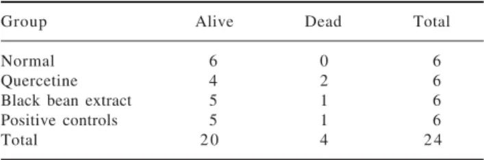
We did not observe any statistically significant differ-ences in the mortality nor survival rates among groups
(Table I).
Histopathological changes
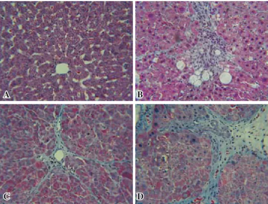
Animals in the normal group showed normal hepatic lobular architecture with central veins and radiating he-patics cords. The positive control, the reference and the test groups all showed evidence of pathological changes, including interstitial fibrosis, inflammatory infiltration, collagen deposition and steatosis. Histological analysis showed that the test group presented a significant reduc-tion of collagen deposireduc-tion (Figure 1).
Quantitative analysis showed that the test group achieved an 18% fibrosis reduction when compared to positive controls (p = 0.006) (Figure 2). The reference group had no significant reduction in fibrosis. The data are showed in Table II.
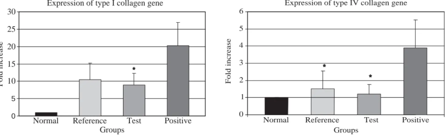
Type I and type IV collagen gene expression
The data regarding type I and IV collagen gene ex-pression are depicted in Table III. Test group showed a decrease of 44.3% in type I collagen gene expression, compared to the positive group (p = 0.030), (Figure 3-A). When we evaluated the expression of type IV collagen gene, the test group had 68.9% less than the positive control group, (p = 0.026) which is statistically signifi-cant, while the reference group also showed a significant decrease (60.8% less) compared to the positive control group (p = 0.049) a difference that is barely significant
(Figure 3-B).
Discussion
Carbon tetrachloride under laboratory conditions is one of the most common and widely used liver in-toxicators today.19 Its action is based on membrane lipid peroxidation and induction of trichloromethyl radical (.CCl3), resulting in severe cell damage.20 An-tioxidants represent a new perspective in liver injury and fibrosis prevention.21 Previous studies have shown that antioxidants, including naringenin, N-acetylcysteine, vitamin E, sylimarin, quercetin and rhein decrease lipid peroxidation and partially ame-liorate liver injury.10,22-26 Extracts obtained using 100% methanol as solvent have demonstrated to pos-sess two of the most important families of antioxi-dants: anthocyanins and tannins.26,27
Table I. Mortality and survival.
Group Alive Dead Total
Normal 6 0 6
4
www.medigraphic.com
Hepatic fibrosis is usually initiated by hepatocyte damage, leading to recruitment of inflammatory cells and platelets, activation of Kupffer cells and subse-quent release of cytokines and growth factors.28 These factors probably link the inflammatory and repair phases of liver cirrhosis by activating hepatic stellate cells (HSC). Previously HSC have been identified as an important cellular source of extracellular matrix (ECM) in liver fibrosis.7 The activated HSC undergo a phenotypic transdifferentiation to contractile myofi-broblast expressing α-smooth muscle actin and ECM.19 It has been shown that lipid peroxidation products in-duce genetic overexpression of fibrogenic cytokines
and increase the synthesis of collagen and initiate the activation of HSC.28
In our experiment, the use of a black bean extract be-longing to a cultivar previously characterized as con-taining a high concentration of antioxidants29 signifi-cantly decreased the expression of type I and IV collagen genes and probably also diminishes the activation of HSC; partially protecting the liver from the CCl4 in-ducedfibrotic effect. Rats treated with the extract had re-duced liver fibrosis by histological examination and a lower expression of type I and IV collagen genes. This effect could be mediated by means of lack of activation of profibrogenic citokine TGFβ, which in turn would not be acting as an activator of the SMAD signaling pathway in HSC, and thereafter the stimulatory effect of this path-way on types I and IV collagen genes would be dimin-ished.30,31 Our results follow the same pattern as the re-sults published in previous works with the use of querce-tin, salacia reticulata and rhein.14,25,26 Since this extract contains a myriad of compounds, and among them is quercetin, we hypothesize that the increased antifibrotic effect found, may be due to the sum of the action of all the flavonoids contained.
Moreover, the cellular response to polyphenols in-volves mainly cell surface receptors and key enzymes as transducers for intracellular signaling, and not a direct physical contact with free radicals. Under such circumstances, the change in redox potential suppos-edly induced by our extract would imply a modifica-tion of the redox state of the hepatocytes, thereby generating changes in the reactions dependent upon
A
A
B
B
C
C
D
D
Figure 1. Histopathological changes. Representative micros-copic photographs, liver stained with Masson Tri-chromic, 40X. A: Normal rat liver, B: Referen-ce (QuerReferen-cetin), C: Test (Black bean extract), D: Positive con-trol (CCl4). Black bean extract
prevented the increase in extra-cellular matrix C, while the in-creased distortion of hepatic ar-chitecture in the positive group D is evident.
Figure 2. Morphological quantitative analysis showed that the use of black bean extract (*) prevented development of fibrosis, when compared against positive group. ANOVA, p < 0.05.
Fibrosis index
Normal Reference Test Positive 0
10 20 30 40 50 60
Groups %
www.medigraphic.com
oxidation-reduction,32 although we did not directly measure this redox potential, and therefore can not assume this as true.
Conclusion
The use of a black bean extract can decrease the ex-pression of type I and IV collagen genes, and reduce the hepatic fibrotic index in a model of CCl4 induced liver fibrosis. Our data warrant further investigation of this ex-tract as a potential treatment in other models of hepatic fibrosis and in the clinical setting.
Competing interests
There were no financial or any other competing inter-est involved in this study.
Authors contributions
ALR conceived and participated in the design of the study, helped and supervised all test and analysis during study, and participated in the writing and corrections of this paper. NAC carried out RNA real time PCR and per-formed CCl4 administration to the study rats. BC carried out the antioxidant extracts administration to the study rats. VJLD performed statistical analysis and participated in the writing and corrections of this paper. GEGS per-formed the organ extraction. JFI perper-formed antioxidant ex-traction system. VMO participated in the RNA exex-traction and the real time PCR. LMG participated in the organ ex-traction and participated in the writing of this paper. GJ and JFGG performed histological preparations and histo-logical analysis. JEMC supervised and participated in the design of the study and the writing of this paper.
Table II. Fibrosis index (%).
Group Mean SD 95% CI Median
Normal 0.85 1.07 0.65, 1.1 0.40 Quercetine 29.80 14.95 26.5, 33.0 27.53 Black bean extract 27.57* 15.31 24.8, 30.3 24.57 Positive controls 33.65 16.76 30.6, 36.7 30.00
* p value < 0.05, one-tailed T test, compared to positive controls.
Table III. Fold increase in collagen genes expression.
Type I Type IV
Group Mean SD 95% CI Mean SD 95% CI Normal 1.00 0.00 NA 1.00 0.00 NA Quercetine 10.39 4.80 5.69, 15.09 1.53* 1.03 0.52, 2.54 Black bean extract 8.93* 3.31 6.03, 11.83 1.21* 0.54 0.74, 1.68 Positive controls 20.16 6.78 14.22, 26.10 3.90 1.63 2.47, 5.33
NA: Not applicable, used as reference to establish fold increase. * p value < 0.05, one-tailed T test, compared to positive controls.
Figure 3 (A, B). Q-PCR analysis of the expression of types I and IV collagen genes. (A) Transcript levels of type I collagen gene (*) p < 0.05, test group compared against positive group; and (B) type IV collagene gene (*) p < 0.05, test and reference group compared against positive group; from the same Q-PCR run using the 2-∆∆CT method. T-test for independent groups, error bars indicate standard de-viation.
Expression of type I collagen gene
Normal Reference Test Positive
0 5
10 15 20 25 30
Groups
Fold
increase *
Expression of type IV collagen gene
Normal Reference Test Positive
0 1 2 3 4 5 6
Groups
Fold
increase
4
www.medigraphic.com
ESTE DOCUMENTO ES ELABORADO POR MEDI-GRAPHIC
Acknowledgements
We express our deepest gratitude to Sergio Serna-Saldivar, PhD, who kindly provided us with the standard-ized extract for this research.
We are indebted to the animal care facilities of the ITESM School of Medicine in charge of Dr. Luis Vazqu-ez-Juarez, for all his support to this project.
References
1. Sokol RJ. Liver cell injury and fibrosis. J Pediatr Gastroenterol Nutr 2002; 35 Suppl 1: S7-10.
2. Tapia G, Pepper I, Smok G, Videla LA. Kupffer cell function in thyroid hormone-induced liver oxidative stress in the rat. Free Radic Res 1997; 26(3): 267-79.
3. Poli G. Pathogenesis of liver fibrosis: Role of oxidative stress.
Mol Aspects Med 2000; 21(3): 49-98.
4. Britton RS, Bacon BR. Intracellular signaling pathways in stellate cell activation. Alcohol Clin Exp Res 1999; 23(5): 922-5. 5. Tsukamoto H, Rippe R, Niemela O, Lin M. Roles of oxidative
stress in activation of kupffer and ito cells in liver fibrogenesis. J Gastroenterol Hepatol 1995; 10 Suppl 1: S50-3.
6. Lee KS, Buck M, Houglum K, Chojkier M. Activation of hepatic stellate cells by TGF alpha and collagen type I is mediated by oxidative stress through c-myb expression. J Clin Invest 1995; 96(5): 2461-8.
7. Friedman SL. Molecular regulation of hepatic fibrosis, an inte-grated cellular response to tissue injury. J Biol Chem 2000; 275(4): 2247-50.
8. Gonzalez-Gallego J, Culebras-Fernández JM, Mataix-Verdú J, Tunon MJ, Sánchez-Campos S. Papel protector de los flavonoides de la cerveza frente a las alteraciones hepáticas inducidas por estrés oxidativo: Estudio de los mecanismos moleculares involucrados. España: Centro de Información cerveza y Salud; 2003 [http:// www.cervezaysalud.com/estudio_12.pdf, cited 01/14/2008]. 9. Middleton Jr E, Kandaswami C, Theoharides TC. The effects of plant
flavonoids on mammalian cells: Implications for inflammation, heart disease, and cancer. Pharmacol Rev 2000; 52(4): 673-751. 10. Lee MH, Yoon S, Moon JO. The flavonoid naringenin inhibits
dimethylnitrosamine-induced liver damage in rats. Biol Pharm Bull 2004; 27(1): 72-6.
11. Beninger CW, Hosfield GL, Nair MG. Flavonol glycosides from the seed coat of a new manteca-type dry bean (phaseolus vulgaris L.). J Agric Food Chem 1999; 47(1): 352.
12. Romani A, Vignolini P, Galardi C, Mulinacci N, Benedettelli S, Heimler D. Germplasm characterization of zolfino landraces (phaseolus vulgaris L.) by flavonoid content. J Agric Food Chem
2004; 52(12): 3838-42.
13. Mazur WM, Duke JA, Wähälä K, Rasku S, Adlercreutz H. Isoflavonoids and lignans in legumes: Nutritional and health as-pects in humans. J Nutr Biochem 1998; 9(4): 193-200. 14. Pavanato A, Tunon MJ, Sanchez-Campos S, Marroni CA, Llesuy
S, Gonzalez-Gallego J, et al. Effects of quercetin on liver damage in rats with carbon tetrachloride-induced cirrhosis. Dig Dis Sci
2003; 48(4): 824-9.
15. Heimler D, Vignolini P, Dini MG, Romani A. Rapid tests to assess the antioxidant activity of phaseolus vulgaris L. dry beans. J Agric Food Chem 2005; 53(8): 3053-6.
16. Garcia L, Hernandez I, Sandoval A, Salazar A, Garcia J, Vera J, et al. Pirfenidone effectively reverses experimental liver fibrosis.
J Hepatol 2002; 37(6): 797-805.
17. Delgado-Rizo V, Salazar A, Panduro A, Armendariz-Borunda J. Treatment with transforming growth factor beta anti-bodies influences an altered pattern of cytokines gene expres-sion in injured rat liver. Biochim Biophys Acta 1998; 1442(1): 20-7.
18. Salazar-Montes A, Delgado-Rizo V, Armendáriz-Borunda J. Dif-ferential gene expression of pro-inflammatory and anti-inflam-matory cytokines in acute and chronic liver injury. Hepatology Research 2000; 16(3): 181-94.
19. Lee KS, Lee SJ, Park HJ, Chung JP, Han KH, Chon CY, et al. Oxidative stress effect on the activation of hepatic stellate cells.
Yonsei Med J 2001; 42(1): 1-8.
20. He SX, Luo JY, Wang YP, Wang YL, Fu H, Xu JL, et al. Effects of extract from ginkgo biloba on carbon tetrachloride-induced liver injury in rats. World J Gastroenterol 2006; 12(24): 3924-8. 21. Mohamed AF, Ali Hasan AG, Hamamy MI, Abdel-Sattar E. An-tioxidant and hepatoprotective effects of eucalyptus maculata.
Med Sci Monit 2005; 11(11): BR426-31.
22. Zhong Z, Froh M, Lehnert M, Schoonhoven R, Yang L, Lind H, et al. Polyphenols from camellia sinenesis attenuate experimental cholestasis-induced liver fibrosis in rats. Am J Physiol Gastrointest Liver Physiol 2003; 285(5): G1004-13.
23. Parola M, Leonarduzzi G, Biasi F, Albano E, Biocca ME, Poli G, et al. Vitamin E dietary supplementation protects against carbon tetrachloride-induced chronic liver damage and cirrhosis.
Hepatology 1992; 16(4): 1014-21.
24. Angulo P, Patel T, Jorgensen RA, Therneau TM, Lindor KD. Silymarin in the treatment of patients with primary biliary cirrho-sis with a suboptimal response to ursodeoxycholic acid.
Hepatology 2000; 32(5): 897-900.
25. Yoshikawa M, Ninomiya K, Shimoda H, Nishida N, Matsuda H. Hepatoprotective and antioxidative properties of salacia reticulata: Preventive effects of phenolic constituents on CCl 4-induced liver injury in mice. Biol Pharm Bull 2002; 25(1): 72-6. 26. Mei-Zi G, Xiao-Sheng L, Hai-Rong X, Zhe-Chuan M, Wei S,
Xiu-Feng Y. Rhein inhibits liver fibrosis induced by carbon tet-rachloride in rats. Acta Pharmacol Sin 2002; 23(8): 739-44. 27. Aparicio-Fernandez X, Yousef GG, Loarca-Pina G, de Mejia E,
Lila MA. Characterization of polyphenolics in the seed coat of black jamapa bean (phaseolus vulgaris L.). J Agric Food Chem
2005; 53(11): 4615-22.
28. Vrba J, Modriansky M. Oxidative burst of kupffer cells: Target for liver injury treatment. Biomed Pap Med Fac Univ Palacky Olomouc Czech Repub 2002; 146(2): 15-20.
29. Salinas-Moreno Y, Rojas-Herrera L, Sosa-Montes E, Pérez-Herrera P. Composición de antocianinas en variedades de frijol negro (Phaseolus vulgaris L.) cultivadas en México. Anthocya-nin composition in black bean (Phaseolus vulgaris L.) varieties grown in Mexico. Agrociencia 2005; 39: 385-394.
30. Said HM, En-Nia A, Mertens P, Gressner AM, Dooley S. YB-1 mediates IFN-ã dependent regulation of TGF-â responsive genes, thereby providing antifibrotic effects. Z Gastroenterol
2004: 42.
31. Chen A. Acetaldehyde stimulates the activation of latent trans-forming growth factor-beta1 and induces expression of the type II receptor of the cytokine in rat cultured hepatic stellate cells.
Biochem J 2002; 368(Pt 3): 683-93.