Carpal-Bone Feature Extraction Analysis in Skeletal Age Assessment Based
on Deformable Model
Pan Lin, Feng Zhang, Yong Yang, Chong-Xun Zheng
Institute of Biomedical Engineering,Xi'an Jiaotong University, Xi’an, China, 710049 [email protected]
ABSTRACT
Skeletal age assessment is one of the important applications of hand radiography in the area of pediatric radiology. Features analysis of the carpal bones can reveal the important information for skeletal age assessment. The present work in this paper faces the problem of the detection of carpal-bone features from its radio-image. A novel and effective segmentation technique is presented in this work with carpal bone image for skeletal age estimation. Carpal bone segmentation is a critical operation of the automatic skeletal age assessment system. This method consists of three procedures. First, the original carpal bone image is preprocessed via anisotropic diffusion. Then, the carpal bone image is segmented by GVF-Snake model. Third, experiments are carried out on images of carpal bone. The results are very promising. In particular the method is able to extract overlapping carpal bones.
Keywords: Carpal-bone radiograph, skeletal age (estimation), Active Contour Model, GVF-Snake.
1. INTRODUCTION
Skeletal age assessment is widely applied to the area of pediatric radiology. There are two respected and widely-used methods for skeletal age assessment from and-wrist radiographs: the GreulichPyle(GP)[1] method and the Tanner-Whitehouse(TW2)method[2]. The doctors must observe the appearance and growth features of specific bones, and hence an empirically derived score, to each bone. The scores are summed to give a net maturity rating, which, by reference to gender-dependent tables, may be converted into a ‘bone age’ or estimated height at adulthood. However, the results of ‘bone age’ assessment highly depend on the doctor expertise and experience. Therefore, it is worthwhile to develop a automatic ‘bone age’ assessment system to assist the doctor in performing a more objective and accurate assessment. Bone age assessment based on image processing can be divided into two phases. Phases one are phalangeal and carpal bone image feature extraction. Phase two is bone age assessment based on the features. Features of the carpal bones (see Fig.1) are important potential information for skeletal age assessment [3,4,5,6] in pediatric radiology. The recognition and separation of
individual carpal bone from the image of the carpal bones is a very difficult task, we face with the presence of several overlapping regions of interest (ROI), the presence of ROI having completely different degree of calcification and overlapping with soft tissue, since different regions are of little grey level variations and the borders of individual carpal bones are hardly visible. A major requirement of the task is an efficient method to segment carpal bones. Many techniques are actually available [3,4]. Techniques based on “classical” segmentation algorithms, such as region growing, edge detection, usually can’t obtain accurate results. The complexity of the skeletal structure in the hand and the variability between different subjects makes very difficult to realize an automatic segmentation of the bones. Therefore, we have explored a new segmentation techniques based on the work of Kass et al [7], which introduces the concept of an active contour model, also commonly called a snakes. Snakes can be represented as energy-minimizing splines guided by external constraint forces and image forces such as lines, edges, subjective contours and region homogeneities found in the image. Furthermore, internal spline forces impose smoothness constrains on the modeled contours. By combining and integrating various types of information found in an image, snakes can lead to results at least comparable to other image segmentation techniques. Later active contours are used extensively in computer vision and image processing applications, particularly to Locate object boundaries. However, this models that have some disadvantages. To make the final result relatively insensitive to the initial conditions, Xu and Prince [8,9] proposed the Gradient Vector Field (GVF) model, which allows for flexible initialization of the snake and helps convergence to boundary concavities. This model has been found to be well adapted to our application, in particular because of this flexible initialization and its good convergence performances compared to other methods. In this paper, we present a novel and effective the carpal bone image segmentation method. This method is able to extract a variety of carpal bones features. In order to improve signal to noise ratio prior to segmentation images anisotropic non-linear diffusion filtering are used. The principle
is to smooth out noise locally by diffusive flow while at the same time prevent flow across object boundaries. Then we use a GVF-Snake model to segment carpal bone from digital X-ray image. The experimental results are very promising.
(a)
[image:2.595.89.278.144.494.2]
(b)
Fig.1 carpal bones image of a hand
The rest of the work is organized as follows: in Section 2 the anisotropic diffusion filter for Image Pre-processing is presented. In section 3 we describe active contour paradigm for image Segmentation and gradient vector flow active contour method are discussed, and finally some experiment results are shown in section 4.
2. CARPAL BONES IMAGE PREPROCESSING
Carpal Bone images can be taken by X-ray. Due to various factors, these images are in general poor in contrast, and image feature in the cross sectional part are often obscured and degraded by artifacts. In order to obtain accurate and reliable feature information we applied image preprocessing to remove artifacts and degradations such as blurring and noise. For this reason we apply an anisotropic diffusion filter that smoothes noisy regions in the image while respecting edge boundaries. The principle is to smooth out noise locally by diffusive
flow while at the same time prevent flow across object boundaries.
Anisotropic diffusion filter was proposed by Perona and Malik(1990)[10].The filtered image is modeled as the solution to the Anisotropic diffusion equation
( , ) div(G(p,t) I(p,t)) t
t p
I = ⋅∇
∂ ∂
(1)
where is the image intensity at position and time .
)
,
(
p
t
I
p
t
∇I is the spatial gradient,∂tI istemporal derivative and is the conductance parameter. If
) , (p t G 1
=
G results in linear diffusion, which has been shown to be equivalent to smoothing the image with a family of Gaussian filter kernels whose scale evolves to t. Instead of this Perona and Malik[9] proposed the use of diffusion coefficients based on a measure of edge strength. The diffusion coefficient then adaptively controls the diffusion strength, smoothing the image within a moderately continuous region while not smoothing across sharp discontinuities. The diffusion process achieves piecewise smoothing while preserving the relevant image edges. The conductivity model suggested in [9] is used in our experiments, thus:
) , (p t G
⎥ ⎥ ⎦ ⎤ ⎢
⎢ ⎣
⎡ ∇
−
=exp ( ( ) ) )
, (
k p I t
p
G (2)
the parameter determines the local behavior of the filter: smoothing if
k
k I ≤
∇ and edge
sharpening if ∇I >k. Edge-preserving smoothing prior to subsequent analysis yielded more robust results. Fig.2 (a) and Fig.2 (b) show the original carpal bone image and the anisotropic smoothing results,respectively.
(a) (b)
Fig.2 anisotropic smoothing of carpal bones image(a) original carpal bone image(b) anisotropic smoothing
results
3. CARPAL BONES IMAGE SEGMENTATION
The goal is to identify the position of each carpal bone. Because the carpal bone image is noisy, the complexity of the skeletal structure in the hand and the variability between different subjects makes very difficult to realize an automatic segmentation of the bones. We propose a novel method, which is based on GVF model, to find the boundary of the carpal bones.
[image:2.595.312.522.507.593.2]3.1 Snakes
A snake in the continuous spatial domain is represented as a 2D parametric contour curve
where )) ( ), ( ( )
(s x s y s
v = s∈[0,1]. In order to fit the snake model to the image data we associate energy terms with the snake and aim to deform the snake in a way that minimizes its total energy. The energy of the snake,
E
, depends on both the shape of the contour and the image data reflected via the internal and external energy terms, and) , (x y I ) (v a ) (v
β , respectively. The total snake energy can be written as: ds s v E s v s v E ext
S2( ( ) ( ) ) ( ( ))
1 ′ 2+ ′′ 2 +
=
∫
α β (3)where and are the first and second derivatives of . The weights
) (s
v′ v′′(s)
) (s
v αandβ control the tension and flexibility of the contour, respectively. The external energy term is computed based on the image information and it drives the active contour to the boundary. In the parametric approach, the general-form energy functional of the active contour. By using the Euler equation it is found that a necessary condition for to minimize the Equation (3) is
v 0 )) ( ( ) ( )
( − 4 −∇ =
′′s v s E v s
v
a β ext (4) 3.2 Gradient Vector Flow Snakes
Despite the snake model is consistency and simplicity, there are still some performance problems. For example, the initial position of the snake must be close to the desired contour in the image, otherwise the snake may not evolve correctly, as it may find local minima away from the contour. This problem can happen every time there are concavities or sharp corners in the gray-level image. To solve this problem several different methods have been proposed. Later Xu and prince [7,8,9] present a new class of external forces for active contour models that address the two problems. This method we called gradient vector flow (GVF). This model produces a field with strong forces near the edges, but also extending the gradient map farther into homogeneous regions using a computational diffusion process, which is also responsible for creating vectors that point into boundary concavities. The gradient vector flow is then defined as the vector field v(x,y)=(u(x,y),v(x,y)) that minimizes the energy functional.
dxdy f v f v v u u
E x y x y
2 2 2 2 2 2 ) ( + + + +∇ −∇ =
∫∫
µ (5)where f(x,y)= ∇(Gσ(x,y)∗I(x,y)) is the edge map which is derived by using an edge detector on the original image convoluted with a Gaussian
kernel. When the gradient of an edge map ∇f is small, the energy is dominated by the sum of the squares of the partial derivatives of the vector field, resulting a slowly varying yet large coverage field. Whereas when ∇f is large, the second term dominates the integral. The parameter µ established the tradeoff between the first term and the second term in the integrand. This parameter should be set directly according to the amount of noise in the image. The GVF can be solved Based on Euler Equations (6a) 0 ) )(
( 2 2
2 − − + =
∇ u u fx fx fy
µ
0 ) )(
( 2 2
2 − − + =
∇ v v fy fx fy
µ (6b)
where is the Laplacian operator, is the
edge image derivative. These equations provide more information behind the GVF formulation. In order to solve Equation (6a) and (6b), such a field is computed as a diffusion of the gradient vectorial field calculated from the edge map derived from the image using the generalized diffusion equations:
2
∇ fx,fy
) ) , ( ) , ( ( )) , ( ) , , ( ( ) , , ( ) , , ( 2 2 2 y x f y x f y x f t y x u t y x u t y x u y x x t + ⋅ − − ∇
=µ (7)
) ) , ( ) , ( ( )) , ( ) , , ( ( ) , , ( ) , , ( 2 2 2 y x f y x f y x f t y x v t y x v t y x v y x y t + ⋅ − − ∇
=µ (8)
From above we can see traditional active contour model external forces vector field direct comprised by based on the gradient vector flow. The
GVF model also improves the problems related to bad initial position without using pressure forces. Besides, this model doesn’t need a previous knowledge about whether the snake will grow or shrink.
y x
f
f
,
4. EXPERIMENTAL RESULTS In this section, we examine the performance of image segmentation method described in previous sections. Several experiments have been done to examine the performance of GVF Snake models for carpal bones image segmentation. The user is required to specify a contour rough demarcation of the object of interest. The whole image segmentation step can be summarized as: edge map calculation, and calculation of the GVF-field. Finally, the program iterates the snake starting from the initialized contour.
Carpal bones image segmentation is important for skeletal age assessment. However, different regions are of little grey level variations and the borders of individual carpal bones are hardly visible. It makes difficult the automatic contour detection procedure. In the experiments the following parameters where used. When initializing the boundaries of carpal bones, σ =2 , α=2 and β =3 . We tested
GVF-Snake algorithm, which was specialized for a set of carpal bones.
(a) (b)
(c ) (d )
[image:4.595.310.522.68.324.2](e)
Fig.3 Trapezoid feature extraction (a) the initial active contour (b) the final contour (c) image GVF field (d) initial snake (e) final result
When two carpal bone overlap it is preferable to initialize snakes inside the carpal bone.Fig.3 illustrates an important aspect about this segmentation method. Fig.3 shows the segmentation process of carpal bones. The experimental results have been shown in Fig.3. The gradient vector flow snake model out performs the traditional snake model in terms of robustness with respect to the real carpal bone image analysis. The edge maps are computed using by Gaussian filter function. Fig.3 (b) shows an edge map computed using
2
)) , ( ) , (
(G x y ∗I x y
∇ σ with σ =1.5 . Fig.3(c) shows original image GVF field. Fig.3 (d) shows the initial snake. The result of the GVF-Snake is shown in Fig.3 (e). In the Fig.4, we illustrate various carpal bones segmentation based on GVF-Snake model.
In the following, we list Carpal bone image segmentation step in detail.
Step 1: Input original image Step 2: Anisotropic diffusion filter Step 3: Edge Map calculation Step 4: calculate GVF-field
Step 5: Initialize contour of carpal bones
Step 6:iterate the snake from the specified initialization contour.
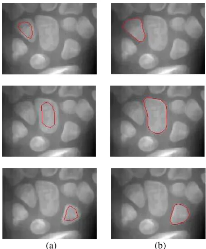
(a) (b)
Fig.4 carpal bones image segmentation (a) initial snake (b) final result
5. CONCLUSIONS
In this paper a deformable model-based approach for carpal bone segmentation from digital X-ray images is proposed. The proposed novel method can correctly extract the carpal bone feature from X-ray image. Anisotropic diffusion filter can effective reduce noise from X-ray image without deteriorating the actual bone boundaries. The complexity of the skeletal structure in the hand and the variability between different subjects makes very difficult to realize an automatic segmentation of the bones. The GVF-Snake provide a powerful framework for carpal bone image segmentation. The experiments are carried out on images of carpal bone and the results are very promising. This method could be extended and applied to other bone structures as well as to other images.
6. REFERENCES
[1] W.W .Greulich, W.W .Pyle, Radiographic atlas of skeletal development of the hand and wrist. 2nd edn. Stanford University Press, Palo Alto, CA, 1959.
[2] J.M.Tanner, R.H .Whitehouse., W.A .Marshall., Healy. M.J.R, Assessment of skeletal maturity and prediction of adult height (TW2 method). 2nd edn. Academic Press, London, 1983. [3] E .Pietka,L.Kaabi, M.L.Kuo, and H. K Huang,
“Feature extraction in carpal-bone analysis,” IEEE Trans. Med. Imag., vol. 12, 1993, pp. 44–49.
[4] C. C .Ko, C.W.Mao, C.J.Lin., and Y.N.Sun, “Image analysis for skeletal evaluation of carpal bones,” in Proc. SPIE, vol. 2501/2, 1995, pp. 951–961.
[image:4.595.70.276.91.376.2][5] D. J. Michael, and A.C.Nelson, “HANDX: A model-based system for automatic segmentation of bones from digital hand radiographs,” IEEE Trans. Med. Imag., vol. 8, no. 1, 1990, pp. 191–193.
[6] N. D. Efford, “Knowledge-based segmentation and feature analysis of hand wrist radiographs,” School of Computer Studies, University of Leeds, Report 94.31, Research Report Series, 1994.
[7] M.Kass, A.Witkin, and D.Terzopoulos, “Snakes: active contour models,” Int’l J.Comp. Vis., vol. 1, no. 4, 1987, pp. 321–331.
[8] C.Xu and J.L.Prince, “Generalized gradient vector flow external forces for active contours,” Signal Processing—An International Journal, vol. 71, no. 2, 1998, pp. 131–139. [9] C.Xu and J.L.Prince, “Snakes, shapes, and
gradient vector flow,” IEEE Trans. Imag. Proc., vol. 7, no. 3, 1998, pp. 359–369.
[10] P.Perona, and J.Malik, “Scale-space and edge detection using anisotropic diffusion”.IEEE Transactions on Pattern Analysis and Machine Intelligence, Vol 12,1990, pp.629-639.