Acute Liver Failure due to Wilson Disease:
Eight Years of the National Liver Transplant
Program in Uruguay
Victoria Mainardi, Karina Rando, Marcelo Valverde, Daniela Olivari, Jorge Castelli, Gabriela Rey, Solange Gerona
Hepatic Biliary and Pancreatic National Center - Teaching and Assistance Unit (UDA) and Bi-Intuitional Unit of Liver Transplantation, Military Hospital, Montevideo, Uruguay.
January-February, Vol. 18 No. 1, 2019: 187-192
The Official Journal of the Mexican Association of Hepatology, the Latin-American Association for Study of the Liver and
the Canadian Association for the Study of the Liver
Manuscript received: Manuscript received:Manuscript received:
Manuscript received:Manuscript received: January 19, 2017. Manuscript accepted:Manuscript accepted:Manuscript accepted:Manuscript accepted:Manuscript accepted: July 04, 2018.
DOI:10.5604/01.3001.0012.7911
A B S T R A C T A B S T R A C T A B S T R A C T A B S T R A C T A B S T R A C T
Introduction and aim. Introduction and aim.Introduction and aim. Introduction and aim.
Introduction and aim. Wilson’s disease (WD) is an uncommon cause of acute liver failure (ALF). Our aim was to describe clini-cal features, diagnostic findings, treatments, and outcomes of patients with ALF due to WD. Material and methods.Material and methods.Material and methods.Material and methods.Material and methods. Retrospec-tive medical record reviews of all patients with ALF due to WD in eight years in Uruguay. Results.Results.Results.Results.Results. WD was the cause of six (15%) of thirty-nine ALF cases. All patients were females, with a mean age of 18 years. Four patients presented with hyperacute liver fail-ure and two with acute failfail-ure. Jaundice was the main complaint of all patients. Mean total bilirubin (TB), alkaline phosphatase (AP), AST, and ALT were 27.5 mg/dL, 45.5 IU/l, 156 IU/L, and 51 IU/L, respectively. Ceruloplasmin levels were low in four patients, uri-nary cooper was high in four, and two had Kayser-Fleischer rings. All patients had Coombs-negative hemolytic anemia, acute kidney injury, histochemical identifiable copper, and advanced fibrosis on liver histology. The average MELD score was 36. All patients were treated with d-penicillamine and listed for urgent liver transplantation (LT). Prometheus® was performed in one patient. Three patients
died: two without LT and one after LT. Three patients survived: one without LT (New Wilson Index < 11) and two with LT. The refer-ral time to the program and the total time (referrefer-ral plus waiting list time) were longer for non-survivors than for survivors (14 vs. 3 days and 23 vs. 8 respectively). Conclusion.Conclusion.Conclusion.Conclusion. All cases had typical clinical, analytical and histopathology characteristics. Early re-Conclusion. ferral was determinant of prognosis.
Key words. Key words.Key words. Key words.
Key words. Chelation therapy. Liver transplantation. Referral time. Waiting list time. Mortality.
INTRODUCTION
Wilson’s disease (WD) has low incidence and preva-lence (1 per 30,000 inhabitants),1 and is an uncommon
cause of acute liver failure (ALF) (3% of cases in United States,2 6 to 12% in Europe,3 and no cases were reported in
Argentina4). It is an autosomal recessive inherited
disor-der of copper metabolism caused by mutation of the ATP7B gene on chromosome 13. This gene encodes a protein expressed in hepatocytes, which is responsible for transporting copper to the bile (for excretion) and to the blood attached to ceruloplasmin (for its use). The deficit of ATP7B results in reduced biliary copper excretion, re-duced incorporation of copper into ceruloplasmin, and the release of free copper into the blood. In consequence, copper accumulates in the liver or other organs such as the brain and cornea.1,5
ALF is an unusual presentation of WD, accounting only for 5% of all cases.6 It is defined by the presence of
coagu-lopathy with an International Normalized Ratio (INR) ≥ 1.5, plus any degree of encephalopathy, in a patient with an illness of < 26 weeks duration, despite having unrecog-nized cirrhosis in the majority of cases of WD.7 ALF may
be the initial clinical presentation of WD or may be a complication in patients who have already been diagnosed and abandoned the treatment. Most of the patients are women in the second decade of life.3,7
There is not a single specific test for WD, so the diag-nosis is based on a combination of clinical and analytical criteria:
2. Decrease of serum ceruloplasmin level (< 20 mg/dL, usually < 10 μg/dL).
3. Increase in urinary copper excretion (> 100 ug/24 h = 1, 6 μmol/24 h).
4. Presence of Kayser–Fleischer rings (KF) in the slit-lamp eye exam.
5. Coombs-negative hemolytic anemia. 6. ATP7B gene mutation.
7. Increased hepatic parenchymal copper concentration (> 250 μg/g of dry weight of copper in the liver biopsy).3,7
In Uruguay, this last technique is not available, but it is possible to detect copper in hepatocytes by histochemical examination with special stains (orcein and rhodamine).
ALF due to WD (ALF-WD) has a high mortality with-out emergency liver transplantation (LT). Therefore, es-tablishing a rapid diagnosis is critical for patient management as well as for family screening. However, in the setting of ALF, copper metabolism parameters became less sensitive and specific, while KF rings are only identi-fied in 50% of cases. Most patients have a characteristic pattern of laboratory findings that include: high levels of total bilirubin (TB), normal or subnormal serum alkaline phosphatase (AP) typically < 40 IU/L, modest elevation of serum aminotransferases with aspartate aminotransferase (AST) > alanine aminotransferase (ALT), as well as Coombs-negative hemolytic anemia and rapid progression of acute kidney injury (AKI) with altered urinary sedi-ment.3,7,8 Different diagnostic ratios have been considered
for quick diagnosis of WD in the setting of ALF. In 1991, Berman described six patients in which the AP/TB ratio < 2 and AST/ALT ratio > 4 had a high sensitivity and spe-cificity for the diagnosis of ALF-WD.9 However, these
parameters could not be validated by other reports.10,11 A
subsequent study by Korman, et al. in 2008 in a cohort of 16 patients with ALF-WD showed that an AP/TB ratio < 4 had a sensitivity of 94% and a specificity of 96% for diagno-sis. In addition, an AST/ALT ratio > 2.2 had a sensitivity of 94% and a specificity of 86%, and the combination of both provided a sensitivity and specificity of 100%.12 Eisenbach,
et al.11 showed that the indexes proposed by Berman were
not applicable to patients with an average MELD score of
18. Also the AP/TB ratio is not always helpful in children due to the increase of bone AP.13 Therefore, even though
these parameters are a guide for diagnosis, they must be used in combination with the conventional diagnostic cri-teria that we mentioned previously.3
Pharmacological chelation therapy is rarely effective in patients presenting with ALF-WD, mainly due to the time required to remove toxic copper from the organism. How-ever, there are cases of patients surviving without LT, with chelation therapy plus supportive care.11 Plasma exchange
and albumin dialysis have been use to rapidly lower serum copper levels in some reports.14,15 A specific prognosis
in-dex was developed by Nazer16 in 1986 and subsequently
modified by Dhawan, et al.,17 the Wilson Disease Index
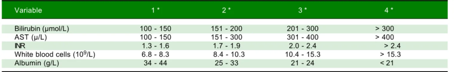
(WDI). A prognostic score ≥ 11 is associated with a high probability of death without LT (Table 1).3,16
Uruguay is a country with a population of 3,432,000 habitants18 that has a single Liver Transplant Program,
which operates in the Military Hospital, and is an adult only program. Children under 16 years of age are sent to the Hospital Italiano in Buenos Aires-Argentina for LT. The objective of the present study is to describe the clini-cal features, diagnostic findings, implemented treatments, and the outcomes of patients with ALF-WD in the first eight years of the National Liver Transplant Program in our country.
MATERIAL AND METHODS
We performed a retrospective, descriptive study. Thir-ty-nine patients were diagnosis with ALF between April 2009 and April 2017, thirty-three adults and six children. WD was the cause in 6 patients (15%), being the fourth most frequent cause after viral hepatitis, indeterminate cause and autoimmune hepatitis.
WD was diagnosed based on typical symptoms and the presence of conventional biochemical indicators, as previ-ously published.3,7 In all cases, other causes of ALF
be-sides WD were ruled out: consumption of toxic substances was discarded, viral markers and liver autoanti-bodies were negative, and the abdominal Doppler ultra-sound showed no vascular alterations or hepatic infiltration.
Table 1. Index forecast in Wilson’s disease, modified by Dhawan, et al.15
Variable 1 * 2 * 3 * 4 *
Bilirubin (μmol/L) 100 - 150 151 - 200 201 - 300 > 300
AST (μ/L) 100 - 150 151 - 300 301 - 400 > 400
INR 1.3 - 1.6 1.7 - 1.9 2.0 - 2.4 > 2.4
White blood cells (109/L) 6.8 - 8.3 8.4 - 10.3 10.4 - 15.3 > 15.3
Albumin (g/L) 34 - 44 25 - 33 21 - 24 < 21
Chart report information was collected for study pur-poses, and a database with the studied variables was creat-ed: age, sex, personal and family history of hepatic-biliary-pancreatic diseases, referral time defined by the time between the first consultation and the contact with the LT center, main compliant, time between the installation of the jaundice and the development of en-cephalopathy to classify as hyperacute, acute, or subacute impairment according to O’Grady’s classification,19
de-gree of encephalopathy according to the West Haven cri-teria,20 complementary tests, treatments, time on waiting
list for LT, and outcomes.
Institutional Board permission was obtained for re-viewing the clinical charts for research purposes.
A statistical analysis was performed to compare referral times, waiting list times and MELD scores between survi-vors and deceases patients. Wilcoxon rank test was per-formed with a CI (confident interval) of 95% due to the small size of the sample.
RESULTS
The 6 patients were women, with a mean age of 18 years (minimum 12 and maximum 22 years). Only one patient
had a family history of hepatic-biliary-pancreatic disease (one brother died due to acute liver failure, and another brother had an undiagnosed liver disease). Two patients had history of altered liver function, with negative viral bi-omarkers in the past. The mean referral time to the LT center was 8.5 days (range: 0-15 days).
Clinical features and complementary tests (Table 2)
Jaundice was the main compliant in all cases. All pa-tients had encephalopathy grade 1 - 2 at the admission to the LT center. Encephalopathy progressed to grade 3 - 4 in three patients. According to the O’Grady classification,18
two cases were hyperacute while the others were acute. The average MELD score was 36 (range: 20 to 49), and five patients had WDI > 11.
• Biochemical liver tests. The mean values were: AST 156 (range: 83 - 250), ALT 51 (range: 15 - 119), TB 27.5 (range: 5.2 - 44), AP 45.5 (range: 11 - 102), and INR 4.2 (range: 2 - 7). Four patients had a ratio AP/BT < 4 and AST/ALT > 2.2; the remaining two did not meet any of these criteria.
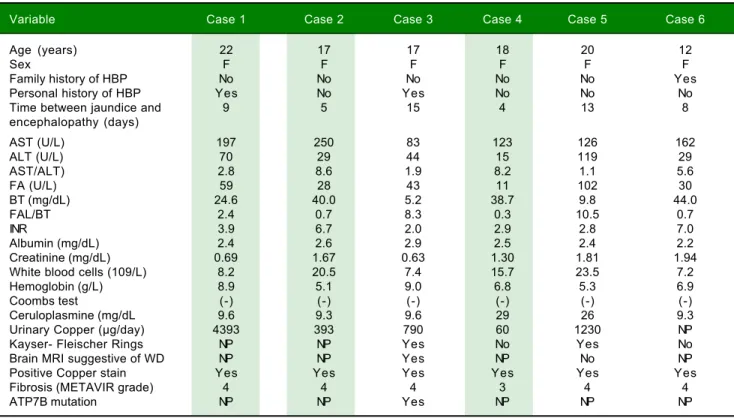
Table 2. Clinical characteristics and complementary test of patients with acute liver failure due to Wilson’s disease.
Variable Case 1 Case 2 Case 3 Case 4 Case 5 Case 6
Age (years) 22 17 17 18 20 12
Sex F F F F F F
Family history of HBP No No No No No Yes
Personal history of HBP Yes No Yes No No No
Time between jaundice and 9 5 15 4 13 8
encephalopathy (days)
AST (U/L) 197 250 83 123 126 162
ALT (U/L) 70 29 44 15 119 29
AST/ALT) 2.8 8.6 1.9 8.2 1.1 5.6
FA (U/L) 59 28 43 11 102 30
BT (mg/dL) 24.6 40.0 5.2 38.7 9.8 44.0
FAL/BT 2.4 0.7 8.3 0.3 10.5 0.7
INR 3.9 6.7 2.0 2.9 2.8 7.0
Albumin (mg/dL) 2.4 2.6 2.9 2.5 2.4 2.2
Creatinine (mg/dL) 0.69 1.67 0.63 1.30 1.81 1.94
White blood cells (109/L) 8.2 20.5 7.4 15.7 23.5 7.2
Hemoglobin (g/L) 8.9 5.1 9.0 6.8 5.3 6.9
Coombs test (-) (-) (-) (-) (-) (-)
Ceruloplasmine (mg/dL 9.6 9.3 9.6 29 26 9.3
Urinary Copper (μg/day) 4393 393 790 60 1230 NP
Kayser- Fleischer Rings NP NP Yes No Yes No
Brain MRI suggestive of WD NP NP Yes NP No NP
Positive Copper stain Yes Yes Yes Yes Yes Yes
Fibrosis (METAVIR grade) 4 4 4 3 4 4
ATP7B mutation NP NP Yes NP NP NP
• Hematological profile. All patients had anemia (de-fined by a concentration of hemoglobin below the ex-pected value based on sex and age), hemolysis parameters (increased LDH), and negative Coombs test. The average hemoglobin was 7.0 g/L (range: 5.1 - 9.0). • Kidney failure (defined as serum creatinine > 1.2
mg/dL) was present in four patients as well as urinary sediment alterations. The rest of them presented kid-ney failure in the follow-up.
• Copper metabolism studies. Four patients had ceru-loplasmin levels < 20 mg/dL. Urinary copper was > 100 μg/day in four of five patients in whom it was de-termined.
• Liver histology. Transyugular liver biopsies were performed in four patients. In one patient, the histolo-gy was obtained from the explanted liver and in the rest one from the necropsy. All patients had histochemical identifiable copper and advanced fibrosis.
• Slit lamp examination. It was performed in four pa-tients; two of them had KF rings.
• Brain magnetic resonance imaging (MRI). It was performed in two cases, one of them showed hyperin-tense lesions of the deep gray matter at the basal ganglia (Globus pallidus), which are characteristic of copper de-posits.
• Genetic studies. Determination of ATP7B was per-formed in only one patient, in which two heterozygote mutations were found in factors c.3207 > A (p.His1069Gin) and c.3809A > G.
Treatments and outcome (Table 3)
The six patients were treated with d-penicillamine che-lation and enlisted for LT. The average waiting list time was 7 days (range 1 to 14).
Two patients died on waiting list: one due to infection (on the 8th day) and the other due to multiple organ dys-function (on 14th day). One patient improved and was delisted on the 10th day, being alive at forty-five months of follow-up. This patient had a WDI < 11.
LT was performed in three patients. Two adults re-ceive a deceased donor liver transplant at day 1 and 6 on waiting list. One of them survived, being alive at thirty-four months of follow-up, and the other died immediately post-surgery. The child receive a living-donor liver trans-plant (LDLT) at day 9; artificial liver replacement by method of Prometheus® (Fresenius) as a bridge to the LT
was performed. She survived with a follow up of seventy-eight months (Figure 1).
Comparison between survivors and non-survivors
The referral time to the LT center was significantly higher for the non-survivors than the survivors (14 vs. 3 days, p = 0.04). The mean waiting time on list for non-survivors was higher in comparison to non-survivors (9 vs. 5 days) although there were not statistical significance (p = 0.25). Nevertheless, the total time (referral time plus waiting list time) was significantly longer (23 vs. 8 days, p = 0.05). The mean MELD score was higher in the non-survivors than in the non-survivors (39 vs. 34) although it presented a non-significant p value (0.35). All non-survi-vors patients reached encephalopaty grade 4, while sur-vivors highest grade of encephalopathy reached was 2.
DISCUSSION
The diagnosis of ALF-WD is challenging and is based on limited evidence from descriptive studies involving a small number of patients.9-12
Table 3. Referral time and waiting list time, prognostic indices and outcome.
Variable Case 1 Case 2 Case 3 Case 4 Case 5 Case 6
Referral time (days) 10 17 0 15 9 0
Waiting list time (days) 8 6 1 14 6 9
Total time (referral time plus 18 23 1 29 15 9
waiting list time, days)
Grade of encephalopathy II/IV II/IV I/I II/IV I/II I/I
at admission/highest achieved
MELD 34 47 20 35 32 49
Wilson disease index 14 16 8 15 14 14
Liver transplantation No Yes No No Yes Yes
Despite the low prevalence of ALF-WD in internation-al series,2-4 in our country WD is the fourth-leading cause
of ALF, along with hepatitis B, autoimmune hepatitis, and indeterminate cause.
The clinical presentation was typical: women in the second decade of life, with characteristic biochemical profiles.3,7,8 The four patients who met the diagnostic
cri-teria proposed by Korman12 had the highest MELD scores.
All patients met more than one classic diagnostic criteri-on. Histology obtained from transyugular biopsy con-firmed the diagnosis, emphasizing the importance of this procedure in our center.
The mortality was 50%: three of six patients, due to multiorgan dysfunction. Two patients died on waiting list, and one died in the immediate post-LT period. Mortality was associated with the delay in transfer the patients to the LT center. Rivera-Penera, et al. in 1997 previously reported in child patients with ALF that delays in transferring pa-tients to a transplant center significantly affected the like-lihood of survival.21 Patients referred late had higher
grades of encephalopathy, bilirubin levels, and proportion of subacute evolution,22 which are associated with worst
survival.2,4,23 Early referral in patients with ALF may be a
key factor in improving patients’ survival because it allows specific and/or highly complex treatment, so it is recom-mended by international clinical practical guidelines.23
The duration of waiting time on list for LT also can significantly affect patient survival, especially for pa-tients with ALF.24-26 A china study showed that waiting
duration was independently correlated with increased mortality with a cut-off value of 5 days for determining the mortality.27 We were not able to show this in our
study, potentially due to limitations of the cohort group size, however the total time since the beginning of the symptoms to the outcome (referral plus waiting list time) was associated with mortality.
The two patients who died without LT, waited on list more than 7 days. The two patients transplanted in our center waited less than 7 days. The child patient that re-ceived a LDLT was on list more than seven days and sur-vived. However, in this case an artificial liver support system (Prometheus®) was performed. To date, case re-ports are encouraging that therapeutic plasma exchange and albumin dialysis can either delay or prevent the need for liver transplantation in patients with fulminant hepatic failure due WD. However, these case reports are mainly from the pediatric or young adult population, and thus further studies in adults are warranted.14,15
The patient who survived without LT had the lowest MELD score and a WDI < 11, supporting the utility of the score.
In conclusion, all cases of ALF-WD had typical clini-cal, analytical and histopathology characteristics. Early re-ferral was determinant of prognosis.
ABBREVIATIONS
• AKI: acute kidney injury. • ALF: acute liver failure. • ALT: alanine aminotransferase. • AP: alkaline phosphatase. • AST: aspartate aminotransferase. • INR: international normalized ratio. • KF: Kayser–Fleischer rings.
• LT: liver transplantation.
• MRI: magnetic resonance imaging. • TB: total bilirubin.
• WD: Wilson’s disease.
CONFLICT OF INTEREST
The authors declares that there is no conflict of interest regarding the publication of this article.
FINANCIAL SUPPORT
This study was not supported by grants or external founds.
REFERENCES
1. Frydman M. Genetic aspects of Wilson’s disease. J Gastro-enterol Hepatol 1990; 5: 483-90.
2. Ostapowicz G, Fontana RJ, Schiodt FV, Larson A, Davern TJ, Han SH, McCashland TM, et al. Results of a prospective study of acute liver failure at 17 tertiary care centers in the United States. Ann Intern Med 2002; 15: 947-55.
3. European Association for the Study of the Liver. EASL Clini-cal Practice Guidelines: Wilson’s disease. J Hepatol 2012; 56: 671-85.
4. Mendizabal M, Marciano S, Videla MG, Anders M, Zerega A, Balderramo DC, Chan D, et al. Changing etiologies and out-comes of acute liver failure: Perspectives from 6 transplant centers in Argentina. Liver Transplant 2014; 20: 483-9. 5. Scheinberg IH. Wilson’s disease. J Rheumatol Suppl 1981;
7: 90-3.
6. Merle U, Schaefer M, Ferenci P, Stremmel W. Clinical presen-tation, diagnosis and long-term outcome of Wilson’s disease: A cohort study. Gut 2007; 56: 115-20.
7. Lee WM, Stravitz RT, Larson AM. Introduction to the revised American Association for the Study of Liver Diseases position paper on acute liver failure 2011. Hepatology 2012; 55: 965-7. 8. Roberts EA, Schilsky ML. AASLD practice guidelines:
Diag-nosis and treatment of Wilson disease: An update. Hepatol-ogy 2008; 47: 2089-111.
9. Berman DH, Leventhal RI, Gavaler JS, Cadoff EM, Van Thiel DH. Clinical differentiation of fulminant Wilsonian hepatitis from other causes of hepatic failure. Gastroenterology
1991; 100: 1129-34.
Diagnos-tic criteria for acute liver failure due to Wilson Disease.
World J Gastroenterol 2007; 13: 1711-4.
12. Korman J, Volenberg I, Balko J, Webster J, Schiodt FV, Squires RH, Fontana RJ, et al. Screening for Wilson disease in acute liver failure: A comparison of currently available di-agnostic tests. Hepatology 2008; 48: 1167-74.
13. Catana AM, Medici V. Liver transplantation for Wilson dis-ease. World J Hepatol 2012; 4: 5-10.
14. Kido J, Matsumoto S, Momosaki K, Sakamoto R, Mitsubishi H, Inomata Y, Endo F, et al. Plasma Exchange and chelator therapy rescues acute liver failure in Wilson disease without liver transplantation. Hepatol Res 2017; 47: 359-63. 15. Reynolds HV, Talekar CR, Bellapart J, Leggett BA, Boots RJ.
Copper removal strategies for Wilson’s disease crisis in ICU.
Anaesth Intensive Care 2014; 42: 253-7.
16. Nazer H, Ede RJ, Mowat AP, Williams R. Wilson’s disease: Clinical presentation and use of prognostic index. Gut 1986; 27: 1377-81.
17. Dhawan A, Taylor RM, Cheeseman P, De Silva P, Katsiyian-nakis L, Mieli-Vergani G. Wilson’s disease in children: 37-year experience and revised King’s score for liver transplantation. Liver Transplant 2005; 11: 441-8.
18. Uruguay en cifras 2014. Instituto Nacional de Estadística. http//www.ine.gub.uy
19. O’Grady JG, Schalm SW, Williams R. Acute liver failure: Re-defining the syndromes. Lancet 1993; 342: 273-5.
20. Ferenci P, Lockwood A, Mullen K, Tarter R, Weissenborn K, Blei AT. Hepatic encephalopathy - Definition, nomenclature, diagnosis, and quantification: Final report of the Working Par-ty at the 11th World Congresses of Gastroenterology, Vien-na, 1998. Hepatology 2002; 35: 716-21.
21. Rivera-Penera T, Moreno J, Skaff C, McDiarmid S, Vargas J, Ament ME. Delayed encephalopathy in fulminant hepatic
fail-ure in the pediatric population and the role of liver transplan-tation. J Pediatr Gastroenterol Nutr 1997; 24: 128-34. 22. Sturm E, Lexmond WS, Verkade HJ. Pediatric acute liver
fail-ure: variations in referral timing are associated with disease subtypes. Eur J Pediatr 2015; 174: 169-75.
23. European Association for the Study of the Liver. EASL Clini-cal PractiClini-cal Guidelines on the management of acute (fulmi-nant) liver failure. J Hepatol 2017; 66: 1047-81.
24. Park SJ, Lim YS, Hwang S, Heo NY, Lee HC, Suh DJ, Yu E, Lee SG. Emergency adult-to-adult living-donor liver trans-plantation for acute liver failure in a hepatitis B virus endem-ic area. Hepatology 2010; 51: 903-11.
25. Liu CL, Fan ST, Lo CM, Wei WI, Yong BH, Lai CL, Wong J. Live-donor liver transplantation for acute-on-chronic hepati-tis B liver failure. Transplantation 2003; 76: 1174-9.
26. Everhart JE, Lombardero M, Detre KM, Zetterman RK, Wiesn-er RH, Lake JR, Hoofnagle JH. Increased waiting time for liv-er transplantation results in highliv-er mortality. Transplantation
1997; 64: 1300-6.
27. Yuan D, Liu F, Wei Y-G, Li B, Yan LN, Wen TF, Zhao JC, et al. Adult-to-adult living donor liver transplantation for acute liver failure in China. World J Gastroenterol 2012; 18: 7234-41.
Correspondence and reprint request:
Ana Evelyn Karina Rando, M.D., Associated Professor 8 de Octubre 3020,
Tel.: +598-9369-7867,