Otras secciones de este sitio:
☞ ☞ ☞ ☞
☞ Índice de este número
☞ ☞ ☞ ☞
☞ Más revistas
☞ ☞ ☞ ☞
☞ Búsqueda
Others sections in this web site:
☞ ☞ ☞ ☞
☞ Contents of this number ☞
☞ ☞ ☞
☞ More journals ☞
☞ ☞ ☞ ☞ Search Artículo:
Intraoperative US-guided large volume ethanol injection for hepatocellular carcinoma greater than 4 cm
Copyright © 2005: Mexican Association of Hepatology
ANNALS OF HEPATOLOGY
Number 3 July-September 2 0 0 5
Volume 4
Annals of Hepatology 4(3) 2005: 200-203
MG
200
edigraphic.com
Annals of Hepatology 2005; 4(3): July-September: 200-203
Annals of Hepatology
1 Departments of Gastroenterology. 2 Departments of Epidemiology.
3 Instituto Nacional de Cancerología, Mexico City. Division of
Surgery. Centro Médico ISSEMyM, Metepec, México.
Address for correspondence:
Dr. Ricardo Mondragón-Sánchez, FACS. Av. Hidalgo # 411 Col. Centro
C.P. 50000
Toluca, Estado de México, México Phone: (52 722) 2134214 Fax: (52 722) 2153538
E-mail: [email protected]
Manuscript received and accepted: 9 and 28 March, 2005.
Abstract
Background: Percutaneous ethanol injection has been successfully used for hepatocelular carcinomas (HCC) smaller than 5 cm in size. For larger lesions large vol-ume ethanol injection has not been well explored. Aim: Evaluate the results of intraoperative Ultrasonograph-ic-guided large volume ethanol injection for HCC larg-er than 4 cm in size. Patients and methods: Ten patients were candidates for this treatment between June 1999 and July 2003. A retrospective review of the clinical files was performed. Absolute ethanol, average of 100 mL (range 80-120 mL) was administered intraopera-tively. Follow-up evaluation included alpha-fetoprotein (AFP) and ultrasound or computed tomography. Re-sults: There were six women and four men, the median age was 62 years (range 56-80). The median lesion size was 8 cm (range 4-15 cm). Hepatitis C liver cirrhosis was the most common associated chronic liver disease (70%). A significant reduction of AFP levels after treat-ment was observed (Initial 966 ng/dL, post treattreat-ment levels: 42 ng/dL) US and CT scan showed tumor necro-sis. Morbidity was 40%. No operative mortality was re-corded. The one and four year survival rate was 60% and 20%. Conclusion: Intraoperative US-guided large volume ethanol injection is a safe palliative therapy for cirrhotic patients with HCC lesions greater than 5 cm in size. The impact on survival should be compared in a controlled double blind study.
Original Article
Intraoperative US-guided large volume
ethanol injection for hepatocellular
carcinoma greater than 4 cm
Ricardo Mondragón-Sánchez;1,3 Ana Lilia Garduño-López;1 Elvira Gómez-Gómez;1 Mauricio Frias-Mendivil;2
Alejandro Mondragón-Sánchez;3 Rigoberto Bernal-Maldonado1
Key words: Large hepatocellular carcinoma, percuta-neous ethanol injection, nonresectional therapies.
Introduction
Percutaneous ethanol injection (PEI) is considered the most effective form of direct ablation therapy for HCC.1-3
It has become one of the most widely used procedures for treating Hepatocellular Carcinoma (HCC) in patients with cirrhosis.3-5 It was first advocated by Sugiura and
associ-ates in 1983.6 The main mechanism whereby ethanol
causes cell necrosis remains speculative but it is well known that it causes denaturation of proteins and dehy-dration of the cytoplasm by the diffusion of ethanol with-in the neoplastic cells. The result is coagulative necrosis, as well as necrosis of the vascular endothelium leading to platelet aggregation and small vessel thrombosis fol-lowed by tissue ischemia.7
PEI has traditionally been restricted to the treatment of single small lesions 5 cm or less in size.8-10 However,
there has been a recent trend, toward treating larger tu-mors using higher doses of ethanol or a combination ther-apy using transcatheter arterial embolization associated to PEI.7,10 PEI is usually performed in the radiology suite but
can also be performed in the operating room under gener-al anesthesia. The benefits are obvious because a large dosage can be instilled, under anesthesia, spilling of etha-nol into the peritoneum or extravasation of ethaetha-nol into the capsular surface is recognized, a Pringle maneuver can be performed, which may decrease the blood flow to a hypervascular HCC and bleeding can be easily diag-nosed and controlled.8
The aim of this study was to evaluate the results of in-traoperative US-guided large volume ethanol injection for HCC lesions greater than 5 cm in size.
Patients and methods
unresect-R Mondragón-Sánchez et al. Large volume ethanol injection for HCC 201
edigraphic.com
:rop odarobale FDP
VC ed AS, cidemihparG
arap
acidémoiB arutaretiL :cihpargideM
sustraídode-m.e.d.i.g.r.a.p.h.i.c sustraídode-m.e.d.i.g.r.a.p.h.i.c
cihpargidemedodabor
able lesions who underwent US-guided intraoperative large volume ethanol injection were included for analysis. The time onset of symptoms was estimated and the pres-ence of any previous risk factors for development HCC were reviewed. Ordinary blood and liver function tests were obtained, measurement of alpha-protein (AFP) lev-el, and serologic studies for viral hepatitis (hepatitis C vi-rus antibody and hepatitis B surface antigen) were evalu-ated. Patients were diagnosed with either ultrasonography (US), computed tomography (CT) and in selected cases with magnetic resonance imaging (MRI). Definitive diag-nosis of HCC was established by histopathologic criteria. The selection criteria for this treatment included pa-tients with preoperative detection with US or CT scan of a lesion larger than 5 cm, that were not considered for liver resection because of liver cirrhosis or bad clinical condi-tions or when the lesion was considered unresectable dur-ing operative evaluation. Tumor size ranged from 5-15 cm (median 8 cm). Patients with more than three lesions, portal vein thrombosis, ascites, extrahepatic metastases or with obstructive jaundice were not considered for ethanol injection. These patients had no other alternative treat-ment. All patients gave informed consent in order to
par-ticipate in the study before they received the treatment.
Technique. The procedure was performed under gen-eral anesthesia, with endotracheal intubation and mechan-ical ventilation. Laparotomy, laparoscopy or thoracotomy were used to evaluate the lesions. The indication for each approach was made depending on the location of the le-sion, presence or not of cirrhosis and of previous abdomi-nal scars. During surgery, exploration consisted of careful inspection and bimanual palpation of the liver tissue with and without a Pringle maneuver as well as careful abdom-inal cavity contents examination. Intraoperative ultra-sound (IOU) was performed and direct scanning of the liver was accomplished with a 7.5 mHz intraoperative probes. Normal saline solution was applied to enhance sound transmission. A systemic evaluation of the entire liver was performed by a radiologist after surgical mobili-zation of the liver in both sagital and transverse planes. The liver was scanned starting from the left lobe where the transducer was placed at the inferior, lateral and supe-rior portions. Examination included demonstration of the hepatic veins, inferior vena cava and the right and left portal veins. The lesions known from preoperative studies were identified and their relationship to hepatic and portal veins established. Resection was evaluated by defining the relationship of the lesion to vascular structures such as portal and hepatic veins and inferior vena cava. When unsuspected palpable or non-palpable lesions were de-tected ultrasound-guided biopsies were performed.
To calculate the volume of ethanol required to ablate the lesion we used the formula for a sphere: V = 4/3 π (R + 0.5 cm)3, where V is the total volume of injected
etha-nol and R is the radius of the tumor (in centimeters).7 This
formula is used only as a reference, because it was
de-signed to be used in several sessions. We performed a sin-gle session so ultrasound was added to adequate the dos-age of ethanol. Absolute ethanol (99.5%) is injected into the lesion throughout the entire diameter. An average of 100 mL (range 80-120 mL) was administered depending on lesion size. One single dose should not exceed 120 mL. In one patient with a 4 cm tumor a second percutane-ous injection was needed and this patient received a total dose of 80 mL. The technique required an average time of 30 minutes. No intra-operative complications were de-tected related to this procedure.
Statistics. We described the frequency and the distri-bution of each studied variable, age, the time onset of symptoms, AFP, and dose of alcohol injection were re-ported as median and range. Survival curve was done by the Kaplan-Meier method. The level of AFP was com-pared with the confidence interval at 95%.
Results
Ten patients with HCC were included for analysis. Six were female and 4 male (sex ratio: 1.5:1). Median age at the time of diagnosis was 62 years old (range 56-80)
(Ta-ble I). History of blood transfusion was recorded in three
and alcoholism in one. Abdominal pain in the right upper quadrant was the main clinical symptom and was present in all patients, a palpable abdominal mass was present in 7 (70%), enlargement of the liver was diagnosed in 3 (30%). Seven patients had morphologic changes usually observed in chronic liver disease (70%). The median time of onset of symptoms was 4 months (range 1-12 months). Liver function tests were normal except for serum eleva-tion of transaminases in 3 patients. Seven patients had hepatitis C liver cirrhosis, 2 had hepatitis C and B liver cirrhosis and one patient had no association with chronic liver disease. All patients were classified as Child-Pugh A. The Karnofsky score was greater than 80% in these patients. Histological examination showed a well-differ-entiated pattern HCC in 5 (50%), trabecular type in 2 (20%), tubular pattern in 2 (10%) and a combination of trabecular and tubular in 1 (10%).
Laparotomy was performed in five patients, because a previous surgical operation was performed that included cholecystectomy in three, and liver biopsy in two. Lap-aroscopy was used in three patients that had anterior le-sions in patients without a previous abdominal scar. Tho-racotomy was performed in two cirrhotic patients because the lesions were located in segments VII and VIII of the liver, these patients were initially thought to be eligible for resection. The lesion was located in the right lobe of the liver in six patients, in the left in three, and one patient had lesions in both lobes. The size of HCC was measured by US, CT scan or during surgery, the median size of the lesions was 8 cm (range 5-15 cm).
in-MG
202
edigraphic.com
Before After
0 200 400 600 800 1000 1200 1400 1600 1800
Patients n =10
AFP measurement
toxication in four patients that resolved spontaneously within the immediate postoperative hours. Morbidity was 40%, nevertheless all cases had minor complica-tions. Pleural effusion was diagnosed in one patient that was operated through a thoracotomy and was re-solved with a thoracic tube. One patient presented tran-sient low grade fever that was attributed to tumor ne-crosis, one patient presented mild hypotension after treatment that responded to intravenous fluids. One
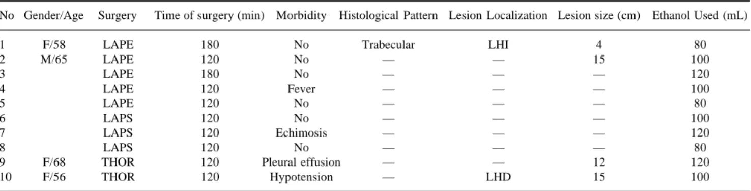
pa-Table 1. Patients with large HCC treated by intraoperative US- guided large volume ethanol injection.
No Gender/Age Surgery Time of surgery (min) Morbidity Histological Pattern Lesion Localization Lesion size (cm) Ethanol Used (mL)
1 F/58 LAPE 180 No Trabecular LHI 4 80
2 M/65 LAPE 120 No — — 15 100
3 LAPE 180 No — — — 120
4 LAPE 120 Fever — — — 100
5 LAPE 120 No — — — 80
6 LAPS 120 No — — — 100
7 LAPS 120 Echimosis — — — 120
8 LAPS 120 No — — — 80
9 F/68 THOR 120 Pleural effusion — — 12 120
10 F/56 THOR 120 Hypotension — LHD 15 100
LAP: Laparotomy, LAPS: Laparoscopy, THOR: Thoracotomy
Figure 1. One year survival of patients with HCC treated with intrao-perative large volume ethanol injection.
Figure 2. Alpha-fetoprotein levels before and after treatment.
tient showed 48 hours after treatment lumbar pain and ecchymosis on the posterior abdominal wall was diag-nosed. No operative mortality was recorded in this se-ries. Median hospital stay was two days but ranged from one to five days. The cumulative survival rate af-ter treatment was 60 and 20% at one and four years
(Figure 1).
Follow-up was performed in all patients until the end of the study or until death. Physical examination was per-formed and the presence of symptoms recorded. Mea-surement of serum AFP levels were also recorded. The median initial AFP level was 966 ng/mL and three months after treatment it was 42 ng/mL (Figure 2). Ab-dominal CT scan performed three and six months after treatment showed tumor necrosis in all patients, reduction in size in one case but the rest showed no modification in the size of the lesion. No recurrence of the primary lesion was diagnosed but in three patients new lesions were di-agnosed in the liver or the lungs.
Discussion
Surgical resection is generally accepted as the treat-ment of choice for HCC. Recent advances of surgical technique, anesthesia and postoperative care have re-sulted in a reduction of operative morbidity and mortal-ity even in cirrhotic patients.10 Unfortunately, it is
esti-mated that only about 20% of HCC can be resected at the initial presentation.11 Prognosis without treatment is
poor and other non-resective forms of treatment have been described.12 Mexico is not considered a high
en-demic area for HCC but the incidence of this neoplasm in the last 25 years has been rising steadily.13 At our
In-stitution most of patients are diagnosed in late clinical stages or with advanced liver cirrhosis and less than 15% of the cases are candidates for liver resection.14
R Mondragón-Sánchez et al. Large volume ethanol injection for HCC 203
edigraphic.com
Percutaneous ethanol injection is a safe treatment and has been used in this and other hospitals for small lesions usually less than 3 cm in size with excellent re-sults.1,3,4,8 Experience though a percutaneous approach
for larger lesions has been scarce and it can be done
ultrasound guided with similar benefits.15 However, it
is generally considered as time-consuming because it requires a great number of sessions and morbidity is related to the dosage of ethanol and the number of
ses-sions. Pain has been the principal side-effect.16
US-guided intraoperative injection of large volume of eth-anol offers the “theoretical” advantage of total tumoral ablation in a single session. Ultrasound is highly rec-ommended because the amount of ethanol needed to produce tumor ablation can be determined more pre-cisely and when a complete morphologic change of the lesion is observed the treatment is finished. This initial experience shows that this procedure can be performed with minimal and minor morbidity and without mortal-ity. Pain was not observed because general anesthesia was used as well as postoperative medications. Tumor-al necrosis was confirmed due to a consistent reduction of serum AFP levels observed as well as changes in the CT scan. Recurrence of the originally treated tumor was not found, but three patients developed satellite le-sions in the liver or lung metastases at eight to 12 months after treatment. Other authors have reported a similar observation.17 Much has to be investigated in
terms of prognostic factors for long term survival. We believe that solitary encapsulated and smaller than 10 cm lesions have a better response than diffuse non-en-capsulated lesions.
Conclusion
Intraoperative US-guided large volume ethanol injec-tion is a safe palliative therapy for patients with HCC le-sions greater than 5 cm in size. We believe that this pro-cedure could increase survival of cirrhotic patients with unresectable HCC. However, prospective and random-ized studies are needed to demonstrate this observation.
References
1. Livraghi T, Festi D, Monti F, Salmi A, Vettori C. US-guided percuta-neous alcohol injection of small hepatic and abdominal tumors.
Ra-diology 1986; 161: 309-312.
2. Williams R, Rizzi P. Treating Small Hepatocellular Carcinomas. N
Eng J Med 1996; 334: 728-729.
3. Schafer D, Sorrell M. Hepatocellular Carcinoma. Lancet 1999; 353: 1253-1257.
4. Marcos-Alvarez A, Jenkins R, Washburn W, Lewis W, Stuart K, Gor-don F, Kane R, Clouse M. Multimodality treatment of Hepatocellu-lar carcinoma in a hepatobiliary Specialty Center. Arch Surg 1996; 131; 292-298.
5. Mor E, Kaspa R, Sheirner P, Schwartz M. Treatment of Hepatocellu-lar Carcinoma Associated with Cirrhosis in the Era of Liver Trans-plantation. Ann Intern Med 1998; 129: 643-653.
6. Liu, Chin Leung, Fan Sheung-Tat. Nonresectional Therapies for Hepatocellular Carcinoma. Am J Surg l997; 173: 358-365. 7. Douglas G, Rosove M, Shaked A, Busuttil R. Current treatment
Modalities for hepatocellular Carcinoma. Ann Surg 1994; 219: 236-247.
8. Okada S. Local Ablation Therapy for Hepatocellular Carcinoma.
Seminars in Liver Disease 1999; 19: 323-328.
9. Régimbeau J, Farges O, Shen B, Sauvanet A, Belghiti J. Is surgery for large hepatocellular carcinoma justified? J Hepatol 1999; 31: 1062-1068.
10. Okuda, Kunio. Hepatocellular carcinoma. J Hepatol 2000; 32: 225-237. 11. Usatoff V, Habib N. Hepatic malignancy: challenges and
opportuni-ties for the surgeon. J R Coll Surg Edinb 2000; 45: 99-109 12. Bartolini F, Gianini A, Napoli P. Hepatocellular carcinoma and
cir-rhosis. A review of the relative incidence in a 25 year period in the Florence area. Hepato-Gastroenterol 1984; 21: 215-217.
13. Cortes-Espinosa T, Mondragón-Sánchez R, Hurtado-Andrade H, Sánchez-Cisneros R. Hepatocellular Carcinoma and Hepatic Cirrhosis in Mexico: A 25 year Necroscopy Review. Hepato-Gastroenterol l997; 44: 1401-1403.
14. Mondragón-Sánchez R, Ochoa-Carrillo FJ, Ruiz-Molina JM, Herrera-Goepfer R, Oñate-Ocaña L, Aiello-Crocifoglio V. Carcinoma hepa-tocellular. Experiencia en el Instituto Nacional de Cancerología. Rev
Gastroenterol Mex 1997; 62: 34-40.
15. Livraghi T, Benedini V, Lazzoroni S, Meloni F, Torzilli G, Vettori C. Long Term Results of Single session percutaneous Ethanol In-jection in Patients with large Hepatocellular Carcinoma. Cancer 1998; 83(1): 48-57.
16. Tapani E, Soiva M, Lavonen J, Ristakari S, Vehmas T. Complica-tions following high-dose percutaneous ethanol injection into he-patic tumors. Act Radiol 1996; 37: 655-659.