High coffee intake is associated with lower grade nonalcoholic
fatty liver disease: the role of peripheral antioxidant activity
Ylse Gutiérrez-Grobe,* Norberto Chávez-Tapia,*Vicente Sánchez-Valle,* Juan Gabriel Gavilanes-Espinar,* Guadalupe Ponciano-Rodríguez**
Misael Uribe,* Nahum Méndez-Sánchez*
* Biomedical Research Unit, Liver Unit, Medica Sur Clinic and Foundation. Mexico City, Mexico.
**Department of Public Health Investigation, Faculty of Medicine, National Autonomous University of Mexico (UNAM), Mexico City, Mexico.
ABSTRACT
Background & aims. Some phytochemicals present in coffee have a potential antioxidant role which seems to protect the human body against cardiovascular diseases, liver disease and malignancies. Nonalcoholic fatty liver disease is a common disease with limited therapeutic options. This study investigated the an-tioxidant effect of coffee by measuring anan-tioxidant enzymes and lipid peroxidation markers in patients with nonalcoholic fatty liver disease. Material and methods. We performed a case-control study at the Univer-sity Hospital, Mexico City. Anthropometric, metabolic, dietary and biochemical variables of all patients were determined and compared. The presence of nonalcoholic fatty liver disease was established by ultra-sonography. All patients completed a dietary questionnaire in order to determine their of coffee con-sumption. Catalase, superoxide dismutase and thiobarbituric acid reactive substances were measured in all of the patients. Results. Seventy-three subjects with and 57 without nonalcoholic fatty liver disease were included. Patients with nonalcoholic fatty liver disease had significantly higher body mass index, blood glu-cose, homeostasis model of assessment–insulin resistance and insulin values in comparison to patients without nonalcoholic fatty liver disease. On the one hand, there was a significant difference in coffee in-take between the groups (p < 0.05, for all comparisons). There was no significant difference between groups in catalase (0.39 ± 0.74 vs. 0.28 ± 0.69 nM/min/mL), superoxide dismutase (5.4 ± 3.45 vs. 4.7 ± 2.1 U/mL) or thiobarbituric acid-reactive substances (4.05 ± 1.87 vs. 3.94 ± 1.59 µM/mL). Conclusions. A high intake of coffee has a protective effect against nonalcoholic fatty liver disease however there was no significant difference in the antioxidant variables analyzed.
Key words. Fatty liver. Epidemiology. Caffeine.
CCorrespondence and reprint request: Nahum Méndez-Sánchez, M.D., Ph.D. Liver Research Unit. Medica Sur Clinic & Foundation
Puente de Piedra, Núm. 150. Col. Toriello Guerra, Mexico City, Mexico Tel.: +5255 5424-7200 (4215). Fax: +5255 5666-4031
E mail: [email protected]
Manuscript received: January 31, 2012. Manuscript accepted: February 13, 2012
INTRODUCTION
Coffee is one of the most frequently consumed be-verages in the world, and it is known to be a psy-choactive beverage with stimulating effects on the central nervous system. Caffeine, one of the main constituents of coffee, has been shown to have a wide spectrum of activities in several biological systems including glucose metabolism and smooth muscle. In addition to caffeine, coffee contains
chlorogenic acid, which has antioxidant, antimuta-tion, anticarcinogenic, antibiotic, antihypercholes-terolemic, antihypertensive and anti-inflammatory actions.1
In 1992, Klatsky & Armstrong reported an in-verse relation between coffee drinking and the risk of liver cirrhosis in a 10-year cohort follow-up. In that study, they reported that coffee drinking ap-peared to protect against cirrhosis attributed to alcoholic liver disease but not to nonalcoholic di-sease.2 Recently, high-level coffee consumption has
been associated with reduced progression of pre-existing liver disease and lower risk of hepatocellular carcinoma.3 It has also been related to a better
response to hepatitis C treatment4 and suggested as
potential treatments for a wide spectrum of featu-res of metabolic syndrome.5
Nonalcoholic fatty liver disease (NAFLD) is a common disease worldwide, and is considered the most frequent chronic liver disease. Public health measures have focused on prevention.6 This is most
important considering the limited therapeutic options7
and the expected increase in this common liver disease.8
The potential role of coffee as a treatment or pre-ventive for hepatic diseases has been widely discus-sed; however, as far as we know there are no studies of the probable antioxidant effect of coffee as a fac-tor in preventing liver disease progression in popu-lations with a high prevalence of NAFLD. In this study, we investigate the potential antioxidant role of coffee in the Mexican population and its associa-tion with NAFLD by measuring the level of the an-tioxidant enzymes superoxide dismutase (SOD) and catalase (CAT) and lipoperoxidation activity by measuring thiobarbituric acid-reactive species (T-BARS).
MATERIAL AND METHODS
Patient population
We conducted a cross-sectional study in the checkup unit of the Diagnostic Clinic at the Medica Sur Clinic & Foundation between February 2010 and December 2010. This hospital provides care for mainly middle- and high-income individuals from Mexico City and surrounding metropolitan areas.
Our sample population was selected from a conse-cutive series of asymptomatic subjects who were referred to the checkup unit by their companies as an annual employment requirement, not for sympto-matic disease, and who had no knowledge of having a chronic disease. Exclusion criteria were an alco-hol intake of > 20 g/d, known liver disease or current use of medication. For liver disease, sub-jects who tested positive for hepatitis B antigen or hepatitis C antibody and those who reported a his-tory of known liver disease, including viral, genetic, autoimmune or drug-induced liver disease, were also excluded. The study comprised 130 patients, 73 with NAFLD and 57 without NAFLD, categorized accor-ding to the degree of steatosis measured by ultraso-nography.
The study was approved by the Human Subjects Committee at the Medica Sur Clinic & Foundation and conformed to the ethical guidelines of the 1983 Declaration of Helsinki. Written informed consent
was obtained from all participants before entry into the study.
NAFLD diagnosis
The diagnosis of NAFLD was based on the pre-sence of a bright liver at ultrasound scanning. Real-time ultrasonographic studies were performed while the subjects were fasting. A 3.5 MHz transducer (Elegra; Siemens Medical Systems, Mountain Grove, CA) was used to obtain the following images: sagittal view of the right lobe of the liver and right kidney, transverse view of the left lateral segment of the liver and spleen, transverse view of the liver and pancreas, and any focal areas of altered echotexture. The severity of echogenicity was graded as follows:9
• Grade 0. Normal echogenicity.d .e
• Grade 1. A slight, diffuse increase in fine echoes in liver parenchyma with normal visualization of the diaphragm and intrahepatic vessel borders. • Grade 2. A moderate, diffuse increase in finee 2
echoes with slightly impaired visualization of in-trahepatic vessels and the diaphragm; and • Grade 3. A marked increase in fine echoes withd 3
poor or no visualization of the intrahepatic ves-sel borders, diaphragm and posterior right lobe of the liver.
Sonographic patterns were classed as:
• 0, homogeneous, normal. • 1, hyperechoic nodules.
• 2, multiple, confluent hyperechoic lesions. • 3, hypoechoic skip nodules.
• 4, irregular hyperechoic and hypoechoic areas. • 5, diffuse involvement.
Dietary history questionnaire
from the questionnaire with known nutrient con-tents of the foods to estimate daily nutrient in-takes.10
Analytical procedures
Insulin concentrations were measured using an immunoenzymometric assay (MEIA; Abbott Diag-nostics. Illinois, USA), with inter- and intra-assay coefficients of variation < 3%. Fasting plasma glu-cose was measured in duplicate with an automated analyzer. The coefficient of variation for a single determination was 1.5%. Total cholesterol, high-density lipoprotein (HDL-C) and triglyceride concentrations were measured by enzymatic colori-metric methods, using CHOL, HDL-C plus (second generation) and TG assays (Roche Diagnostics Co., Indianapolis, IN), respectively. Low-density lipopro-tein cholesterol (LDL-C) concentrations were calcu-lated using the Friedewald formula. Assessment of insulin resistance was made using the homeostasis model assessment (HOMA–IR) originally described by Matthews, et al.:11
HOMA–IR = [(fasting insulin (U/L) x fasting glucose (mmol/L))/22.5].
Determination of antioxidant enzymes and lipid peroxidation in serum was determined in duplicate. Commercial kits from Cayman Chemical (Michigan, USA), which use a colorimetric reaction detected by spectrophotometry, were used as outlined below.
• SOD detection. Cayman’s SOD assay kit (No. te t n 706002) utilizes a tetrazolium salt for detection of superoxide radicals generated by xanthine oxi-dase and hypoxanthine. One unit of SOD is defi-ned as the amount of enzyme needed to exhibit 50% dismutation of the superoxide radical. The SOD assay measures all three types of SOD (Cu/ Zn, Mn and FeSOD). To SOD standard and sam-ple wells were added 200 µL of radical detector and 10 µL of standard or sample. Reactions were initiated by adding 20 µL xanthine oxidase and carefully shaking the 96-well plate for a few se-conds to mix. Plates were covered and incubated on a shaker for 20 min at room temperature and the absorbance was read at 460 nm.
• CAT detection. Cayman’s CAT assay kit (No.A o 707002) utilizes the peroxidase function of CAT to determine enzyme activity. The method is based on the reaction of the enzyme with methanol in the presence of an optimal concentration of H2O2.
The formaldehyde produced is measured colori-metrically with 4-amino-3-hydrazino-5-mercapto-1,2,4-triazole (Purpald) as the chromogen. Purpal specifically forms a bicyclic heterocycle with aldehydes, which upon oxidation changes from colorless to a purple color. The assay can be used to measure CAT activity in serum. To duplicate sample and standard wells was added 100 µL of assay buffer, 30 µL of methanol and 20 µL of standard or sample. The reaction was initiated by adding 20 µL of hydrogen peroxide to all wells, and then the plate was covered and in-cubated on a shaker for 20 min at room tempera-ture. Potassium hydroxide (30 µL) was added to each well to terminate the reaction, followed by 30 µL of Purpal (chromogen). The plate was co-vered and incubated for 10 min at room tempera-ture on a shaker. Finally, 10 µL of potassium periodate was added to each well, and the plate was covered and incubated for 5 min at room temperature before the absorbance was read at 540 nm.
• T-BARS detection. Cayman’s T-BARS assay- te t n kit (No. 10009055), for assaying lipid peroxida-tion in serum, measures colorimetrically at 540 nm the malondialdehyde (MDA)- thiobarbi-turic acid (TBA) adduct formed by the reaction of MDA and TBA under acidic conditions at high temperature (90-100 °C). One hundred microli-ters of sample or standard was added to appro-priately labeled 5 mL vials, to which 100 µL of sodium dodecyl sulfate solution was added and swirled to mix, before the forceful addition of 4 mL of color reagent down the side of each vial. Vials were capped and placed in a holder to keep them upright during boiling, and then placed in vigorously boiling water for 1 h. After 1 h, vials were removed and placed in an ice bath to stop the reaction, incubated on ice for 10 min, then centrifuged for 10 min at 1,600 x g at 4 °C. After warming the vials to room temperature, duplicate 150 µL aliquots from each vial were added to a clear plate and the absorbance read at 540 nm.
Statistical analysis
the probability of NAFLD being associated with the level of caffeine intake. All analyses were carried out with SPSS/PC v 16.0 (Chicago IL). Differences were considered significant with P values of < 0.05.
RESULTS
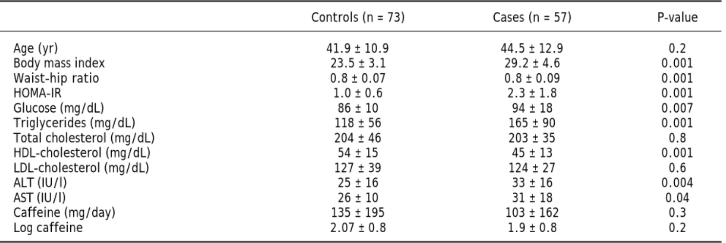
Of the 130 patients included, 57 had NAFLD and 73 did not. There were no differences in age (41.9 ± 10 vs. 44.6 ± 12.9 years, P = 0.2), total cholesterol (204 ± 46 vs. 203 ± 35 mg/dL, P = 0.8) or LDL-C (127 ± 39 vs. 124 ± 27 mg/dL, P = 0.6). Neither ca-ffeine intake (135 ± 195 vs. 103 ± 162 mg/day, P = 0.3) nor log caffeine intake (2.07 ± 0.8 vs. 1.9 ± 0.8, P = 0.2) differed between the groups. Other characte-ristics associated with obesity and altered liver func-tion were higher in those with NAFLD (Table 1).
The severity of steatosis was assessed against ca-ffeine consumption. Caca-ffeine intake in those with se-vere steatosis (log caffeine 0.15 ± 0.05) differed from intakes in patients with moderate steatosis (log ffeine 1.58 ± 0.72, P = 0.05), mild steatosis (log
ca-ffeine 1.61 ± 0.79, P = 0.04) and without steatosis (log caffeine 1.75 ± 0.70, P = 0.01).
There were no significant differences between pa-tients with and without NAFLD in the antioxidant variables assessed by SOD and CAT and the lipope-roxidation activity measured by T-BARS (Table 2). A subanalysis was performed based on steatosis seve-rity and sex, but it showed no significant differences (data not shown).
The caffeine consumption and oxidative stress markers was analyzed according the presence of overweight and obesity, and not differences across the groups were founded (data not shown).
DISCUSSION
The consumption of coffee is more than a cultural issue. Caffeine is a purine alkaloid, acting through the antagonism of adenosine receptors A1 and A2A, with its main effect being observed at therapeutic concentrations of 10-100 µM. However many other potential effects have been described.12 Coffee
con-Table 1. General characteristics of the participants divided according to the presence of NAFLD.
Controls (n = 73) Cases (n = 57) P-value
Age (yr) 41.9 ± 10.9 44.5 ± 12.9 0.2
Body mass index 23.5 ± 3.1 29.2 ± 4.6 0.001
Waist-hip ratio 0.8 ± 0.07 0.8 ± 0.09 0.001
HOMA-IR 1.0 ± 0.6 2.3 ± 1.8 0.001
Glucose (mg/dL) 86 ± 10 94 ± 18 0.007
Triglycerides (mg/dL) 118 ± 56 165 ± 90 0.001
Total cholesterol (mg/dL) 204 ± 46 203 ± 35 0.8
HDL-cholesterol (mg/dL) 54 ± 15 45 ± 13 0.001
LDL-cholesterol (mg/dL) 127 ± 39 124 ± 27 0.6
ALT (IU/l) 25 ± 16 33 ± 16 0.004
AST (IU/l) 26 ± 10 31 ± 18 0.04
Caffeine (mg/day) 135 ± 195 103 ± 162 0.3
Log caffeine 2.07 ± 0.8 1.9 ± 0.8 0.2
HOMA-IR: homeostasis model assessment of insulin resistance. HDL: high-density lipoprotein. LDL: low-density lipoprotein. ALT: alanine aminotransferase. AST: aspartate aminotransferase. Data expressed as mean ± standard deviation
Table 2. Antioxidant characteristics according to the presence of NAFLD.
Control (n = 73) Cases (n = 57) P-value
Mean SOD (UA/mL) 5.4 ± 3.4 4.7 ± 2.1 0.157
SOD log 0.65 ± 0.28 0.62 ± 0.2 0.544
Catalase mean (nM/min/mL) 0.3 ± 0.7 0.2 ± 0.7 0.391
Catalase log -0.4 ± 0.4 -0.6 ± 0.4 0.240
T-BARS mean (µM/mL) 4.0 ± 1.8 3.9 ± 1.5 0.842
T-BARS log 0.55 ± 0.22 0.56 ± 0.16 0.925
GPO mean 226 ± 85 231 ± 144 0.848
tains numerous substances, including caffeine, chlo-rogenic acid, quinides, trigonelline and lignan, that have been shown to affect glucose metabolism in animals or metabolic studies.13
In this study, we analyzed the effects of caffeine consumption on the prevalence and severity of NAFLD in the general population. Focusing on the antioxidant capacity of coffee, we observed a dose-dependent reduction in the consumption of caffeine with increasing severity of steatosis. This effect has been observed in animal models of fatty liver, in which caffeine intake improves insulin resistance and reduces inflammatory cytokine production. In addi-tion, the weight of the animals and the intrahepatic levels of glucose were reduced with coffee con-sumption.13 Similar findings were obtained in a
hu-man case-control study in which coffee consumption was an independent protective risk factor against the severity of fatty liver disease, although no corre-lation between coffee consumption and insulin resis-tance was observed.14 This lack of association with
insulin resistance markers was also observed in pa-tients with biopsy-proven nonalcoholic steatohepati-tis (NASH); there was a significant association with the probability of fibrosis in patients with NASH, but not with the presence of NASH itself.14
These findings are consistent with those of our study. We were interested in the hypothesis that coffee has antioxidant properties. The antioxidant activity of coffee is generally attributed to Maillard reaction products formed during roasting, in addi-tion to certain natural phenolic compounds such as chlorogenic acid, caffeic acid, ferulic acid and p-coumaric acid.15 In our work, the antioxidant
acti-vity was assessed in a case-control study by measuring the end products of oxidant activity. This could partially explain the lack of differences between the groups, because it is not clear how the acute consumption of coffee can affect a quick pro-cess such as antioxidant activity. However, the dose-dependent effect observed on the severity of fatty liver, together with the previously mentioned studies, suggests an important role of coffee con-sumption as a protective risk factor against not only steatosis but also fibrosis associated with NASH. In experimental studies, this protective effect was also demonstrated to participate in the regulation of genes involved in the fibrogenic res-ponse.16 This antifibrogenic effect was also
descri-bed in other chronic liver diseases including hepatitis C virus infection17 and cirrhosis resulting
from alcohol abuse.18 In addition, the general risk
of liver cirrhosis is diminished in people who drink
coffee;19 this could in part be explained by its
pro-tective effect in NAFLD.
This study provides epidemiological evidence of the protective effects of coffee consumption. Howe-ver, the association of these effects with the antioxi-dant activity of coffee is not clear and requires further research.
CONCLUSION
In conclusion, this study demonstrates a reduction in the severity of NAFLD in those subjects with a higher consumption of coffee, but it does not show an association between NAFLD and the antioxidant pro-perties of coffee. More studies are necessary to define the mechanisms involved in such protective effects.
WHAT IS CURRENT KNOWLEDGE
There is negative association between coffee in-take and severity of non-alcoholic fatty liver disease. The anti-oxidant properties are the putative me-chanism involved in the protective effect of coffee.
WHAT IS NEW HERE
We observe protective effect in patients with high intake of caffeine.
There is no association of this protective effect with markers of oxidative stress.
CONFLICT OF INTEREST
None.
FINANCIAL SUPPORT
None.
ABBREVIATIONS
• NAFLD: non-alcoholic fatty liver disease.A L • SOD: superoxide dismutase.
• CAT: catalase.T
• T-BARS: thiobarbituric acid-reactive species.: • HDL-C: high-density lipoprotein cholesterol.D C: • LDL: low-density lipoprotein.L:
• HOMA-IR: homeostasis model assessment forR insulin resistance.
• MDA: malondialdehyde.D : • TBA: thiobarbituric acid.A: • SD: standard deviation.
REFERENCES
1. Sugiyama K, He P, Wada S, et al. Teas and other beverages suppress D-galactosamine-induced liver injury in rats. J Nutr 1999; 129: 1361-7.
2. Klatsky AL, Armstrong MA. Alcohol, smoking, coffee, and cirrhosis. Am J Epidemiol 1992; 136: 1248-57.
3. Johnson S, Koh WP, Wang R, et al. Coffee consumption and reduced risk of hepatocellular carcinoma: findings from the Singapore Chinese Health Study. Cancer Causes Con-trol 2011; 22: 503-10.
4. Freedman ND, Curto TM, Lindsay KL, et al. Coffee con-sumption is associated with response to peginterferon and ribavirin therapy in patients with chronic hepatitis C. Gastroenterology 2011; 140: 1961-9.
5. Cherniack EP. Polyphenols: planting the seeds of treatment for the metabolic syndrome. Nutrition 2011; 27: 617-23. 6. Mendez-Sanchez N, Chavez-Tapia NC, Zamora-Valdes D, et
al. Hepatobiliary diseases and insulin resistance. Curr Med Chem 2007; 14: 1988-99.
7. Mendez-Sanchez N, Arrese M, Zamora-Valdes D, et al. Treating nonalcoholic fatty liver disease. Liver Int 2007; 27: 1157-65.
8. Mendez-Sanchez N, Villa AR, Chavez-Tapia NC, et al. Trends in liver disease prevalence in Mexico from 2005 to 2050 through mortality data. Ann Hepatol 2005; 4: 52-5. 9. Saadeh S, Younossi ZM, Remer EM, et al. The utility of
ra-diological imaging in nonalcoholic fatty liver disease. Gas-troenterology 2002; 123: 745-50.
10. Hernandez-Avila M, Romieu I, Parra S, et al. Validity and reproducibility of a food frequency questionnaire to assess dietary intake of women living in Mexico City. Sal Pub Mex 1998; 40: 133-40.
11. Matthews DR, Hosker JP, Rudenski AS, et al. Homeostasis model assessment: insulin resistance and beta-cell function from fasting plasma glucose and insulin concentrations in man. Diabetologia 1985; 28: 412-9.
12. Kot M, Daniel WA. Caffeine as a marker substrate for tes-ting cytochrome P450 activity in human and rat. Pharma-col Rep 2008; 60: 789-97.
13. Yamauchi R, Kobayashi M, Matsuda Y, et al. Coffee and caffeine ameliorate hyperglycemia, fatty liver, and in-flammatory adipocytokine expression in spontaneously diabetic KK-Ay mice. J Agric Food Chem 2010; 58: 5597-603.
14. Catalano D, Martines GF, Tonzuso A, et al. Protective role of coffee in non-alcoholic fatty liver disease (NAFLD). Dig Dis Sci 2010; 55: 3200-6.
15. Delgado-Andrade C, Rufian-Henares JA, Morales FJ. Asses-sing the antioxidant activity of melanoidins from coffee brews by different antioxidant methods. J Agric Food Chem 2005; 53: 7832-6.
16. Shin JW, Wang JH, Kang JK, et al. Experimental evidence for the protective effects of coffee against liver fibrosis in SD rats. J Sci Food Agric 2010; 90: 450-5.
17. Freedman ND, Everhart JE, Lindsay KL, et al. Coffee in-take is associated with lower rates of liver disease pro-gression in chronic hepatitis C. Hepatology 2009; 50: 1360-9.
18. Klatsky AL, Morton C, Udaltsova N, et al. Coffee, cirrho-sis, and transaminase enzymes. Arch Intern Med 2006; 166: 1190-5.