33
Hepatitis E virus serum antibodies and RNA prevalence. , 2016; 15 (1): 33-40
Hepatitis E virus serum antibodies and RNA prevalence
in patients evaluated for heart and kidney transplantation
Alberto Unzueta, Riccardo Valdez, Yu-Hui H. Chang, Yvonne M. Desmarteau, Raymond L. Heilman, Robert L. Scott, David D. Douglas, Jorge Rakela
Mayo Clinic, Scottsdale, Arizona, USA.
A B S T R A C T A B S T R A C T A B S T R A C T A B S T R A C T A B S T R A C T
Background. Background.Background. Background.
Background. Acute hepatitis E virus (HEV) infection in solid organ transplant recipients is rare, but can cause severe hepatic and extrahepatic complications. We sought to identify the pretransplant prevalence of HEV infection in heart and kidney candidates and any associated risk factors for infection. Material and methods.Material and methods.Material and methods.Material and methods.Material and methods. Stored frozen serum from patients undergoing evaluation for transplant was tested for HEV immunoglobulin G (IgG) antibodies and HEV RNA. All patients were seen at Mayo Clinic Hospital, Phoenix, Arizona, with 333 patients evaluated for heart (n = 132) or kidney (n = 201) transplant. HEV IgG antibodies (anti-HEV IgG) were measured by enzyme-linked immunosorbent assay, and HEV RNA by a noncommercial nucleic acid amplification assay. Re-Re-Re-Re- Re-sults.
sults.sults. sults.
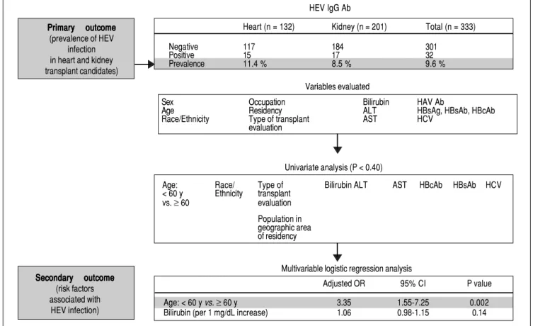
sults. The prevalence of anti-HEV IgG was 11.4% (15/132) for heart transplant candidates and 8.5% (17/201) for kidney transplant candidates, with an overall seroprevalence of 9.6% (32/333). None of the patients tested positive for HEV RNA in the serum. On multivariable analysis, age older than 60 years was associated with HEV infection (adjusted odds ratio, 3.34; 95% CI, 1.54-7.24; P = 0.002). Conclusions.Conclusions.Conclusions.Conclusions.Conclusions. We conclude that there was no evidence of acute HEV infection in this pretransplant population and that old-er age seems to be associated with positive anti-HEV IgG.
Key words. Key words. Key words. Key words.
Key words. Risk Factors. Epidemiology. Diagnosis. Enzyme-linked immunosorbent assay. Nucleic acid amplification techniques. January-February, Vol. 15 No. 1, 2016: 33-40
ORIGINAL ARTICLE
INTRODUCTION
Solid-organ transplant (SOT) recipients are a popula-tion at risk for development of severe complicapopula-tions asso-ciated with hepatitis E virus (HEV) infection that include acute and chronic hepatitis, graft dysfunction and cirrhosis. In SOT recipients, chronic HEV can develop in 60 to 80% of patients, with acute infection.1-3 Accelerated
pro-gression of liver fibrosis and cirrhosis also has been re-ported in approximately 15% of patients.1 Most of these
patients have been described in locally acquired HEV in-fection secondary to genotype 3.4 Recent evidence
demon-strates that monotherapy with ribavirin is effective to achieve sustained virologic response after 3 months of therapy in 78% (46/59) of SOT recipients with acute HEV infection.5
In a series of 274 heart transplant recipients, the anti-HEV immunoglobulin G (IgG) seroprevalence was 11% (31/274), compared with 2% (11/537) in healthy controls, and the prevalence of chronic infection was 2% (4/274).6 In
contrast, the reported prevalence of chronic infection in kidney transplant patients may be as high as 80% (12/15).7
In the US general population, the anti-HEV IgG sero-prevalence has been reported as high as 21% (3,925/18,695), with a higher prevalence in men, non-Hispanic whites, Midwest residents, and metropolitan residents.8 However
a recent study using a high performance assay reported a much lower seroprevalence of 6% (529/8,814).9 Risk
fac-tors associated with positive seroprevalence include pet ownership and consumption of organ meats such as liv-er.8,10 In a French study of HEV infection in SOT
recipi-ents, the only risk factor associated with HEV infection was consumption of wild game.3
The cost benefit of routine pretransplant screening for HEV infection remains to be defined. Data are also limited on the prevalence of HEV infection in the pretransplant population in SOT candidates.
Pretransplant seroprevalence has been evaluated in 700 kidney and liver transplant recipients screened on the day of their transplant.10 Positive anti-HEV IgG and anti-HEV
The Official Journal of the Mexican Association of Hepatology, the Latin-American Association for Study of the Liver and
the Canadian Association for the Study of the Liver
Manuscript received: Manuscript received:Manuscript received:
Manuscript received:Manuscript received: April 06, 2015. Manuscript accepted:Manuscript accepted:Manuscript accepted:Manuscript accepted:Manuscript accepted: May 02, 2015.
immunoglobulin M (IgM) were found in 14% (99/700). No association between demographic or clinical factors and HEV serum antibodies was identified, and none of the patients tested positive for HEV RNA.
We evaluated serum from heart and kidney transplant candidates in our tertiary care center as part of the pre-transplant evaluation. The main outcome of our study was to identify the prevalence of serum anti-HEV IgG and HEV RNA in heart and kidney transplant candidates.
MATERIAL AND METHODS
Patients
We evaluated 337 adult heart and kidney transplant can-didates for anti-HEV IgG antibodies and HEV RNA at Mayo Clinic Hospital, Phoenix, Arizona, between March 1, 2011, and August 31, 2013. Mayo Clinic Arizona is part of the United Network for Organ Sharing (UNOS) re-gion 5 that includes Arizona, California, Nevada, New
Mexico and Utah. Most of the heart and kidney candidates had residence in the west of the United States. The number of patients that was born in other countries was not analyzed, however historically the transplant popula-tion served at Mayo Clinic Arizona is mainly from US born citizens with a very few number of patients from other countries which is limited by UNOS regulations.
Archived frozen serum was tested, all patients were older than age 18 years, and informed consent was ob-tained for clinical evaluation prior to transplant. The study protocol had approval from the Mayo Clinic Institutional Review Board. Demographic and clinical characteristics were obtained from the electronic medical record for each patient (Figure 1).
Anti-HEV IgG and HEV RNA testing
A commercially available qualitative in vitro enzyme-linked immunosorbent assay (ELISA) to detect IgG anti-bodies against HEV (recomWell; Mikrogen; Neuried,
Figure 1. Figure 1. Figure 1. Figure 1.
Figure 1. Summary of study results and outcomes of the pretransplant prevalence of hepatitis e virus infection in a solid-organ transplant population of 333 patients. None of the 333 patients included in the analysis tested positive for hepatitis E virus RNA in the serum. Ab: indicates antibody. ALT: alanine ami-notransferase. AST: aspartate amiami-notransferase. HAV Ab: hepatitis A virus antibody. HBcAb: hepatitis B core antibody. HBsAb: hepatitis B surface antibody. HBsAg: hepatitis B surface antigen. HCV: hepatitis C virus. HEV: hepatitis E virus. HEV IgGAb: hepatitis E virus IgG immunoglobulin G antibody. OR: odds ratio. y: years.
HEV IgG Ab
Primary outcome Primary outcome Primary outcome Primary outcome Primary outcome (prevalence of HEV
infection in heart and kidney transplant candidates)
Heart (n = 132) Kidney (n = 201) Total (n = 333)
Negative 117 184 301
Positive 15 17 32
Prevalence 11.4 % 8.5 % 9.6 %
Variables evaluated
Secondary outcome Secondary outcome Secondary outcome Secondary outcome Secondary outcome
(risk factors associated with HEV infection)
Sex Occupation Bilirubin HAV Ab
Age Residency ALT HBsAg, HBsAb, HBcAb
Race/Ethnicity Type of transplant AST HCV
evaluation
Adjusted OR 95% CI P value
Age: < 60 y vs. ≥ 60 y 3.35 1.55-7.25 0.002
Bilirubin (per 1 mg/dL increase) 1.06 0.98-1.15 0.14 Univariate analysis (P < 0.40)
Multivariable logistic regression analysis
Age: Race/ Type of Bilirubin ALT AST HBcAb HBsAb HCV
< 60 y Ethnicity transplant vs. ≥ 60 evaluation
35
Hepatitis E virus serum antibodies and RNA prevalence. , 2016; 15 (1): 33-40
Germany) was used. This test is based on the principle of an indirect sandwich ELISA and can identify antibodies against HEV genotypes 1 and 3. A positive result indicates a previous or an active primary infection. A qualitative in vitro nucleic acid assay system for the detection of HEV RNA in serum was used (Procleix HEV Assay; Hologic Gen-Probe Inc, San Diego, CA). This assay is currently under development and is not available for commercial use. The assay involves 3 main steps:
• Sample preparation using a magnetic-based specific target capture method.
• HEV RNA target amplification by transcription-medi-ated amplification, and
• Detection of the amplification products (amplicon) using chemiluminescent nucleic acid probes.
Development data indicate that the assay has a 95% limit of detection of approximately 10 IU/mL (HEV World Health Organization [WHO] Standard 6329/10) and is ca-pable of detecting the 4 known HEV genotypes with simi-lar sensitivity. Preliminary testing showed specificity of 99.95%. A positive result indicates active infection.
Sera from 337 heart and kidney transplant candidates were tested using the Procleix HEV assay on the fully au-tomated Procleix Panther System (nucleic acid test sys-tem) for the presence of HEV RNA. The serum samples were initially collected for anti-HLA antibody screening tests routinely performed on all SOT candidates, had been retained frozen (-80°C) for the required period of time, and were ready to be discarded. The approximate volume of the samples was between 1 and 2 mL. Samples were transferred to proprietary tubes designed for low-volume samples and barcoded for tracking and data analysis. Sam-ples were then run on the Procleix Panther System using a research-use-only notebook lot of the reformulated Pro-cleix HEV assay reagent kit. For complete testing, a total of 2 runs was performed. Appropriate negative calibrators and positive calibrators (in vitro transcript of HEV 3a whose sequence was derived from the HEV WHO Standard 6329/ 10 [nucleotide sequence]) were included in each run. Af-ter a run, the result reports were printed and the raw data were exported for further analysis using Microsoft Excel (Microsoft Corp, Redmond, Washington) software.
Statistical analysis
Descriptive statistics were used to summarize patient demographic and clinical characteristics. Median and range were reported for continuous variables, and count and percentage were reported for nominal variables. Char-acteristics were compared by type of transplant (heart or kidney) and by negative or positive anti-HEV IgG using
the Wilcoxon rank-sum test or the χ2 test. Possible factors
associated with HEV infection were evaluated by applying logistic regression to model the probability of positive anti-HEV IgG. For the univariate analysis comparing posi-tive and negaposi-tive anti-HEV IgG, a variable with P < 0.40 was considered in the multivariable model. The higher cutoff for significance was chosen because of the small sample size for those with positive anti-HEV IgG. Back-ward elimination was conducted to select the set of varia-bles, and any variable with P < 0.15 was retained in the model. The adjusted odds ratios (ORs), the corresponding 95% CIs, and P values were reported. All analyses were performed in SAS 9.2 (SAS Institute Inc). Two-sided tests were used, and statistical significance was defined as P < 0.05.
RESULTS
We studied 337 heart and kidney transplant candidates to determine the prevalence of HEV infection. Four patients were excluded from analysis because of duplicated or miss-ing clinical data. We therefore evaluated demographic char-acteristics and routine laboratory tests of 333 patients (132 heart and 201 kidney) (Tables 1 and 2; Figure 1).
The prevalence for HEV IgG antibodies was 11.4% (15/ 132) for heart transplant candidates and 8.5 % (17/201) for kidney transplant candidates, with an overall seropreva-lence of 9.6% (32/333). None of the 333 patients tested positive for HEV RNA in the serum.
To evaluate possible risk factors associated with HEV infection, we compared the characteristics of patients with positive results for HEV IgG antibodies to those of pa-tients with negative results for HEV IgG antibodies. We also compared heart vs. kidney transplant groups and found statistically significant differences associated with age, race/ethnicity, population in geographic area of residence, total bilirubin, alanine aminotransferase (ALT), aspartate aminotransferase (AST), hepatitis A virus (HAV) antibod-ies, and hepatitis B surface antibodies (HBsAb) (Tables 1 and 2; Figure 1).
Heart transplant candidates were more frequently white (P < 0.001) and had higher total bilirubin, ALT, and AST levels (all P < 0.001) than kidney transplant candi-dates. Compared to heart transplant candidates, kidney transplant candidates had a higher percentage of positive HAV antibodies (47.9 vs. 28.3%; P = 0.001) and HBsAb (61.6 vs. 15.2%; P < 0.001).
Unzueta A, et al.
, 2016; 15 (1): 33-40
Variable Heart transplant Kidney transplant P valueb Negative Positive P valueb Total (n = 333)
candidates (n = 132) candidates (n = 201) anti-HEV IgG anti-HEV IgG (n = 301) (n = 32)
Sex 0.23 0.59
Female 39 (29.5) 72 (35.8) 99 (32.9) 12 (37.5) 111 (33.3)
Male 93 (70.5) 129 (64.2) 202 (67.1) 20 (62.5) 222 (66.7)
Age, yc 0.47 0.009
20-29 6 (4.5) 9 (4.5) 15 (5.0) 0 (0) 15 (4.5)
30-39 12 (9.1) 22 (10.9) 34 (11.3) 0 (0) 34 (10.2)
40-49 21 (15.9) 46 (22.9) 64 (21.3) 3 (9.4) 67 (20.1)
50-59 39 (29.5) 47 (23.4) 78 (25.9) 8 (25.0) 86 (25.8)
≥ 60 54 (40.9) 77 (38.3) 110 (36.5) 21 (65.6) 131 (39.3)
Race/ethnicityd < 0.001 .03
White 94 (71.2) 89 (44.3) 163 (54.2) 20 (62.5) 183 (55.0)
Hispanic 17 (12.9) 31 (15.4) 39 (13.0) 9 (28.1) 48 (14.4)
Native American 1 (0.8) 50 (24.9) 50 (16.6) 1 (3.1) 51 (15.3)
Black 16 (12.1) 21 (10.4) 36 (12.0) 1 (3.1) 37 (11.1)
Asian 4 (3.0) 10 (5.0) 13 (4.3) 1 (3.1) 14 (4.2)
Occupation typee 0.68 > 0.99
Professional 26 (19.7) 46 (22.9) 65 (21.6) 7 (21.9) 72 (21.6)
Managerial/technical 18 (13.6) 22 (10.9) 37 (12.3) 3 (9.4) 40 (12.0)
Skilled nonmanual 38 (28.8) 61 (30.3) 88 (29.2) 11 (34.4) 99 (29.7)
Skilled manual 26 (19.7) 37 (18.4) 57 (18.9) 6 (18.8) 63 (18.9)
Environmental manual 12 (9.1) 11 (5.5) 21 (7.0) 2 (6.3) 23 (6.9)
Other 12 (9.1) 24 (11.9) 33 (11.0) 3 (9.4) 36 (10.8)
Residence by US regionf 0.27 > 0.99
West 130 (98.5) 190 (94.5) 289 (96.0) 31 (96.9) 320 (96.1)
Other 2 (1.5) 11 (5.5) 12 (4.0) 1 (3.1) 13 (3.9)
Residence by populationg < 0.001 0.39
Metropolitan 92 (69.7) 103 (51.2) 174 (57.8) 21 (65.6) 195 (58.6)
Non-metropolitan 40 (30.3) 98 (48.8) 127 (42.2) 11 (34.4) 138 (41.4)
Anti-HEV IgG: anti-HEV immunoglobulin G antibody. aValues are number (percentage) unless indicated otherwise. bBy the χ2 test or the Fisher exact test. cAge category percentages for heart transplant
candidates (n = 132) and for all transplant candidates (n = 333) total < 100% due to rounding. dRace/ethnicity category percentages for negative anti-HEV IgG transplant candidates (n = 301) total >
100% due to rounding; and race/ethnicity category percentages for positive anti-HEV IgG transplant candidates (n = 32) total < 100% due to rounding. eOccupation type category percentages for all
transplant candidates (n = 333) total < 100% due to rounding. fGeographic region was defined by standard US Census Bureau categories: Northeast, Midwest, South, and West. gMetropolitan residence
37
Hepatitis E virus serum antibodies and RNA prevalence. , 2016; 15 (1): 33-40
residence region because these variables have been associ-ated with HEV IgG antibodies in the general US popula-tion8 but only white race was statistically significant in the
univariate analysis.
DISCUSSION
The pre-transplant HEV seroprevalence (9.6%) was higher for heart candidates (11.4%) than for kidney candi-dates (8.5%), with no evidence of acute infection based on HEV RNA test results.
Previously, a few studies have evaluated pretransplant seroprevalence of HEV infection.6,10 A French study found
HEV seroprevalence of 14.1% (99/700) in a group of kidney (14.5% [77/529]) and liver (12.9% [22/171]) transplant can-didates.10 A German study found a prevalence of 1.5% (4/
274) in heart transplant candidates and a prevalence of 7.3% (10/137) in nontransplanted cardiac patients.6 Acute HEV
reactivation was not confirmed in SOT in a study that evaluated patients with positive HEV IgG antibodies but negative HEV RNA at transplant and at 1-year follow-up.10,12
Studies of seroprevalence of HEV infection in devel-oped countries have variable results, most likely because of differences in the prevalence and incidence of HEV in-fection within and among different countries.11 Another
possible explanation for these differences is the use of as-says with poor sensitivity for detecting IgG and IgM HEV antibodies, resulting in underestimation of seropreva-lence.9,12,13
In our study, we used a commercially available ELI-SA test for detection of HEV IgG antibodies (recomWell; Mikrogen, Neuried, Germany) that has a good analytical sensitivity compared with other assays.14
Currently, there are no US Food and Drug Administra-tion-approved serologic assays in the US, which may have limited the diagnosis of HEV infection. Until re-cently, molecular assays were not standardized and had substantially varied performance among laboratories. WHO developed an international standard strain (code number 6329/10) using a genotype 3a HEV strain to im-prove interlaboratory results for detection and quantifi-cation of HEV RNA.15
The higher prevalence of HEV IgG antibodies found in heart transplant candidates is consistent with that reported from a previous study that evaluated heart transplant recip-ients.6 A possible explanation for this finding could be the
higher exposure to blood products in patients with ad-vanced cardiac disease.
All patients who had significantly elevated bilirubin were heart transplant candidates, and this finding could be secondary to their more severe disease compared to kid-ney transplant patients.
We found that kidney transplant candidates compared with heart transplant candidates had a higher prevalence of antibodies for HAV (47.9 vs. 28.3%, p = 0.001) and anti-HBs (61.6 vs. 12.2%, p < 0.001) this could represent a higher rate of vaccination in this population since a signif-icant number of kidney transplant candidates are usually on hemodialysis and could have been screened more ex-tensively for HAV and HBV than heart transplant candi-dates.
In our study previous exposure to HBV infection with positive HBcAb was similar in heart and kidney candi-dates (4.4 vs. 5.8%) and only one kidney transplant candi-date had a positive HBsAg. Current guidelines recommend vaccination for HAV in heart and kidney can-didates and recipients that have increase risk of exposure. Solid organ transplant candidates that have a negative HB-sAb should receive vaccination for HBV since the risk in-creases due to long-term immunosuppression.16 In heart
transplant patients it has been reported that up to 20% of patients acquired hepatitis B after transplantation.17
Re-garding HCV antibodies, the prevalence was similar in heart and kidney transplant candidates (6.4 vs. 7.8%). The seroprevalence of HCV infection in patients that are on hemodialysis has been estimated as 13.5% compared with 3% in the general population18 and is even higher after
kid-ney transplantation (11 to 49%).19 In heart transplant
recip-ients the prevalence of HCV has been reported from 7 to 18%.20
Our study had several limitations, and our results should be interpreted in the context of the study design. Considering that in the US, the incidence rate of HEV in-fection has been calculated as 7/1,000 persons per year us-ing a model that estimates incidence based on seroprevalence data from NHANES III,21 our sample size
is small for detecting new cases of HEV infection. We did not assess HEV markers after transplantation and the prob-ability of detecting HEV RNA in a small number of non-immunosuppressed candidates for SOT is quite low and does not predict the outcome or the possible complica-tions after transplantation. In the context of acute HEV in-fection, the period of viremia may be brief, and negative HEV RNA does not exclude the diagnosis, in that case, testing serum for anti-HEV IgM antibodies would have been more accurate to detect patients with acute HEV and HEV RNA below the level of detection. We used a molec-ular assay to detect HEV RNA that has not yet been vali-dated, although preliminary testing has shown it to have a specificity of 99.95% (95% CI, 99.88-99.98%).
addi-Unzueta A, et al.
, 2016; 15 (1): 33-40
Variable Heart transplant Kidney transplant P value Negative Positive P value Total candidates (n = 132) candidates (n = 201) anti-HEV IgG anti-HEV IgG (n = 333)
(n = 301) (n = 32)
Bilirubin, mg/dL < 0.001a 0.08a
Median (range) 0.7 (0.1-38.7) 0.4 (0.1-1.4) 0.4 (0.1-38.7) 0.5 (0.1-30.6) 0.4 (0.1-38.7)
ALT, IU/L < 0.001a 0.31a
Median (range) 30 (6-2,036) 19.5 (10-86) 22 (6-2,036) 25.5 (10-86) 23 (6-2,036)
AST, IU/L < 0.001a 0.05a
Median (range) 30 (12-5,739) 21 (9-95) 23 (9-5,739) 27.5 (11-95) 24 (9-5,739)
HAV Ab, n (%) (n = 92) (n = 190) 0.001b (n = 253) (n = 29) 0.98b
Negative 66 (71.7) 99 (52.1) 148 (58.5) 17 (58.6) 165 (58.5)
Positive 26 (28.3) 91 (47.9) 105 (41.5) 12 (41.4) 117 (41.5)
HBcAb, n (%) (n = 91)d- (n = 191) 0.78c (n = 253) (n = 29) 0.38c
Negative 87 (96.5) 180 (94.2) 238 (94.1) 29 (100) 267 (94.7)
Positive 4 (4.4) 11 (5.8) 15 (5.9) 0 (0) 15 (5.3)
HBsAg, n (%) (n = 92) (n = 200) > 0.99c (n = 263) (n = 29) > 0.99c
Negative 92 (100) 199 (99.5) 262 (99.6) 29 (100) 291 (99.7)
Positive 0 1 (0.5) 1 (0.4) 0 1 (0.3)
HBsAb, n (%) (n = 92) (n = 185) < 0.001b (n = 248) (n = 29) 0.35
b-Negative 78 (84.8) 71 (38.4) 131 (52.8) 18 (62.1) 149 (53.8)
Positive 14 (15.2) 114 (61.6) 117 (47.2) 11 (37.9) 128 (46.2)
HCV Ab, n (%) (n = 94) (n = 192)d 0.66b (n = 257) (n = 29) 0.71c
Negative 88 (93.6) 177 (92.2) 237 (92.2) 28 (96.6) 265 (92.7)
Positive 6 (6.4) 15 (7.8) 20 (7.8) 1 (3.4) 21 (7.3)
ALT: alanine aminotransferase. anti-HEV IgG: hepatitis E virus immunoglobulin G antibody. AST: aspartate aminotransferase. HAV Ab: hepatitis A virus antibody. HBcAb: hepatitis B core antibody. HBsAb: hepatitis B surface antibody. HBsAg: hepatitis B surface antigen. HCV Ab: hepatitis C virus antibody. a By the Wilcoxon rank-sum test. bBy the χ2 test. c By the Fisher exact test. d Percentages
39
Hepatitis E virus serum antibodies and RNA prevalence. , 2016; 15 (1): 33-40
tional investigation. We conclude that the prevalence of HEV IgG antibodies was higher in heart than in kidney transplant candidates and that there was no evidence of ac-tive infection (HEV RNA) in any of the patients. The only variable associated with HEV-positive IgG was age ≥ 60 years.
ABBREVIATIONS
• ALT: alanine aminotransferase.
• anti-HEV IgG: hepatitis E virus IgG antibody. • AST: aspartate aminotransferase.
• ELISA: enzyme-linked immunosorbent assay. • HAV: hepatitis A virus.
• HB: hepatitis B.
• HBsAb: hepatitis B surface antibody. • HEV: hepatitis E virus.
• IgG: immunoglobulin G. • IgM: immunoglobulin M.
• NHANES: National Health and Nutrition Examina-tion Survey.
• OR: odds ratio.
• SOT: solid-organ transplant. • WHO: World Health Organization.
DISCLOSURE
The authors declare no conflicts of interest.
Part of this study was presented at the 2014 World Transplant Congress, San Francisco, California, July 30, 2014 as a poster presentation.
AUTHORS CONTRIBUTIONS
Concept/design: Dr. Unzueta, Dr. Valdez, Dr. Rakela. Data analysis/statistics: Dr. Chang.
Drafting article: Dr. Unzueta.
Critical revision of article: Dr. Rakela, Dr. Heilman, Dr. Scott, Dr. Douglas.
Data collection: Dr. Valdez, Ms. Desmarteau.
REFERENCES
1. Kamar N, Garrouste C, Haagsma EB, Garrigue V, Pischke S, Chauvet C, Dumortier J, et al. Factors associated with chronic hepatitis in patients with hepatitis E virus infection who have received solid organ transplants. Gastroenterolo-gy 2011; 140: 1481-9.
2. Moal V, Motte A, Kaba M, Gerolami R, Berland Y, Colson P. Hepatitis E virus serological testing in kidney transplant re-cipients with elevated liver enzymes in 2007-2011 in south-eastern France. Diagn Microbiol Infect Dis 2013; 76: 116-8.
3. Legrand-Abravanel F, Kamar N, Sandres-Saune K, Garrou-ste C, Dubois M, Mansuy JM, Muscari F, et al.
Characteris-tics of autochthonous hepatitis E virus infection in solid-or-gan transplant recipients in France. J Infect Dis 2010; 202: 835-44.
4. Unzueta A, Rakela J. Hepatitis E infection in liver transplant recipients. Liver Transpl 2014; 20(1): 15-24.
5. Kamar N, Izopet J, Tripon S, Bismuth M, Hillaire S, Dumortier J, Radenne S, et al. Ribavirin for Chronic Hepatitis E Virus In-fection in Transplant Recipients. N Engl J Med 2014; 370: 1111-20.
6. Pischke S, Stiefel P, Franz B, Bremer B, Suneetha PV, Heim A, Ganzenmueller T, et al. Chronic hepatitis e in heart trans-plant recipients. Am J Transplant 2012; 12: 3128-33. 7. Moal V, Legris T, Burtey S, Morange S, Purgus R, Dussol B,
Garcia S, et al. Infection with hepatitis E virus in kidney transplant recipients in southeastern France. J Med Virol
2013; 85: 462-71.
8. Kuniholm MH, Purcell RH, McQuillan GM, Engle RE, Wasley A, Nelson KE. Epidemiology of hepatitis E virus in the United States: results from the Third National Health and Nutrition Examination Survey, 1988-1994. J Infect Dis 2009; 200: 48-56.
9. Ivo D, Fausta D, Pardha D, Calistus D, Patrick SK, Michael C. Current epidemiology of hepatitis E virus infection in the United States: Low seroprevalence in the National Health and Nutrition Evaluation Survey. Hepatology 2014; 60: 815-22.
10. Legrand-Abravanel F, Kamar N, Sandres-Saune K, Lhomme S, Mansuy JM, Muscari F, Sallusto F, et al. Hepatitis E virus infection without reactivation in solid-organ transplant recipi-ents, France. Emerg Infect Dis 2011; 17: 30-7.
11. Nelson KE, Kmush B, Labrique AB. The epidemiology of hep-atitis E virus infections in developed countries and among im-munocompromised patients. Expert Rev Anti Infect Ther
2011; 9: 1133-48.
12. Wenzel JJ, Preiss J, Schemmerer M, Huber B, Jilg W. Test performance characteristics of anti-HEV IgG assays strong-ly influence hepatitis e seroprevalence estimates. J Infect Dis 2013; 207: 497-500.
13. Drobeniuc J, Meng J, Reuter G, Greene-Montfort T, Khudyak-ova N, DimitrKhudyak-ova Z, Kamili S, et al. Serologic assays specific to immunoglobulin M antibodies against hepatitis E virus: pangenotypic evaluation of performances. Clin Infect Dis
2010; 51: e24-e27.
14. Pas SD, Streefkerk R, Pronk M, de Man RA. Diagnostic per-formance of selected commercial HEV IgM and IgG ELISAs for immunocompromised and immunocompetent patients. J Clin Virol 2013; 58: 629-34.
15. Baylis SA, Blumel J, Mizusawa S, Matsubayashi K, Sakata H, Okada Y, Nubling C, et al. World health organization interna-tional standard to harmonize assays for detection of hepati-tis e virus RNA. Emerg Infect Dis 2013; 19: 729-35.
16. Danziger-Isakov L, Kumar D. AST Infectious Diseases Com-munity of Practice. Vaccination in solid organ transplanta-tion. Am J Transplant 2013; 13(Suppl. 4): 311-7.
17. Wedemeyer H, Pethig K, Wagner D, Flemming P, Oppelt P, Petzold DR, Haverich A, et al. Long-term outcome of chronic hepatitis B in heart transplant recipients. Transplantation
1998; 66: 1347.
18. Fissell RB, Bragg-Gresham JL, Woods JD, Jadoul M, Gillespie B, Hedderwick SA, Rayner HC, et al. Patterns of hepatitis C prevalence and seroconversion in hemodialysis units from three continents: the DOPPS. Kidney Int 2004; 65: 2335-42.
20. Fagiuoli S, Minniti F, Pevere S, Farinati F, Burra P, Livi U, Nac-carato R, et al. HBV and HCV infections in heart transplant recipients. J Heart Lung Transplant 2001; 20: 718-24. 21. Faramawi MF, Johnson E, Chen S, Pannala PR. The
inci-dence of hepatitis e virus infection in the general population of the USA. Epidemiol Infect 2011; 139(8): 1145-50.
Correspondence and reprint request: Alberto Unzueta, M.D. Division of Gastroenterology, Hepatology and Nutrition, University of Florida, 1600 SW Archer Rd P.O. Box 100277,
Gainesville, FL 32610. USA. Ph.: 352-327-3018. Fax: 352-392-7393