Epilt-psia. 29(5):63&634. 1988 Raven Press, Ltd.. New York
0 International League Against Epilepsy
Paroxysmal Aphasias
Alfredo Ardila and Maria Victoria Lopez
Instituto Neurologico de Colombia, Bogota, Colombia
Summary: Forty cases of paroxysmal aphasia were found in a sample of 4,000 patients with epilepsy. Twenty-five had structural brain damage demonstrated by CT scan. Except for two cases, the epileptic focus was located in the left hemisphere. Two patients had a paroxysmal al-
exia associated with the aphasic disorder. Though prelim- inary, our data suggest a correlation of dysphasic com- ponents with posterior lesions and phonatory compo- nents with anterior lesions. Key Words: Epilepsy-Partial seizures-Aphasia.
Paroxysmal language disorders, first described by Jackson in 1874 (1958), are infrequently ob- served and study is frequently limited to history and interictal examination. Language disorders may ap- pear either as a focal form of epilepsy that may or may not progress to a generalized seizure, or during the postictal period when confusion, somnolence, and other symptoms also occur.
Paroxysmal aphasias have been classified into two different groups: dysphasic seizures (paroxys- mal posterior aphasia) in the psychic seizure group, and phonatory seizure (paroxysmal anterior apha- sia) in the motor seizure group (Commission, 1981). Previous studies have identified the focus of ep- ileptic activity usually in the left hemisphere, how- ever, the expressive type of paroxysmal aphasia is more often found in left-handed than in right- handed patients (Hecaen and Piercy, 1956). Ala- jouanine and Sabouraud ( I 960) distinguished three types of speech disorders associated with epilepsy: aphasia, speech arrest, and dysarthria. Hecaen and Angelergues (1960) reported 32 cases of verbal au- tomatisms in 176 cases of speech disorders. There are several reports of aphasic epileptic status (De Pasquet et al., 1976; Hamilton and Matthews, 1979). Wilson et al. (1983) reported a case of par- oxysmal jargon aphasia, and Peled et al. (1984) de- scribed a case of paroxysmal speech arrest associ- ated with damage in the supplementary motor area. Ardila et al. (1986) correlated the topography of
Received September 1987; revision accepted December 1987. Address correspondence and reprint requests to Dr. A. Ardila at Miami Institute of Psychology, 1401 S.W. First Street, Miami,
FL 33135, U.S.A.
cortical damage with the clinical characteristics of paroxysmal aphasias. They suggest distinguishing three kinds of expressive type paroxysmal apha- sias: paroxysmal Broca’s aphasia, language arrest (parasagittal area involving the supplementary
mo-
tor area), and paroxysmal palilalia. In theory, it should also be possible to find variants of posterior paroxysmal aphasias.
METHOD
Subjects
After reviewing approximately 4,000 clinical his- tories of epileptic patients registered in the Neuro- logical Institute .of Colombia during the last 13 years, 40 patients (1%) with language phenomena were identified. All patients had an EEG with an epileptic focus. We assume that the actual number might be much higher since language phenomena are often not considered by the patient or the ex- aminer. Those patients with a CT scan showing fo- cal structural damage were selected for study (the CT scan was introduced in Colombia in 1977). The final sample size was 25 ( 1 1 male, 14 female, aver- age age 30.8, range 12-64 years). Follow-up was between 2 weeks and 7 years. All but one patient were right-handed. Seizures appeared after age 20 in all but one patient.
Instruments
Two types of CT scanner techniques were used: from 1977 to June 1982 a General Electric CT/N scanner with a 160 x 160 matrix and 1.0 cm cuts, and from July 1982 to 1986, a General Electric CTTI 8000 with a 320 x 320 matrix and 0.5 cm cuts. A standard template was designed to interpret the le-
P A R O X Y S M A L APHASIAS 63 1
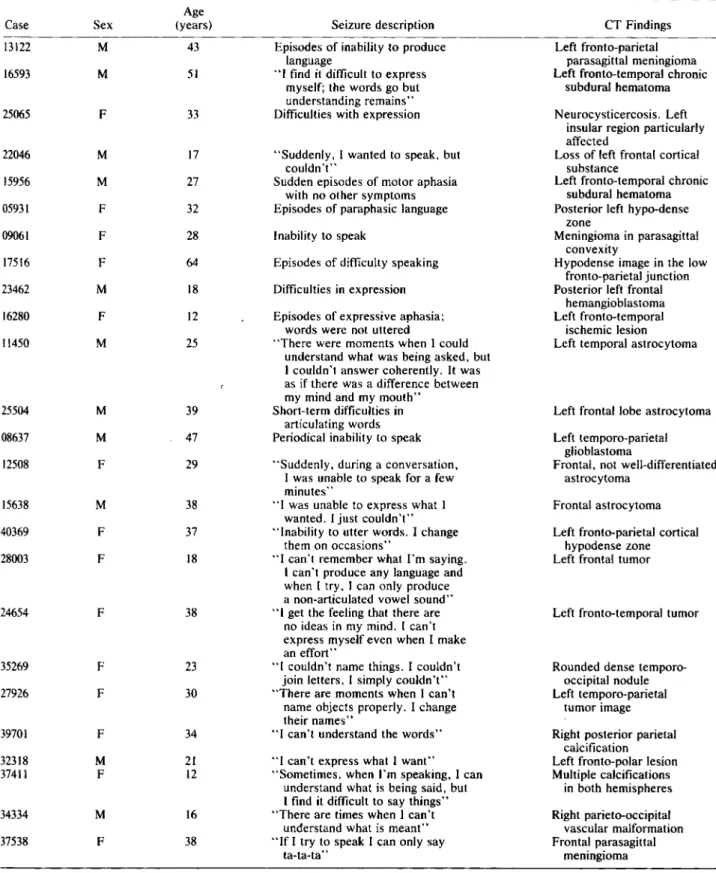
TABLE 1. Paroxysmal phenomena and CT findings
Case 13122 16593 25065 22046 15956
0593 1
0906 1
17516
23462
16280
I1450
Age
Sex (years)
25504 M
08637 M
12508 F
15638 M
40369 F
28003 F
M 43
M 51
F 33
M 17
M 27
F 32
F 28
F 64
M 18
F 12
M 25
24654
35269
27926
3970 1
323 I 8 3741 1 34334 37538 F F F F M F M F 39 47 29 38 37 18 Seizure description
Episodes of inability to produce language
"I find it difficult to express myself; the words go but understanding remains" Difficulties with expression
"Suddenly. I wanted to speak, but
Sudden episodes of motor aphasia
Episodes of paraphasic language
Inability to speak
Episodes of difficulty speaking
Difficulties in expression
Episodes of expressive aphasia;
"There were moments when 1 could couldn't''
with no other symptoms
words were not uttered
understand what was being asked, but I couldn't answer coherently. It was as if there was a difference between my mind and my mouth"
articulating words Short-term difficulties in
Periodical inability to speak
~~
C T Findings
Left fronto-parietal parasagittal meningioma Left fronto-temporal chronic
subdural hematoma
Neurocysticercosis. Left insular region particularly affected
Loss of left frontal cortical substance
Left fronto-temporal chronic subdural hematoma Posterior left hypo-dense
zone
Meningioma in parasagittal convexity
Hypodense image in the low fronto-parietal junction Posterior left frontal
hemangioblastoma Left fronto-temporal
ischemic lesion
Left temporal astrocytoma
Left frontal lobe astrocytoma
Left temporo-parietal
Frontal, not well-differentiated glioblastoma
astrocytoma
Frontal astrocytoma
Left fronto-parietal cortical hypodense zone Left frontal tumor "Suddenly, during a conversation,
I was unable to speak for a few minutes"
"I was unable to express what 1 wanted. 1 just couldn't'' "Inability to utter words. I change
them on occasions"
"I can't remember what I'm saying. I can't produce any language and when I try. I can only produce a non-articulated vowel sound" "1 get the feeling that there are
no ideas in my mind. I can't express myself even when I make an effort"
join letters, I simply couldn't"
name objects properly. I change their names"
38 Left front 0- temporal tumor
23 "I couldn't name things. I couldn't Rounded dense temporo-
30 "There are moments when I can't Left temporo-parietal
34 "1 can't understand the words" Right posterior parietal
21 "I can't express what 1 want" Left fronto-polar lesion 12 "Sometimes, when I'm speaking, I can Multiple calcifications
in both hemispheres
16 "There are times when 1 can't Right parieto-occipital
38
occipital nodule
tumor image
calcification
understand what is being said, but I find it difficult to say things"
understand what is meant" "If I try to speak I can only say
ta-ta-ta" meningioma
vascular malformation Frontal parasagittal
sions in a uniform manner. The template included 10 standard scanner cuts, from the base to the most cortical portions, each corresponding to a standard scanner cut.
Procedure
Identified lesions from the 25 patients were inter- preted using the standard template. Different le- sions were superimposed on the same template and
632 A . ARDlLA A N D M . V . LOPEZ
the topography of the lesion was correlated with each patient’s seizures.
RESULTS
Table I correlates the experiential description of the seizures with the scanner findings. Seizures were divided into two groups: those corresponding to a paroxysmal disorder associated with language production (phonatory seizures, 17 cases); and those corresponding to a posterior paroxysmal aphasia, that is, inability to understand language, paraphasias, difficulties in remembering words, etc. (dysphasic seizures, eight cases). Classification was based on the clinical report of the patient and friends or relatives. The basic classification criteria was whether the seizure resembled an anterior (nonfluent) or posterior (fluent) form of aphasia. In all but two cases the epileptic focus was located in the left hemisphere; one of these patients was left- handed. In these two cases (not included in the su- perimposition), language disorder was character- ized by difficulties in understanding. Similarly, two of the patients presented a paroxysmal alexia asso- ciated with the aphasic disorder.
Figure I displays the topography of the phona- tory seizures. The lesions are grouped into two zones: a parasagittal area corresponding to the sup- plementary motor area; and a premotor area located in front of the primary motor area of the face and tongue, corresponding to Broca’s area. Many of the elements found in paroxysmal phonatory disorders correspond not only to a deficit of an aphasic nature but also to dysarthria. Thus, a third type of parox-
ysmal expressive disorder, characterized by “par- oxysmal palilalia” in which the patient repeats in a reiterative manner elements of a more complex level (words, phrases), should be expected. In the only well-documented case we studied, the damage was found in front of Broca’s area and was, as a consequence, prefrontal.
Figure 2 displays the topography found in dys- phasic seizures. As expected, lesions were located towards the posterior temporal lobe. Patients re- ported difficulties in understanding language and re- membering words, and exhibited paraphasias. In addition, two of the patients presented paroxysmal difficulties in reading (paroxysmal alexia). One showed a calcification in the occipital lobe (CH 37411 and the other a left temporo-occipital rounded nodule (CH 35269).
It is important to observe that language phenom- ena were associated with other types of seizures in
18 cases, the most frequent being autonomic, affec- tive, somatosensory, cognitive, dysmnesic, halluci- nations (visual and auditory), partial motor, adver- sive, and complex partial seizures. In 19 patients, phonatory and dysphasic seizures had evolved, at least on one occasion, to a generalized tonic-clonic seizure.
DISCUSSION
Despite limitations, some interesting conclusions can be drawn from this research. Paroxysmal lan- guage disorders have been associated with left hemisphere discharges (Frederiks, 1985) and it is
FIG. 1. Phonatory seizures. Superimposition of the CT scan images on the standard template.
PAROXYSMAL APHASIAS 633
FIG. 2. Dysphasic seizures. Superimpasition of the CT scan paroxysmal alexia are shown (HC 3741 1 and HC 35269).
usually recognized that they Fan adopt different forms. Curiously enough, two patients in our sam- ple had a focus in the right hemisphere. Paroxysmal language disorders caused by right hemisphere foci have been described by Williamson et al. (1985). Our research confirms the existence of variants in language disorders associated with epilepsy.
The description by one of our patients (CH 24654) who had a paroxysmal motor aphasia is of interest. She said that while alone and without using lan- guage in any way, she has had seizures: “1 know I have had an attack because m y mind goes blank. I know that if 1 tried to speak at that moment, I wouldn’t be able to do
so
properly. This lasts for a few seconds. It seems as though my thoughts have gone.” This could suggest the lack of an inner lan- guage, although there was no actual use of expres- sive language.The two cases of paroxysmal alexia would corre-
spond to a verbal alexia. In both cases, the foci
were posterior and correlated with difficulties in oral speech (“I can’t name things”). They did not constitute two different seizures, just one which was characterized by an inability to name things (paroxysmal anomia) and to read, due to the impos- sibility of joining letters (paroxysmal verbal alexia); if the patient tried to speak, he could not remember names and if he wanted to read, he was unable to join letters.
Phonatory seizures were twice as frequent as dysphasic. Dysphasic phenomena were more fre- quently associated with other types of seizures and were therefore considered secondary in conse-
images on the standard template, The two patients (1, 2) with
quence and were not reported. In dysphasic sei- zures, besides the association found in some cases with paroxysmal alexia, language disorders pre- sented different variants (word forgetfulness, pho- nological and/or semantic paraphasias, difficulties in understanding, etc.).
CT scan abnormalities are difficult to localize precisely. Several patients had infiltrating gliomas. some had multiple cerebral lesions, and some le- sions were quite extensive. Nevertheless, results suggest a correlation of dysphasic components with posterior lesions and phonatory components with anterior lesions. Our results should be considered only preliminary and mainly illustrative of a possi- ble approach to seizure semiology.
Acknowledgment: This research was supported in part
by a grant received from Colciencias (Fondo Colombiano
de lnvestigaciones Cientificas y Proyectos Especiales).
REFERENCES
Alajouanine T, Sabouraud A. Les perturbations paroxystiques de langage dans i’epilepsie. Encepphule 1960;49:95-133. Ardila A, Montanes P, Bernal B, Serpa A, Ruiz E. Partial psy-
chic seizures and brain organization. In1 J Neuroscience I986;30:23-32.
Commission on classification and terminology of the Internation- al League Against Epilepsy. Proposal for revised clinical and electroencephalographic classification of epileptic seizures. Epilepsia 1981 ;22:489-501.
De Pasquet EG. Gaudin ES, Bianchi A, Mendilaharsu SA. Pro- longed and monosymptomatic dysphasic status epilepticus. Neurology 1976;26:244-1.
Frederiks JAM. Paroxysmal neuropsychological disorders. In: Frederiks JAM. ed. Handbook of Clinical Nectrology. vol45:
634 A . ARDILA A N D M . V . LOPEZ
Clinical Ne~irop,psychology. Amsterdam: Elsevier Science Publishers, 1985507- 14.
Hamilton NG. Matthews T. Aphasia: the sole manifestation of focal status epilepticus. Neuroloxy 1979;29:745-8.
Hecaen H, Piercy M. Paroxysmal dysphasia and the problem of cerebral dominance. J N e u r o l Nerirosurg P s y c h i a t r y 1956; 19: 194-201.
Hecaen H, Angelergues RA. Epilepsie et troubles du language.
Jackson JH. On the scientific and empirical investigation of ep- ilepsies. In: Taylor J , ed. Selected writings ofJohn Hughlings Jackson, vol 1 . London: Hodden and Stoughton, 18741
Peled R. Harnes B, Borovich B, Sharf M. Speech arrest and supplementary motor area seizures. Neurology 1984;34:
110-1.
Williamson PD. Spencer DD, Spencer SS, Novelly R. Mattson RH. Episodic aphemia and epileptic focus in nondominant hemisphere: relieved by section of corpus callosum. Neiirol-
0g.y 1985;35: 1069-71.
Wilson A, Petty R, Perry A, Rose CR. Paroxysmal language disturbances in an epileptic treated with clobazam. Neiirolo- g y 1983;33:6524.
1958: 162-273.
&SUME
40 cas d'aphasie paroxystique ont ttt trouvts parmi 4,000 pa- tients tpileptiques. 25 patients prtsentaient une lesion ctrebrale prouvee par la tomodensitometrie. Sauf dans 2 cas, le foyer tp- ileptique se trouvait dans I'htmisphtre gauche. 2 patients prkse- ntaient une alexie paroxystique associte a I'aphasie. Nos con- statations, bien que prdiminaires, suggtrent qu'il existe une cor- rtlation entre les composantes dysphasiques et les ltsions posterieures. et les componsantes phonatoires et les ltsions an- terieures.
(P. Genton, Marseille)
RESUMEN
Se hallaron 40 casos de afasias paroxisticas en una muestra de 4.000 pacientes con epilepsia. Entre ellos, 25 presentaben un
dafio estructural mostrado escanograficamente. Exceptuando dos casos, 10s d e m h pacientes presentaban un dafio hemisfer- ico izquierdo; dos de 10s pacientes presentaban una alexia par- oxistica. Se analizan 10s diferentes tipos de alteraciones parox- isticas del lenguaje. Aunque nuestra informaci6n es preliminar, sugiere la existencia de una correlacion de 10s componenetes disfAsicos con posibles lesiones posteriores y del componente fonatorio con lesiones anteriores.
(Translation supplied by authors)