Impact of the severity of end-stage
liver disease in cardiac structure and function
Odilson Marcos Silvestre,** Fernando Bacal,** Danusa de Souza Ramos,* Jose L. Andrade,*** Meive Furtado,*** Vincenzo Pugliese,* Elisangela Belleti,* Wellington Andraus,* Flair José Carrilho,*Luiz Augusto Carneiro D’Albuquerque,* Alberto Queiroz Farias*
* Department of Gastroenterology. ** Heart Institute. *** Institute of Radiology. University of Sao Paulo School of Medicine. Brazil.
ABSTRACT
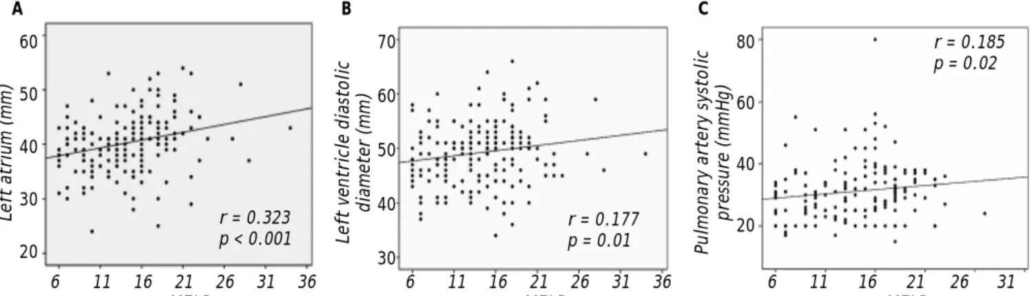
Background. The impact of end-stage liver disease (ESLD) in cardiac remodeling of patients with cirrhosis is unknown. Our aim was to correlate the severity of ESLD with morphologic and functional heart changes. Material and methods. 184 patients underwent a protocol providing data on the severity of ESLD and un-dergoing echocardiography to assess the diameters of the left atrium and right ventricle; the systolic and diastolic diameters of the left ventricle, interventricular septum, and posterior wall of the left ventricle; systolic pulmonary artery pressure; ejection fraction; and diastolic function. Severity of ESLD was assessed by the Model for End-Stage Liver Disease (MELD) score. Results. Left-atrial diameter (r = 0.323; IC 95% 0.190-0.455; p < 0.001), left-ventricular diastolic diameter (r = 0.177; IC 95% 0.033-0.320; p = 0.01) and systo-lic pulmonary artery pressure (r = 0.185; IC 95% 0.036-0.335; p = 0.02) significantly correlated with MELD score. Patients with MELD ≥ 16 had significantly higher left-atrial diameter and systolic pulmonary artery pressure, compared with patients with MELD scores < 16 points. Conclusions. Changes in cardiac structu-re and function corstructu-relate with the severity of ESLD.
Key words. Cirrhosis. Cardiac remodeling. Liver transplantation. MELD score. Cirrhotic cardiomyopathy.
Correspondence and reprint request: Alberto Queiroz Farias, M.D. Hospital das Clínicas da USP. Av. Dr. Eneas de Carvalho Aguiar 255, sala 9117. São Paulo, SP, Brazil. Zip code 05403-000
Ph.: +55-11-30674238
E-mail: [email protected]
Manuscript received: May 02, 2012. Manuscript accepted: June 26, 2012. INTRODUCTION
Advanced liver cirrhosis is associated with seve-ral cardiovascular and pulmonary abnormalities, in-cluding hyperdynamic circulation with decreased effective arterial blood volume, cirrhotic cardiomyo-pathy and arterial pulmonary hypertension.1
High cardiac output and low systemic vascular resistance index are typical features of end-stage liver disease (ESLD), and are related to the common clinical findings of increased heart rate and arterial hypotension.2
The pulmonary consequences of the hyperdyna-mic circulation and portal hypertension are hepato-pulmonary syndrome and portohepato-pulmonary
hypertension. According to the data of the European Respiratory Society Task Force on Pulmonary-He-patic Vascular Disorders, the prevalence of hepato-pulmonary syndrome can approach 20% in some series of patients awaiting liver transplantation, whereas portopulmonary hypertension has its pre-valence in the order of 5%.3 Cirrhotic
cardiomyopa-thy is characterized by an abnormal and blunted response to pathological or pharmacological stress in the absence of any other associated cardiac disea-se. The combination of features including baseline increased cardiac output, attenuated systolic con-traction and diastolic relaxation, electrophysiologi-cal repolarization abnormalities and a reduced response to beta-1 adrenergic stimulation are seen in patients with ESLD.1,4,5
Structural and functional cardiac abnormalities have been reported in patients with cirrhosis, irres-pective of its etiology, including dilation of both heart chambers. In particular, cirrhosis is associa-ted with increase in left atrium and right atrium si-zes and in right ventricle diastolic diameter.6
weighted sum of serum values of bilirubin, interna-tional normalized ratio (INR) and creatinine.7
Collectively, these parameters measure organic dysfunctions in liver cirrhosis and correlate with the severity of ESLD. The liver allocation system for orthotopic liver transplantation (OLT) changed from the classical Child-Pugh system to the MELD score in most of countries of the world after 2002 be-cause direct evidence demonstrated that mortality rates of patients waiting for liver transplantation were lower with the implementation of the MELD scoring system.8,9 We hypothesized that the severity
of ESLD could influence cardiac remodeling in cir-rhosis. The aim of this study was to assess the co-rrelation between echocardiographic abnormalities and severity of ESLD (assessed by the MELD score) in patients waiting for liver transplantation.
MATERIAL AND METHODS
Patients
From May 2009 to January 2011, 220 adult pa-tients with liver cirrhosis who presented consecuti-vely to the Department of Gastroenterology of the University of Sao Paulo Hospital were recruited for the current study. The protocol was approved by the Institutional Ethics Board Review. The informed consent was obtained from each patient. The study protocol conformed to the ethical guide-lines of the 1975 Declaration of Helsinki. All pa-tients were medically stable and currently listed for liver transplantation. The diagnosis of cirrhosis was established in all patients based on clinical and laboratory data, by imaging methods or by liver biopsy in patients with safe coagulation para-meters. The etiology of cirrhosis was hepatitis C virus infection in 81 (44%), hepatitis B in 16 (8.6%), alcohol-induced in 48 (26%), non-alcoholic steatohepatitis in 3 (1.6%), autoimmune hepatitis in 5 (3%), primary biliary cirrhosis in 5 (3%), primary sclerosing cholangitis in 2 (1%) patients and miscellaneous in the remaining 26 patients (14%). All patients with a history of alcohol intake had been abstinent from alcohol for at least 12 months before entering the study.
Twenty eight (12.7%) patients were excluded due to having primary heart disease, of whom 9 had valvar heart disease, 14 coronary artery disease, 3 systolic dysfunction with ejection fraction below 55% (ischemic cardiomyopathy n = 2; hypertensive cardiomiopathy n = 1) and 2 congenital heart disease. Eight patients (3.6%) had well-preserved liver function
and concurrent chronic renal failure requiring chro-nic hemodialysis. Because the MELD calculation is dependent on creatinine levels, patients with chro-nic renal failure were excluded because high MELD scores in patients under hemodialysis may not cor-relate to the deterioration of liver function. A total of 184 patients with ESLD remained after exclusions and were the focus of this study.
Esophageal varices at endoscopy were considered a surrogate marker of significant portal hyperten-sion. Functional renal dysfunction in cirrhosis (serum creatinine > 1.5 mg/dL) was diagnosed ac-cording to the revised criteria of the International Ascites Club.10 Out of the 105 patients on
proprano-lol use, 37 (35%) were Child-Pugh A, 53 (51%) were B, and 15 (14%) were C. These patients were found similarly distributed among all three Child-Pugh groups, without a statistically significant difference. Baseline demographic and clinical features of the included patients are shown in table 1.
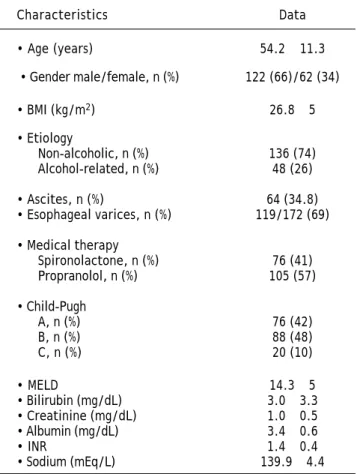
Table 1. Baseline demographic and clinical data of 184 patients with cirrhosis waiting for liver transplantation.*
Characteristics Data
• Age (years) 54.2 ± 11.3
• Gender male/female, n (%) 122 (66)/62 (34)
• BMI (kg/m2) 26.8 ± 5
• Etiology
Non-alcoholic, n (%) 136 (74) Alcohol-related, n (%) 48 (26)
• Ascites, n (%) 64 (34.8) • Esophageal varices, n (%) 119/172 (69)
• Medical therapy
Spironolactone, n (%) 76 (41) Propranolol, n (%) 105 (57)
• Child-Pugh
A, n (%) 76 (42) B, n (%) 88 (48) C, n (%) 20 (10)
• MELD 14.3 ± 5
• Bilirubin (mg/dL) 3.0 ± 3.3 • Creatinine (mg/dL) 1.0 ± 0.5 • Albumin (mg/dL) 3.4 ± 0.6
• INR 1.4 ± 0.4
• Sodium (mEq/L) 139.9 ± 4.4
Cardiac evaluation
A standard protocol, including medical history, physical examination, ECG and chest-X-ray, was performed for all patients by the same cardiologist. All patients underwent two dimensional transthora-cic echocardiography with color Doppler according to the recommendations of the American Society of Echocardiography.11 Measurement of the following
parameters were performed: left-atrial and right-ventricular diameters, left-right-ventricular systolic and diastolic diameters, interventricular septum, left-ventricular posterior wall, ejection fraction and esti-mated pulmonary artery pressure. This former was determined from the peak tricuspid regurgitation, using the simplified Bernoulli equation and combi-ning the values with an estimate of the right atrial pressure.12 The E/A ratio (Early maximal
ventricu-lar filling velocity/Atrial maximal filling velocity) was used as an index of diastolic function, and was considered abnormal when < 1.0, as reported elsewhere.13,14 All parameters were recorded in three
cardiac cycles, and the mean of the measurements was taken for analysis.
In patients with ascites requiring therapeutic pa-racentesis with albumin infusion, echocardiography was performed at least two weeks after the procedu-re to avoid inaccurate measuprocedu-rements due to heart diameters changing transiently in response to volu-me overload. Therapy with beta-blockers was stop-ped before echocardiography exam to avoid hypothetical interferences in the measurements.
MELD score
Previous data showed that the MELD score is an accurate predictor of survival in cirrhotic patients
on the waiting list for OLT7 and has been used
worldwide as a reliable parameter for liver alloca-tion policies because it correlates with the decline of the liver function.9 Three easily assessed variables,
bilirubin, INR and creatinine, are used to calculate a score that continuously ranges from 6 to a capped value at 40. The score is calculated according to the formula MELD = 9.57 (loge creatinine) + 3.78 (loge bilirubin) + 11.2 (loge INR) + 6.43, as reported el-sewhere.7 We stratified the resulting values as < 16
or ≥ 16 points because this a usual cut off point for considering OLT.15
Statistical analysis
Mann-Whitney and Wilcoxon tests were used to compare non-parametric continuous variables. Chi-square and Fisher exact tests were used to compare dichotomous variables when appropriate. P values < 0.05 were considered significant. Spearman rank correlation was used to assess the association bet-ween echocardiography parameters and MELD score. All calculations were performed with the PASW statistical package (SPSS version 18.0, Chicago, IL) software. The results were expressed as the mean ± standard deviation and 95% confidence interval when indicated.
RESULTS
Left-atrial diameter, left-ventricular diastolic dia-meter and systolic pulmonary artery pressure signi-ficantly correlated with the MELD score (Figure 1 and Table 2). Patients with more-severe liver disease (MELD ≥ 16) were found to have higher left atrium diameter and higher values of systolic pulmonary artery pressure when compared with patients who
Figure 1. Correlation between the severity of ESLD as assessed by the MELD score and echocardiography parameters.
A. MELD score and left atrium. B. MELD score and left ventricle diastolic diameter. C. MELD score and pulmonary artery systolic
pressure.ESLD: end-stage liver disease. MELD: model for end-stage liver disease.
A B C
60
50
40
30
20
Left atrium (mm)
6 11 16 21 26 31 36
MELD
r = 0.323 p < 0.001
70
60
50
40
30
Left ventricle diastolic
diameter (mm)
6 11 16 21 26 31 36
MELD
r = 0.177 p = 0.01
80
60
40
20
Pulmonary artery systolic
pressure (mmHg)
6 11 16 21 26 31
MELD
had MELD < 16 points. These results are shown in table 2. 49.3% of patients (77 out of 156) were found to have diastolic dysfunction, which did not correlate with the MELD score (p = 0.193), nor with the Child-Pugh classification (Child-Pugh A and B
vs. Child-Pugh C, p = 0.806).
Although all patients were abstinent from alcohol, a previous history of alcohol intake was present in 24.5% of patients with MELD < 16 and 17.3% of patients with MELD ≥ 16; this difference was not sta-tistically significant (p = 0.2). Diabetes mellitus and arterial hypertension were diagnosed in 39 (21%) and 36 (19%) patients, respectively. Hepatopulmonary syndrome was diagnosed in 5% out of the 184 patients.
DISCUSSION
Our study shows that echocardiographic abnor-malities seen in patients with liver cirrhosis, who are on the waiting list for transplantation, correlate directly with the severity of ESLD assessed by the MELD score. The most striking findings in our stu-dies were an increase in atrial and diastolic left-ventricular diameters and an increase in systolic pulmonary artery pressure. Although diastolic dys-function was seen in 49.3% of the subjects, no correlation was found with the severity of ESLD (MELD score or Child-Pugh class).
The demonstrable morphological and functional heart changes in patients with cirrhosis have been grouped under the term cirrhotic cardiomyopathy,16
whose underlying physiopathology closely resembles the cardiac remodeling seen in conditions such as volume or pressure overload, myocarditis and myo-cardial infarction. Although the other conditions are different, they share biochemical and mechanical events that lead to gross changes in the heart, cha-racterized by changes in geometry, mass, function and wall stress. Continuous increased wall stress produces further dilatation by the stimulation of a number of neuro-hormonal pathways, resulting in chronic heart failure.17
The mechanisms for the cardiac remodeling ob-served in patients with cirrhosis are not fully un-derstood, but may be related to the presence of the hyperdynamic circulation, which is a hallmark of cirrhosis, in accordance to forward flow theory and peripheral vasodilatation hypothesis.18
In an echocardiography study of 24 patients with alcoholic cirrhosis, the enlarged left ventricular dia-meter was found both at end diastole and end systo-le. Increased cardiac output occurred in conjunction with an enlarged ventricle throughout the cardiac
Table 2.
Correlation between echocardiographic parameters and MELD score.*
Parameters
Whole group, n = 184
r value (95% CI)
p
value
MELD< 16, n = 105
MELD
≥
16, n = 79
p
value
Left-atrial diameter (mm)
40.29 ± 5.19 0.323 (0.190-0.455) <0.001 38.96 ± 4.63 42.05 ± 5.40 < 0.001
Right-ventricular diameter (mm)
20.59 ± 3.46 0.074 (-0.089-0.237) 0. 2 20.59 ± 2.96 20.59 ± 4 .0 8 0.615
Diastolic left-ventricular diameter (mm)
49.26 ± 5.66 0.177 (0.033-0.320) 0. 01 48.59 ± 5.25 50.17 ± 6 .0 7 0.062
Systolic left-ventricular diameter (mm)
31.29 ± 3.95 0.125 (-0.023-0.272) 0. 09 30.83 ± 3.57 31.91 ± 4 .3 4 0.067
Interventricular septum (mm)
9. 94 ± 1.50 -0.013 (-0.158-0.132) 0. 6 9.85 ± 1.49 10.06 ± 1 .5 2 0.249
Left ventricular posterior wall (mm)
9. 50 ± 1.38 0.009 (-0.137-0.154) 0. 6 9.49 ± 1.46 9.68 ± 1.28 0.210
Pulmonary artery systolic
pressure (mmHg) 31.14 ± 9.18 0.185 (0.036-0.335) 0. 02 29.84 ± 8.28 32.83 ± 10.06 0.026
Ejection fraction (%)
66.01 ± 4.22 0.040 (-0.105-0.184) 0. 6 66.12 ± 3.91 65.85 ± 4 .6 1 0.662
Diastolic dysfunction, n = 77 (%)
-51 (53.7) 26 (42.6) 0.193
*Results expressed as mean
±
cycle, i.e., the increase in left ventricular end-systo-lic diameter seems not to be related to the diminis-hed afterload, but determined by an increase in vascular volume.19
Recent findings, in accordance with the prevailing hypothesis favoring central hypovolemia and decrea-sed effective arterial blood volume in cirrhosis, have shown that a higher portal pressure and a higher hepatic blood flow independently determines a higher cardiac output.2
However, divergent results regarding the mor-phological changes in the hearts of patients with cirrhosis are reported.6,20,21 Those differences are
probably related to the accuracy of the echocardio-graphic measurements, differences in patient selec-tion and differences in the etiology and severity of the liver disease. Previous publications have found left-atrial, right-atrial and right-ventricular diasto-lic diameters to be significantly greater in cirrhotic patients, compared with controls, but parameters concerning left-ventricular systolic dimensions, sep-tal wall and posterior wall thickness did not show significant differences.6
Diastolic dysfunction, which is considered a ma-jor criterion for cirrhotic cardimiopathy,1 is highly
prevalent among patients with ELVD,22 but in the
current study and previous reports23 it did not
correlate with the severity of liver disease. Further-more, in a retrospective study of 209 patients, the baseline E/A ratio < 1.0 could not predict the decrease in left ventricle stroke work despite an increase in filling pressure, which was observed in approximately up to a quarter of patients with cirrhosis undergoing liver transplantation.22
In the transplant setting, the MELD score is a re-liable parameter to assess the deterioration of liver function because it correlates with mortality.9 In
this regard, the current study demonstrates that the MELD scoring system also correlates with heart changes observed in echocardiographic studies.
It is important to mention that several factors have been reported to influence the size of the left atrium.24 In cardiovascular diseases, particularly
ar-terial hypertension and diabetes mellitus associated with left-ventricular diastolic dysfunction, the hig-her ventricular filling pressure can increase the size of the left atrium.25 It is unlikely that the presence of
concurrent diabetes mellitus has biased our results because no correlation between left-atrial size and diastolic dysfunction was found. We excluded pa-tients with valvar disease. Thus, the enlargement of the left atrium could not be explained by mitral re-gurgitation. Hepatopulmonary syndrome, which is
defined as an arterial oxygenation defect induced by intrapulmonary vascular dilatations associated with hepatic disease, is well recognized as a cause of left atrium volume increase.3,26
Portopulmonary hypertension is a pre-capillary form of pulmonary arterial hypertension (mean arte-rial pulmonary pressure higher than 25 mmHg at rest and normal pulmonary capillary wedged pressure) associated to portal hypertension in the absence of other causes of pulmonary hypertension.3
Although it can be a cause of enlargement of the rig-ht ventricle and increased atrial volume, there is no published data on the influence of pulmonary artery hypertension on the left-side chambers.27
One could argue that a number of patients in this study had previous history of alcohol intake, diabe-tes and arterial hypertension. However, it is unlike-ly that this scenario influenced the results because a similar proportion of cases were found in patients with higher or lower MELD scores.
A confounding effect of beta-blockers on the re-sults is also unlikely. These drugs were withdrawn before echocardiography and the proportion of pa-tients on therapy with propranolol was similar in the different Child-Pugh classes. Furthermore, pro-pranolol has not been reported to influence cardiac remodeling, neither in heart failure nor in cirrhosis. Progressive major cardiac remodeling is conside-red an ominous sign and usually relates to overt heart failure.17 Because low peripheral resistance
re-duces cardiac workload in patients with liver cirrho-sis, the clinical consequences of this process may not be evident unless additional volume overload oc-curs. However, adverse cardiac outcomes may be precipitated by liver transplantation22 or
transjugu-lar intrahepatic portosystemic shunt insertion.28
Pulmonary congestion after albumin infusion during therapeutic paracentesis has also been associa-ted with cirrhotic cardiomyopathy.29 Furthermore,
as many as 50% of cirrhotic patients undergoing liver transplantation will have cardiac dysfunction, and cardiovascular complications may be implicated in 23.8% of deaths after OLT.2,30-32
It is likely that the cardiac remodeling seen in pa-tients with liver cirrhosis is associated with a pro-portion of cardiovascular morbidity and mortality, because small increases in ventricular volume have been associated with independent risk of mortality in patients with coronary artery disease33 or heart
failure.34 However, this phenomenon has not yet
syndrome. Published data suggest that the decrease in cardiac output that occurs during episodes of bac-terial infection reduces renal perfusion pressure and may precipitate the development of hepatorenal syn-drome.35
A potential limitation of the present study is found in the echocardiography protocol. The estima-tion of the pulmonary artery pressure was perfor-med by echocardiography and not by right catheterization. In our previous experience, 18% of patients with presinusoidal portal hypertension had elevated systolic pulmonary pressure at echocardio-graphy, but invasive hemodynamics confirmed pul-monary hypertension in 7%.36
Another potential limitation is the theoretical in-fluence of the medical therapy with propranolol in reversing cardiac remodeling. However, propranolol induced remodeling was not reported neither in pa-tients with heart failure nor with cirrhosis.
Cardiovascular disease has emerged as a leading cause of perioperative morbidity and mortality in the liver transplant setting. It has been suggested that a number of cardiovascular complications in ci-rrhotic patients and in liver transplant recipients are not associated with myocardial ischemia.33
However, it is unclear whether heart remodeling ac-counts for a proportion of cardiovascular mortality and morbidity in patients with cirrhosis that under-go OLT. A better understanding of this process may aid in assessing the predictive value of echocardio-graphy changes as it relates to prognosis because it is known that the greater the extent of the cardiac remodeling, the poorer the prognosis in other condi-tions. Future studies are warranted to explore the impact of heart changes seen by echocardiography in cardiac risk stratification and adverse outcomes during OLT.
ABBREVIATIONS
• ESLD: end-stage liver disease.
• MELD: Model for End-Stage Liver Disease.
• INR: international normalized ratio. • OLT: orthotopic liver transplantation.
ACKNOWLEDGMENTS
None.
FINANCIAL SUPPORT
This study was supported by the Department of Gastroenterology of the University of Sao Paulo.
REFERENCES
1. Zardi EM, Abbate A, Zardi DM, Dobrina A, Margiotta D, Van Tassel BW, Afeltra A, et al. Cirrhotic Cardiomyopathy. J
Am Coll Cardiol 2010; 56: 539-49.
2. Møller S, Hobolth L, Winkler C, Bendtsen F, Christensen E. Determinants of the hyperdynamic circulation and central hypovolaemia in cirrhosis. Gut 2011; 60: 1254-59.
3. Rodríguez-Roisin R, Krowka MJ, Hervé P, Fallon MB. Pulmo-nary-hepatic vascular disorders: a task force report. Eur
Respir J 2004; 24: 861-80.
4. Ma Z, Lee SS. Cirrhotic cardiomyopathy: getting to the heart of the matter. Hepatology 1996; 24: 451-9.
5. Møller S, Henriksen JH. Cardiovascular complications of ci-rrhosis. Gut 2008; 57: 268-78.
6. Valeriano V, Funaro S, Lionetti R, Riggio O, Pulcinelli G, Fiore P, Masini A, et al. Modification of cardiac function in cirrhotic patients with and without ascites. Am J
Gas-troenterol 2000; 95: 3200-5.
7. Malinchoc M, Kamath PS, Gordon FD, Peine CJ, Rank J, Borg PC. A model to predict poor survival in patients un-dergoing transjugular intrahepatic portosystemic shunts.
Hepatology 2000; 31: 864-71.
8. Huo TI, Lee SD, Lin HC. Selection an optimal prognostic system for liver cirrhosis: the model for end-stage liver di-seases and beyond. Liver Int 2008; 28: 606-13.
9. Freeman RB Jr, Wiesner RH, Harper A, McDiarmid SV, Lake J, Edwards E, Merion R, Wolfe R, et al. The new liver allo-cation system: moving toward evidence-based transplan-tation policy. Liver Transpl 2002; 8: 851-8.
10. Arroyo V, Ginès P, Gerbes AL, Dudley FJ, Gentilini P, Laffi G, Reynolds TB, et al. Definition and diagnostic criteria of refractory ascites and hepatorenal syndrome in cirrhosis. International Ascites Club. Hepatology 1996; 23: 164-76. 11. Douglas PS, DeCara JM, Devereux RB, Duckworth S, Gardin
JM, Jaber WA, Morehead AJ, et al. Echocardiographic imaging in clinical trials: American Society of Echo-cardiography Standards for EchoEcho-cardiography core laboratories. J Am Soc Echocardiogr 2009; 22: 755-65. 12. Lang RM, Bierig M, Devereux RB, Flachskampf FA, Foster E, Pellikka PA, Picard MH, et al. Recommendations for chamber quantification: a report from the American Socie-ty of Echocardiography’s Guidelines and Standards Com-mittee and the Chamber Quantification Writing Group, developed in conjunction with the European Association of Echocardiography, a branch of the European Society of Cardiology. J Am Soc Echocardiogr 2005; 18: 1440-63. 13. Nagueh SF, Appleton CP, Gillebert TC, Marino, Oh JK,
Smi-seth AO, Waggoner AD, et al. Recommendations for the evaluation of left ventricular diastolic function by echo-cardiography. J Am Soc Echocardiogr 2009; 22: 107-33. 14. Rabie RN, Cazzaniga M, Salerno F, Wong F. The use of E/A
ratio as a predictor of outcome in cirrhotic patients trea-ted with transjugular intrahepatic portosystemic shunt.
Am J Gastroenterol 2009; 104: 2458-66.
15. Merion RM. When is a patient too well and when is a pa-tient too sick for a liver transplant? Liver Transpl 2004; 10: S69-S73.
16. Møller S, Henriksen JH. Cirrhotic cardiomyopathy: a pa-thophysiological review of circulatory dysfunction in liver disease. Heart 2002; 87: 9-15.
18. Schrier RW, Arroyo V, Bernard N, Epstein M, Henriksen JH, Rodes J. Peripheral arterial vasodilation hypothesis: a proposal for the initiation of renal sodium and water re-tention in cirrhosis. Hepatology 1988; 8: 1151-7.
19. Lewis FW, Adair O, Rector WG Jr. Arterial vasodilation is not the cause of increased cardiac output in cirrhosis.
Gastroenterology 1992; 102: 1024-9.
20. Pozzi M, Carugo S, Boari G, Pecci V, de Ceglia S, Maggiolini S, Bolla GB, et al. Evidence of functional and structural cardiac abnormalities in cirrhotic patients with and without ascites. Hepatology 1997; 26: 1131-7.
21. Wong F, Liu P, Lilly L, Bomzon A, Blendis L. Role of cardiac structural and functional abnormalities in the pathogene-sis of hyperdynamic circulation and renal sodium reten-tion in cirrhosis. Clinical Science 1999; 97: 259-67. 22. Ripoll C, Catalina MV, Yotti R, Olmedilla L, Perez-Pena J, Lo
Iacono O, Rincón D, et al. Cardiac dysfunction during li-ver transplantation: incidence and preoperative predic-tors. Transplantation 2008; 85: 1766-72.
23. De BK, Majumdar D, Das D, Biswas PK, Mandal SK, Ray S, Bandopadhyay K, et al. Cardiac dysfunction in portal hy-pertension among patients with cirrhosis and non-ci-rrhotic portal fibrosis. J Hepatol 2003; 39: 315-9. 24. Gottdiener JS, Kitzman DW, Aurigemma GP, Arnold AM,
Ma-nolio TA. Left atrial volume, geometry and function in sys-tolic and diassys-tolic heart failure of persons=65 years of age (the cardiovascular health study). Am J Cardiol 2006; 97: 83-9.
25. Benjamin EJ, D’Agostino RB, Belanger AJ, Wolf PA, Levy D. Left atrial size and the risk of stroke and death. The Fra-mingham Heart Study. Circulation 1995; 92: 835-41. 26. Zamirian M, Aslani A, Shahrzad S. Left atrial volume: a
no-vel predictor of hepatopulmonary syndrome. Am J
Gas-troenterol 2007; 102: 1392-6.
27. Krowka MJ, Mandell MS, Ramsay MAE, Kawut SM, Fallon MB, Manzarbeitia C, Pardo M Jr, et al. Hepatopulmonary syn-drome and portopulmonary hypertension: A report of the
multicenter liver transplant database. Liver Transpl 2004; 10: 174-82.
28. Braverman AC, Steiner MA, Picus D, White H. High-output congestive heart failure following transjugular intrahepa-tic portal-systemic shunting. Chest 1995; 107: 1467-9. 29. Ginès P, Uriz J, Beymer C, Calahorra, Garcia-Tsao G,
Ka-math PS, Del Arbol LR, et al. Transjugular intrahepatic por-tosystemic shunting versus paracentesis plus albumin for refractory ascites in cirrhosis. Gastroenterology 2002; 123: 1839-47.
30. Snowden CP, Hughes T, Rose J, Roberts JRD. Pulmonary edema in patients after liver transplantation. Liver Trans-pl 2000; 6: 466-70.
31. Safadi A, Homsi M, Maskoun W, Lane KA, Singh I, Sawada SG, Mahenthiran J. Perioperative Risk Predictors of Car-diac Outcomes in Patients Undergoing Liver. Circulation
2009; 120: 1189-94.
32. Fouad TR, Abdel-Razek WM, Burak KW, Bain VG, Lee S. Pre-diction of cardiac complications after liver transplanta-tion. Transplantation 2009; 87: 763-70.
33. Hammermeister KE, DeRouen TA, Dodge HT. Variables pre-dictive of survival in patients with coronary disease. Se-lection by univariate and multivariate analyses from the clinical, electrocardiographic, exercise, arteriographic and quantitative angiographic values. Circulation 1979; 59: 421-30.
34. Pfeffer MA, Braunwald E. Ventricular remodeling after myocardial infarction. Experimental observations and cli-nical implications. Circulation 1990; 81: 1161-72.
35. Ruiz-del-Arbol L, Monescillo A, Arocena C, Valer P, Ginès P, Moreira V, Milicua JM, et al. Circulatory function and he-patorenal syndrome in cirrhosis. Hepatology 2005; 42: 439-47.