Genetic, genomic and epigenetic
alterations in congenital malformations:
implications in genetic counseling
Clara Serra Juhé
DOCTORAL THESIS UPF / 2012
THESIS SUPERVISOR
Prof. Luis A. Pérez Jurado
iii
Als meus pares,
a l’Alba, en Pau i l’Aniol,
v
ACKNOWLEDGEMENTS
Al llarg d’aquests anys de doctorat, he tingut la sort de conèixer i d’aprendre de moltíssimes persones i m’agradaria no deixar-me’n cap en aquests agraïments.
En primer lloc m’agradaria agrair a en Luis, no només la oportunitat que em va donar de fer el doctorat, sinó també la oportunitat de conèixer, acabant la carrera, el món de la genètica, que em va fascinar des del primer moment. Per tot el que he pogut aprendre durant aquests anys i per la seva dedicació.
A tot el grup de genètica de la UPF (Mariví, Ivon, Cris, Tina, Marta, Aïda, Raquel, Débora, Maria, Gaby, Andreu, Fátima, Verena, Olaya, Benja, Anna...) per haver collaborat de manera MOLT important a què aquests anys de tesi hagin estat fantàstics entre sopars, make-up parties, partits de vòlei i tantes altres coses que hem compartit!
A en Benja, per ser el meu primer mestre al laboratori. Per ensenyar-me a fer MLPA i (intentar) ensenyar-me a fer anar l’R però, sobretot, per estar sempre disposat a donar un cop de mà, fins i tot el dia abans de marxar de vacances!
A la Ivon, per tenir tanta paciència com per a contestar totes les preguntes i dubtes de tot aquest temps i per ajudar-me molt els meus primers mesos al laboratori, intentant esbrinar què se suposava que havia de fer amb tants arrays...
A la Raquel, per tota l’ajuda durant aquests últims mesos i per tot el que m’ha ajudat a descobrir a part de la ciència (com trobarem a faltar l’Esprit!).
A l’Olaya, per tots els bons consells sobre la tesi, per animar-me i ajudar-me.
A la Maria, per tocar sempre de peus a terra i fer-nos veure tot sovint que tot s’ha de relativitzar… I pels seus ànims i ajuda (ai, les quantis…) durant aquests últims dies.
A la Gaby, per haver-me fet entendre la importància dels “brownie points” i per les llargues xerrades a la terrassa i al passadís. Per totes les correccions de l’anglès i per intentar aprendre plegades l’embriologia del cor...
A l’Aïda, per totes les hores barallant-nos amb els MLPAs de metilació i amb el Genome Studio, per ser tan detallista, per la visita a Nijmegen.
A Cris, por ser la mejor compañera de mesa que se puede tener! Y por enseñarme a recitar una secuencia mientras leía otra y escribía la complementaria…
A tot l’equip de genètica de Vall d’Hebron (a la Teresa, en Miguel, l’Alberto, l’Asun, l’Anna, la Mercè, la Beth, la Mati, la Leo, la Carmen, la Neus, la Montse, la Beni…) per haver-nos acollit tan bé en les primers pràctiques que hi vam fer amb la Irene i per totes les que van seguir. I especialment a la Teresa per tota l’ajuda durant la tesi.
Als Q-genomics (en Manel, la Cristina, la Sonia, en Lluís, en Xavi, la Ma Jesús), perquè tot i estar al “lado oscuro” i tenir pipetes més guais que les nostres són uns grans companys de laboratori.
A la junta de SEAGen, especialment a l’Estela, perquè sense les nostres reunions aquests últims temps no haguessin estat el mateix!
I also would like to thank all the people in the Genomic Disorders in Nijmegen (Michael, Peer, Ilse, Christian, Konny, Irene, Lisenka, Marloes, Petra, Thessa, Sabine... ). It was really nice to work with all of you and I learned a lot (about exome sequencing and about The Netherlands, after travelling a lot!). I would like to thank Joris for the opportunity to go there and for his comments and suggestions writing the thesis. And specially, I would like to thank Alex for his help before going there, for the six months in Nijmegen and also for his contribution and support writing the thesis. It was gezellig!
I a en Joan Marc, la Laia, en Joan, l’Eva, la Carme, la Marta, l’Anabel, l’Anna, la Mireia, l’Ivan... per totes les estones divertides al llarg d’aquests anys. A l’Isa, per tots els anys compartint pis, per les trucades al tren i per les tardes de rebaixes. I també a en Jesús pels vespre a Laietana.
A la Rosa Ma i en Pepe, per acollir-nos tot sovint a Barcelona, i també a Canet, per cuidar-nos tan bé i per fer-me croquetes!
Als meus pares, per haver-nos donat la oportunitat d’aprendre i formar-nos i per haver-nos ensenyat a valorar-ho, i pel seu recolzament en tots els reptes que emprenem. A l’Alba, en Bosco, en Pau, la Laura i l’Aniol per totes les estones junts, els caps de setmana a l’Armentera, els dinars a Salt, Badalona, el Carmel o Caldes (on sigui per trobar-nos!) i pels tips de riure que ens fem quan estem plegats. A la Berta, per ser la nebodeta més maca i riallera que es pot tenir, i a l’Aleix que està a punt d’arribar. I als tius Pitus i a l’àvia.
I finalment a en Carles, pel seu suport incondicional durant aquests quatre anys (que no sempre ha estat fàcil!). Per no dubtar en marxar cap a Nijmegen tot i el fred, els frikandels i la falta de muntanyes... Per la seva paciència durant aquests últims quinze dies i per tantes altres coses.
Moltes gràcies a tots!
vii
ABSTRACT
Mechanisms underlying congenital malformations are largely unknown
despite its high incidence, affecting 2-3% of liveborn infants. A broader
knowledge about the causes of birth defects would provide valuable
information regarding the outcome and prognosis of the anomaly, the
development and establishment of diagnostic protocols, the design of
therapeutic strategies and genetic counseling to the family. Different
approaches have been used in the present thesis regarding technologies and
model diseases to elucidate the contribution of genetic and epigenetic
alterations in the etiopathogenesis of congenital malformations. Copy
number variations, methylation patterns, as well as point mutations have
been explored. Moreover, a study to analyze genetic counseling in relation to
one of the new molecular techniques used has been performed. Obtained
data reveal a relevant role of genetic and epigenetic alterations in congenital
malformations, in some cases as a unique cause to explain the disease and in
others as part of an oligogenic or multifactorial model.
RESUM
ix
PROLOGUE
The eclosion in recent years of new molecular techniques in genetic and
epigenetic research has provided an amazingly large amount of data
regarding the genome and its alterations leading to disease. The application
of those techniques in several disorders has supplied valuable information
that contributes to the understanding of genetic and epigenetic mechanisms
involved in the etiopathogenesis of several diseases.
This thesis presents the results of the application of new molecular
techniques, such as chromosomal microarray analysis, exome sequencing and
methylation arrays in the study of congenital malformations, a group of
severe disorders affecting 2-3% of liveborn infants. The clinical application
of one of those techniques and its implications in genetic counseling was also
explored due to the relevance of translating research into clinical practice.
This thesis is divided in several chapters following the classical structure.
In the introduction a general overview regarding congenital malformations,
possible causes for the disorder and model diseases used in this thesis are
included, as well as a brief description of those techniques.
In the main body of the thesis, the articles describing the different studies
and approaches used are included, as well as a short explanation of the
reasons to design each study.
A general
discussion contrasting all the results obtained and possible
xi
CONTENTS
ABSTRACT ... VII
PROLOGUE ... IX
LIST OF FIGURES ... XV
LIST OF TABLES ... XVIII
INTRODUCTION ... 1
1 CONGENITAL MALFORMATIONS ... 3
1.1 Incidence ... 4
1.2 The relevance of identifying the cause of congenital defects ... 5
1.3 Causes ... 6
1.3.1 Teratogenic agents: medication and drug exposures ... 6
1.3.2 Maternal diseases ... 7
1.3.3 Genetic causes ... 7
1.3.3.1 Genetic rearrangements ... 8
1.3.3.2 Methylation alterations ... 9
1.3.3.3 Point mutations ... 9
1.4 The relevance to study fetal tissues ... 10
2 MODEL DISEASES ... 11
2.1 Multiple malformations ... 11
2.2 Congenital Heart Defects ... 11
2.2.1 Hypoplasia of the heart ... 13
2.2.2 Obstruction defects ... 14
2.2.3 Septal defects ... 14
2.2.4 Cyanotic defects ... 14
2.3 Central Nervous System malformations ... 15
2.3.1 Holoprosencephaly ... 15
2.3.2 Ventriculomegaly ... 16
2.3.3 Neural tube defects ... 16
2.3.3.1 Anencephaly ... 17
2.3.3.2 Encephalocele ... 17
2.3.3.3 Spina bifida ... 17
2.3.3.4 Prevention of neural tube defects ... 19
2.4.1 Renal agenesis ... 20
2.4.2 Renal dysplasia ... 20
2.5 Down syndrome... 20
3 TECHNIQUES TO DETECT THE CAUSE OF THE
MALFORMATION ... 22
3.1 Techniques to study genetic rearrengements ... 22
3.1.1 MLPA ... 22
3.1.2 CMA ... 23
3.1.3 SNP arrays ... 24
3.2 Techniques to study methylation alterations ... 26
3.2.1 Methylation arrays ... 26
3.2.2 Methylation specific MLPA ... 27
3.2.3 EpiTyper ... 28
3.3 Techniques to study point mutations ... 28
3.3.1 Sanger sequencing ... 28
3.3.2 Next generation sequencing ... 28
4 GENETIC COUNSELING ... 31
HYPOTHESIS ... 35
OBJECTIVES ... 39
CHAPTER 1 ... 43
Contribution of rare copy number variants to isolated human malformations
CHAPTER 2 ... 69
Clinical utility of chromosomal microarray analysis in invasive prenatal
diagnosis
CHAPTER 3 ... 85
Genetic counseling with high throughput prenatal screening methodologies:
identification of relevant factors in decision making
CHAPTER 4 ... 99
xiii
CHAPTER 5 ...117
The role of point mutations in fetuses with left heart hypoplasia
CHAPTER 6 ...141
Exome sequencing in neural tube defects
DISCUSSION ...161
CONCLUSIONS ...175
REFERENCES ...181
LIST OF ACRONYMS ...199
LIST OF FIGURES
xv
LIST OF FIGURES
Introduction
Figure 1. Picture of a heart with left heart hypoplasia ... 13
Figure 2. Picture of NTD: myelomeningocele and meningocele ... 18
Figure 3. Overview of MLPA steps ... 22
Figure 4. Schematic overview of CMA ... 24
Figure 5. SNP array ... 25
Figure 6. Principle of methylation array ... 26
Figure 7. MS-MLPA overview ... 27
Figure 8. Scheme of the steps in EpiTYPER technology ... 28
Figure 9. Schematic overview of Sanger sequencing procedure ... 29
Chapter 1
Figure 1. Strategy followed to study samples of fetuses with congenital
malformations ... 49
Figure 2. Detection, validation and inheritance of the two imbalances in
case 57 ... 53
Chapter 3
Figure 1. Anxiety level of pregnant women depending on reasons to
extend prenatal studies and indication for invasive prenatal sampling ... 92
Chapter 4
Figure 1. Proportion of hypomethylated, hemimethylated and
hypermethylated CpG per sample ...104
Figure 2. Manhattan dendograms...105
Figure 3. Hypermethylation of 8 CpGs located in gene
MSX1
...107
LIST OF FIGURES
xvi
Chapter 5
Figure 1: Heterozygous mutation in
USP32
identified in case 14 ...124
Figure 2: Heterozygous mutation in
NCAPD3
identified in case 13 ...124
Figure 3: Genes identified in the overlap analysis ...126
Supplementary figure 1: Analysis workflow ...137
Chapter 6
Figure 1: Heterozygous mutation in
PRICKLE1
identified in case 12 ...147
Figure 2: Heterozygous mutations in
FZD1
and
DACT1
identified in
case 13 ...151
Figure 3: Heterozygous mutation in
PAMR1
...152
LIST OF TABLES
xvii
LIST OF TABLES
Introduction
Table 1. Incidence of the most common malformations in Spain ... 5
Table 2. Incidence of the most common congenital heart malformations
reported in England ... 12
Table 3. Recurrence risk of heart malformations ... 12
Chapter 1
Table 1. Overview of malformations in the 95 analyzed fetuses ... 47
Table 2. Summary of copy number variations detected in 95 samples
of fetuses with congenital malformations ... 51
Table 3. Comparisons of rare copy number changes >100kb detected
in the fetuses with congenital malformations and controls ... 52
Supplementary table 1. List of heart malformations present in the
cohort of 33 studied fetuses with isolated congenital heart defect ... 61
Supplementary table 2. Overview of the central nervous malformations
in 26 of the analyzed fetuses ... 62
Supplementary table 3. Type of renal malformations observed in 9 of
the studied fetuses ... 63
Supplementary table 4. Overview of the affected organs and systems
in fetuses with multiple malformations ... 64
Supplementary table 5. MLPA probes used to discard well-known
genetic alterations related to MCA / MR ... 65
Supplementary table 6. MLPA probes used to validate the alterations
detected by CMA and to study parental samples ... 66
Supplementary table 7. Summary of rare copy number variations
>100kb detected in samples of 168 control subjects... 67
Chapter 2
Table 1 Indications for sampling and abnormality detection rates ... 75
Table 2 Chromosomal alterations identified ... 77
LIST OF TABLES
xviii
Table 4 Diagnostic accuracy measures of the different techniques ... 80
Chapter 3
Table 1. Social and demographic characteristics of participants in
the survey ... 90
Table 2. Significant correlations in anxiety levels between different
socio-demographic characteristics ... 91
Chapter 4
Table 1. List of heart malformations present in the cohort of 22
studied fetuses ...109
Table 2. DMCpG identified comparing individually fetuses with
isolated CHD with the control group ...110
Table 3. DMCpG identified comparing individually fetuses with
DS-CHD and two control groups (fetuses with normal development
and fetuses with DS without CHD) ...111
Table 4. DMCpG identified in more than one sample with CHD
when compared to controls ...112
Table 5. DMCpG identified comparing DS and DS-CHD patients as
a group with control samples ...112
Table 6. DMCpG identified comparing DS, DS-CHD and CHD
patients as a group with control samples ...113
Table 7. Results of over representation analysis ...114
Supplementary table 1. MLPA probes used to validate the alterations
detected by Illumina Infinium Human Methylation arrays Platform ...115
Chapter 5
Table 1. List of heart malformations present in the cohort of 14
studied fetuses with left heart hypoplasia ...121
Table 2. Exome sequencing statistics ...122
Table 3.
De novo
mutations identified in analyzed patient-parent trios ...124
Table 4. Variants identified by using the overlapping analysis ...125
Table 5. Nonsense and frameshift variants identified in genes with
none nonsense variants described in left heart hypoplasia samples ...127
LIST OF TABLES
xix
Table 7. Overview of candidate variants identified per fetus with left
heart hypoplasia ...129
Supplementary table 1. Overview of exome-sequencing performance
per sample ...138
Supplementary table 2. List of the genes included in the candidate
gene list for left heart hypoplasia ...139
Chapter 6
Table 1. List of neural tube defects present in the cohort of 14 fetuses ...145
Table 2. Exome sequencing statistics ...146
Table 3. Variants identified by using the overlapping analysis ...148
Table 4. Nonsense and frameshift variants identified in genes with
none nonsense variants described in samples of neural tube defects ...148
Table 5. Variants identified in candidate genes for neural tube defects ...149
INTRODUCTION
3
1 CONGENITAL MALFORMATIONS
The term anomaly or defect refers to structural abnormalities that, when
present at birth, are called congenital anomalies or birth defects. Anomalies
and defects may be classified into two main categories:
-Major defect: A structural abnormality that requires medical
and/or surgical treatment or creates significant problems for the
patient. Neural tube defects or renal dysplasia are examples of major
defects.
-Minor defect: A structural abnormality that does not require
medical or surgical treatment but might be an indicator of some
problem during development. Minor defects can also be defined as
the features that vary from those that are most commonly seen in
the population but do not increase morbidity. Hypotelorism and
single palmar crease are minor defects.
When a major defect is detected, it is relevant to search for associated minor
defects in order to define whether the major defect is part of a more
complex syndrome affecting more organs or systems.
Four categories may be considered for major birth defects (1, 2):
-Malformation:
fetal growth and development do not proceed
normally due to underlying genetic, epigenetic or environmental
factors which alter a particular structure. Holoprosencephaly and
transposition of great arteries are examples of malformations in the
central nervous system and heart, respectively.
-Deformation: an abnormal external force during in utero
development results in abnormal growth or formation of a fetal
structure. For example, fetuses that grow in a uterine environment
where not enough amniotic fluid is present may have a flattened face
due to compression of the face against the uterine wall.
INTRODUCTION
4
-Dysplasia: abnormal tissue structure because of an alteration in
size, shape and/or organization of the cells. Many skeletal
syndromes and some renal diseases such as multicystic kidney are
due to dysplasia in the developing bone or renal tissue.
It is important to properly classify major defects into the four mentioned
categories because the causes underlying them are substantially different.
Deformations and disruptions do not have a genetic origin, although they
can be the consequence of a genetic disease. As mentioned before, a low
quantity of amniotic fluid may cause some deformations in fetuses;
oligohydramnios could be caused by a genetic disease like multicystic kidney.
Otherwise, dysplasies and malformations might have a genetic cause (1, 2).
From now on, the term congenital malformation will be used referred to
major birth defects with a potentially genetic basis included in two of the
categories described above, malformation as well as dysplasies.
Congenital malformations often occur in the setting of multiple congenital
anomalies, including dysmorphic facial features, or in association with
different organ malformations, developmental abnormalities, or growth
abnormalities (3, 4). When a combination of multiple birth defects occurs
together it is known as a polymalformation syndrome. On the other hand, in
an important proportion of cases, only one malformation is detected without
the presentation of other minor or major defects.
1.1 Incidence
A potentially lethal or disabling major defect occurs in 2–3% of liveborn
infants (5). Congenital malformations have become the main cause of infant
mortality during the first years of life (6) and are associated with long term
morbidity (7, 8).
In the next table, most common major malformations and their incidence in
Spain between 1980 and 1995 are shown (9).
INTRODUCTION
5
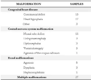
Table 1. Incidence per 10,000 liveborns of the most common malformations reported in Spain between 1980 and 1995 by the registers for congenital malformations existing in the conuntry.1.2 The relevance of identifying the cause of congenital defects
[image:25.499.90.417.72.403.2]INTRODUCTION
6
Moreover, the identification of the basis of genetic diseases is essential to
provide an accurate definition of disease risk, a critical element to ensure
proper genetic counseling and disease prevention. Genetic counseling has
become more relevant in this area considering that more individuals with
congenital malformations are living into adulthood due to advances in
medical and surgical care and may have the opportunity to reproduce (3).
1.3 Causes
Congenital disorders vary widely in causation. A congenital disorder may be
the result of a non-appropriate intrauterine environment, medication or drug
exposures or maternal infections during pregnancy, or the result of a genetic
abnormality.
It is hypothesized that in a significant percentage of congenital
malformations, especially in cases where the malformation is isolated and
none dimorphic features are identified, the etiology might be multifactorial
with variable contribution of genetic and environmental factors that alter
developmental pathways.
1.3.1 Teratogenic agents: medication and drug exposures
Some medications and drugs may cause congenital malformations by
disturbing the development of the embryo or fetus.
Alcohol intake during pregnancy has been strongly related to a
well-established disease called Fetal Alcohol Spectrum Disorder. The main
features of this disorder include affectation of the central nervous system
-causing intellectual disability and behavioral problems-, distinctive facial
features and growth retardation. Some malformations, like congenital heart
defects (10) or genitourinary malformations also have an increased incidence
in children with Fetal Alcohol Spectrum Disorder (11).
INTRODUCTION
7
Not only drugs but also medications have been linked to birth defects.
Nearly 50% of pregnant women are exposed to at least one medication
during gestation (18), while 52% of 200 individuals referred for genetic
counseling had been exposed to more than one potential teratogen (19).
Some antiepileptic medications, such as valproic acid and carbamazepine,
have been implicated in congenital malformations, mainly in neural tube
defects. The use of valproic acid monotherapy in the first trimester has been
associated with significantly increased risk for other congenital
malformations such as atrial septal defect, cleft palate, hypospadias,
polydactyly and craniosynostosis (20).
Systemic retinoids, used for several
diseases such as skin conditions or some types of cancer, are related to an
increased risk of spontaneous miscarriage, dysmorphic features, congenital
heart defects, limb defects, hydrocephaly and microcephaly.
1.3.2 Maternal diseases
Some infections and other diseases suffered by the mother during pregnancy
might cause birth defects. Diabetes (21, 22), hyperthyroidism (23-25),
hypothyroidism (24, 25), phenylketonuria (26, 27) and epilepsy (28, 29) are
some of the diseases that may affect pregnancy outcome. Several maternal
infections such as rubella, cytomegalovirus, varicella-zoster, toxoplasmosis,
syphilis or parvovirus B19 may contribute to alterations of fetal development
(30-33). The severity of birth defects depends on the developmental stage
when the infection occurs; usually the outcome is worse if the infection takes
place at earlier developmental stage. Infection for cytomegalovirus is one of
the most frequent infections during pregnancy that may cause deafness,
mental retardation or microcephaly to the fetus.
1.3.3 Genetic causes
Genetic factors are clear contributors to congenital malformations. Several
genetic and genomic alterations have been reported in patients with multiple
congenital anomalies, with or without developmental delay or growth
abnormalities. Deletions and duplications, corresponding to recurrent
genomic disorders and other genomic regions, can be detected in 15-24%
(34, 35) of patients with multiple congenital anomalies and/or intellectual
disability. Point mutations in genes such as
TBX1
(DiGeorge syndrome) (36)
INTRODUCTION
8
The role of genetic factors in isolated congenital malformations is not as
clear as in MCA/ID. Epidemiological studies have shown that siblings and
offspring of individuals with congenital malformations have an increased risk
of this type of anomalies (38-40) pointing out the high heritability of this
type of diseases. Nevertheless, when considering isolated congenital
malformations it is hypothesized that, in an important proportion of cases,
genetic factors are relevant included within a multifactorial model (41-43). As
an example, for some malformations like left heart hypoplasia an oligogenic
model has been proposed (44). In other cases, genetic alterations related to
the disease are identified in patients but inherited from a healthy parent,
meaning that other factors –genetic or environmental ones- are also related
to the disease.
1.3.3.1 Genetic rearrangements
Birth defects are often caused by chromosomal imbalances, especially when
associated with dysmorphic features or developmental delay. Nevertheless,
there are many syndromes and diseases related to developmental
abnormalities as well as major malformations without a known molecular
cause. It is likely that some of these syndromes are due to genomic
alterations involving small chromosomal regions not identified so far. Several
techniques, such as standard karyotyping, MLPA (Multiplex
ligation-dependent probe amplification), aCGH (array Comparative Genomic
Hybridization) or SNP (Single Nucleotide Polymorphism) array are used in
order to detect genetic rearrangements.
INTRODUCTION
9
Concerning isolated congenital malformations, few data is available. In those
cases, the detection of genomic rearrangements could represent the
identification of the genetic basis of the disease, perhaps as part of a more
complex syndrome without other recognizable manifestations at this stage of
development, or the finding of genetic susceptibility factors contributing to
mutational load in a multifactorial model. Therefore, submicroscopic
deletions and duplications may play a significant role in the etiology of this
condition, either as direct cause or as possible genetic risk factor for isolated
congenital anomaly (55).
1.3.3.2 Methylation alterations
Epigenetic mechanisms seem to contribute to many physiological processes
as development or aging. Therefore, its alterations could have an important
role in several diseases. Epigenetics includes those changes in the regulation
of gene expression not caused by modification of the nucleotide sequence.
DNA methylation and histone modification are both epigenetic processes.
DNA methylation refers to the addition of a methyl group in 5’ carbon of
citosines, which alters the secondary interactions of the DNA molecule
modifying the gene expression pattern. Aberrant DNA methylation may
result in changes in transcription and subsequently in gene expression. As an
example, hypermethylation of CpG islands located in promoter regions has
been described as a mechanism to prevent the transcription of some genes
leading to an abnormal gene expression profile related to tumor
development, for example.
Genetic diseases with a well-established cause such as Prader-Willi or
Angelman syndrome are related to a misregulation of epigenetic mechanisms
(56). Regarding methylation, some studies have shown its relevance in
diseases with a genetic component such as schizophrenia or bipolar
disorders. Furthermore, some data is already available regarding the role of
methylation in congenital anomalies, including correlation of genetic variants
in genes regulating methylation, such as the folate pathway, and the risk for
heart malformations in Down syndrome patients (57). Folate-pathway has
also been related in some articles with isolated heart defects without Down
syndrome, as in a meta-analysis with polymorphisms in the
MTHFR
gene
(58).
1.3.3.3 Point mutations
INTRODUCTION
10
technologies developed in the last few years have increased the number of
known genetic diseases caused by point mutations.
Point mutations in some genes have also been reported as the cause of
congenital malformations. In MCA/MR mutations in several genes, such as
DHCR7
(59),
FOXF1
(60) and
NIPBL
(61) have been reported. In isolated
congenital heart malformations mutations in genes with a crucial role in
heart development have been detected, such as mutations in
GATA4
(62) or
NKX2-5
(63). Also in other types of malformations, as holoprosencephaly or
microcephaly, point mutations are strongly related to the phenotype.
Otherwise, in neural tube defects point mutations detected in genes related
to planar cell polarity have been described in affected patients, although an
apparently healthy progenitor was also a carrier of the same variant. In those
cases, point mutations might be a mutation with incomplete penetrance or a
susceptibility factor that requires other genetic or environmental factors to
cause the malformation.
1.4 The relevance to study fetal tissues
INTRODUCTION
11
2 MODEL DISEASES
2.1 Multiple malformations
Congenital malformations often occur in the setting of multiple congenital
anomalies, including dysmorphic facial features, or in association with
different organ malformations, developmental defects, or growth
abnormalities (3, 4). In these cases in which there is a more complex
syndrome, chromosomal aberrations are a frequent cause of disease,
although point mutations in specific genes have also been described. As an
example, Wolf-Hirschhorn (64) syndrome is a polymalformation syndrome
caused by a recurrent microdeletion in 4p16.3. The most common clinical
features in Wolf-Hirschhorn syndrome are growth retardation, microcephaly,
skeletal anomalies, hypotonia, severe developmental delay and congenital
heart defects. Other polymalformation syndromes are DiGeorge (65)
syndrome or 1p36 deletions. In other diseases polymalformation syndromes
are caused by a point mutation as Kabuki syndrome (66) or Rubinstein-Taybi
(67).
Nevertheless quite often when multiple congenital anomalies are identified is
not possible to recognize a known syndrome. In those cases, screening
methodologies such as MLPA for subtelomeric rearrangements or recurrent
genomic disorders or CMA are useful to diagnose the cause. In those cases,
CMA leads to a detection rate of 15-24% of causative segmental aneusomies
in patients with MCA/MR (34, 35).
2.2 Congenital Heart Defects
INTRODUCTION
12
Table 3.
Recurrence risk of heart malformations and type of CHD in relatives
of patients with left heart defects.
About half of congenital cardiovascular malformations are severe and require
one or more surgical procedures in the neonatal period or during childhood
(71). Because of its severity, congenital heart disease is a leading cause of
morbidity and mortality during infancy; about 8% of all infant deaths is due
to congenital heart defects (72).
Table 2. Incidence per million live births of the most
common congenital heart malformations reported in England
INTRODUCTION
13
There are several classification systems for CHD. In 2000 the International
Congenital Heart Surgery Nomenclature was developed to provide a generic
classification system.
2.2.1 Hypoplasia of the heart
Hypoplasia of the heart refers to an underdevelopment of one side of the
heart and is the most severe form of CHD. It may compromise the right
ventricle or the left one resulting in an ineffective blood pumping. If the
hypoplasia affects the right ventricle, the blood circulation to the lungs is not
functioning properly; hypoplasia of the left ventricle prevents the correct
blood pumping to the body. Hypoplasia of the heart has an approximate
incidence of 3/10.000 pregnancies. Regarding left heart hypoplasia, often
associated with obstruction to left ventricle outflow when is severe, surgery
is required for long-term survival due to the inability of the left ventricle to
support the systemic circulation. There are two possible surgical treatments:
neonatal cardiac transplantation or a sequence of complex open-heart
operations in infancy in order to achieve a univentricular circulation in which
the right ventricle supports the systemic circulation and pulmonary blood
flow is passive. When hypoplasia is mild, the outflow obstruction repair may
prevent further complications since the left ventricle may be capable of
supporting the systemic circulation (73).
It is though that heart hypoplasia has an important genetic background,
taking into consideration the proven high heritability. Nevertheless, the
mechanisms causing this type of malformations are poorly known.
Figure 1.Picture of a normal developed heart (left) and a
heart with left heart hypoplasia (right). Note the
INTRODUCTION
14
2.2.2 Obstruction defects
Obstruction defects refer to malformations of heart valves, arteries or veins,
which difficult blood circulation because of its narrowing or blocking. When
it occurs, the heart has to pump harder to achieve a proper circulation,
causing heart hypertrophy.
Common obstructive defects include pulmonic stenosis, aortic stenosis, and
coarctation of the aorta. Bicuspid aortic valve stenosis and subaortic stenosis
are also obstructive defects with a lower incidence. Some studies have shown
a high heritability of some of these conditions, hypothesizing an important
genetic component in the etiology of this type of heart defects (74).
2.2.3 Septal defects
The septum is the partition wall that separates the left heart from the right
heart. A defect in the septum allows blood to circulate from the left side of
the heart to the right, reducing heart efficiency. Depending on where the
defect is placed, it is called atrial septal defect or ventricular septal defect.
Ventricular septal defects (VSD) are the most common type of CHD,
affecting approximately 0.44-0.48 per 1.000 elementary school children (75).
VSD
is an abnormal opening in the interventricular septum which allows
communication between the right and left ventricles. VSD might be part of a
more complex heart malformation or an isolated malformation. Severity of
VSD may depend on the size, location and conjunction with other heart malformations.Point mutations in GATA4, GATA6 and JAG1 have been identified in patients
with VSD, as well as deletions in 8p23.11, which have been related to VSD and auricular septal defects (ASD).
2.2.4 Cyanotic defects
Cyanotic defects include those heart malformations causing cyanosis because
of a lack of oxygen in the body. The most frequent cyanotic defect is
Tetralogy of Fallot, a complex heart malformation which consists of
pulmonary infundibular stenosis, overriding aorta, VSD and right ventricular
hypertrophy. This conjunction of heart malformations implies a low
oxygenation of blood because of the mixing of oxygenated and
deoxygenated blood. Tetralogy of Fallot has a high incidence in patients with
22q11.2 deletion syndrome (22%). In addition, point mutations in some
genes such as
FOG2
and
Nkx2-5
have been identified related to the same
INTRODUCTION
15
2.3 Central Nervous System malformations
There are several types of central nervous system malformations. Some of
the most frequent ones are described below.
.2.3.1 Holoprosencephaly
Holoprosencephaly is a complex brain malformation in which the
prosencephalon fails to develop into two hemispheres, affecting the
forebrain and the face. Prevalence is estimated to be 1/10,000 live and
stillbirths
and
1/250
of
pregnancies.
Mechanisms
underlying
holoprosencephaly are partially understood and it seems that
HOX
genes,
which may guide the placement of embryonic structures, do not function
properly along the midline of the head. Activation of
HOX
genes prevents
the merge of the left and right side of prosencephalon by the division of that
embryonic structure (77).
Depending on the severity, three forms have been described: lobar,
semi-lobar and asemi-lobar. Another form, the less severe one, has also been described
and it is called microform and is characterized by midline defects without the
typical holoprosencephaly brain malformation. Often it is not easy to classify
this type of malformations into those categories taking into account that
holoprosencephaly is a continuous spectrum of abnormal separation of the
hemispheres (77).
The cases with cyclopia, proboscis, premaxillary agenesis or median or
bilateral cleft lip/palate with also a severe brain malformation leading to
miscarriages or stillbirths in some cases are at the most severe end of the
spectrum. In less severe cases, patients might present a normal or
near-normal brain development and facial defects such as hypotelorism, solitary
maxillary median incisor or even normal face. In affected patients, a wide
range of associated clinical manifestations is reported:
developmental delay,
hydrocephalus,
motor
impairment,
feeding
difficulties,
oromotor
dysfunction, epilepsy or hypothalamic dysfunction. Prognosis depends on
the severity and the associated complications.
INTRODUCTION
16
genes have been implicated:
SHH
,
ZIC2, SIX3, GIF, PTCH1, GLI2,
FOXH1, TDGF1 , DISP1 , NODAL, FGF8, GAS1, DLL1, and CDON
(78,
79). Environmental factors such as maternal diabetes or hypocholesterolemia
during gestation might be also related to that brain malformation.
2.3.2 Ventriculomegaly
Ventriculomegaly is one of the most common abnormal brain findings on
prenatal ultrasound, occurring in around 1–2 per 1000 pregnancies.
Ventriculomegaly might be an isolated malformation or part of a more
complex syndrome. Ventriculomegaly is a brain malformation which occurs
when the lateral ventricles are abnormally dilated or enlarged.
Ventriculomegaly is diagnosed when the width of the atrium of the lateral
ventricle measures more than 10 mm. Depending on that measurement,
ventriculomegaly is described as mild to moderate (between 10 and 15 mm)
or severe (greater than 15mm). In many cases of mild ventriculomegaly,
there is resolution during the pregnancy. Ventriculomegaly may affect also
the third and fourth ventricle. Enlargement of the ventricles may occur for a
number of reasons, such as loss of brain volume or impaired outflow or
absorption of cerebrospinal fluid from the ventricles. The underlying
mechanisms for the decreased of brain volume or the accumulation of
cerebrospinal fluid may include genetic alterations, maternal infections or
tumors located in the fetal brain. Some genetic syndromes, like
Walker-Walburg syndrome, include ventriculomegaly as one of their main features.
Mutations in
L1CAM
have been reported in families with X-linked
aqueductal stenosis causing ventriculomegaly. Often, however, there is no
identifiable cause (80).
2.3.3 Neural tube defects
Neural tube defects (NTDs) are one of the most common birth defects,
occurring in approximately one in 1,000 live births. The neural tube is a
narrow channel that folds and closes during the third and fourth weeks of
pregnancy to form the brain and spinal cord. An NTD is an opening in the
spinal cord or brain that occurs very early in human development as a result
of the failure in neural tube closure.
INTRODUCTION
17
bifida. Closed NTDs occur when the spinal defect is covered by skin;
common examples of closed NTDs are lipomeningocele and tethered cord
(81-84).
2.3.3.1 Anencephaly
Anencephaly is a neural tube defect that occurs when the head end of the
neural tube is not closed properly resulting in an absence of a major portion
of the brain and skull, including the cranial vault and the covering skin. As a
consequence of this malformation, the brain mass is reduced and infants
with this condition are born without the main part of the forebrain. Infants
are either stillborn or usually die within a few hours or days after birth. Its
prevalence at birth ranges from 1 in 5000 to 1 in 2000 (81).
2.3.3.2
Encephalocele
Sac-like protrusions of the brain and the membranes that cover it through
opening in the skull are known as encephalocele. Encephalocele might be
located in different parts of the skull as in the middle of it, between the
forehead and nose or on the back side of the skull. The severity of
encephalocele varies depending on its location and depending on the
quantity of brain tissue affected (82).
2.3.3.3
Spina bifida
Spina bifida is another type of neural tube defect in which the opening of the
tube is located along the spinal cord and therefore one or more vertebral
arches may be incomplete. The most common locations of those
malformations are the lumbar and sacral areas. Spina bifida malformations
fall into three categories: spina bifida occulta, spina bifida cystica with
meningocele, and spina bifida cystica with myelomeningocele (83).
INTRODUCTION
18
Meningocele is the protrusion of the meninges of the spinal cord through a
defect in the spinal column forming a cyst filled with cerebrospinal fluid that
does not contain neural tissue. As the nervous system remains undamaged,
individuals with meningocele are unlikely to suffer long-term health
problems. They are treated surgically.
Myelomeningocele is the most significant form of spina bifida and it leads to
disability in most affected individuals. In individuals with myelomeningocele,
the unfused portion of the spinal column allows the spinal cord to protrude
through an opening. The meningeal membranes that cover the spinal cord
form a sac enclosing neural tissue. The neural tissue enclosed in the
protrusion and the nerves that originate at that level of the cord are damaged
or not properly developed.
As a result, there is usually some degree of
paralysis and loss of sensation below the affected area of the spine.
Therefore, the higher the level of the myelomeningocele, the more severe the
nerve dysfunction may be. Affected individuals may have loss of sensation,
deformities of the hips, knees or feet, difficulty walking or inability to walk,
loss of muscle tone, bladder and bowel incontinence, urinary tract infections
and impaired renal function.
Many individuals with spina bifida will have an associated abnormality of the
cerebellum, called the Arnold Chiari II malformation. In affected individuals,
the cerebellar tonsils are displaced from the back of the skull down thorough
Figure 2.
Picture of NTD: myelomeningocele andmeningocele. Note the opening of the tube and the protrusion of the sack in both pictures. On the left side, the spinal cord protrudes as well as the meninges, so the neural tissue might be damaged; this image corresponds to a myelomeningocele. On the right side, a meningocele is shown: the meninges protrude trough the opening but not neural tissue is included within the
[image:38.499.144.360.148.292.2]INTRODUCTION
19
foramen magnum. In an important proportion of patients with
Arnold-Chiari malformation, hydrocephalus will also occur because the displaced
cerebellum interferes with the normal flow causing an accumulation of
cerebrospinal fluid.
Other central nervous system malformations such as
corpus callosum abnormalities or cortex anomalies are also seen in patients
with spina bifida.
The elected treatment for neural tube defect varies depending on the type of
the malformation. For instance, some mild versions of spina bifida require
minimal treatment. Otherwise, more severe forms require surgery to correct
the problems and, despite the operation, the normal function to the affected
part of the spinal cord might not be restored. Intrauterine surgery for spina
bifida has also been performed and the safety and efficacy of this procedure
is currently being investigated (85).
2.3.3.4 Prevention of neural tube defects
Regarding the cause of this malformation, it is hypothesized that neural tube
defects present a multifactorial inheritance, the result of gene-environment
interactions. Mutations in some genes related to planar cell polarity have
been described (86-90) although in the majority of cases mutations were
inherited from a healthy parent. Those results arise doubts about the role of
these mutations in the etiology of the disease, probably being one of the
contributing factors, but not the only one, to neural tube defects.
Nonetheless, familial cases with a seemingly autosomal recessive mode of
inheritance have been reported.
Since several years ago, the relation between folic acid intake before and
during pregnancy and neural tube defect has been proven. The incidence of
spina bifida may decrease by up to 70% when daily folic acid supplements
are taken prior to conception and during the pregnancy. However, the
mechanism underlying the role of folic acid in neural tube defects is poorly
understood (91, 92).
2.4 Renal malformations
Congenital anomalies of the kidney and urinary tract occur in 1 in 500 births
and are a major cause of morbidity and mortality in children. Some renal
anomalies are part of a more complex syndrome, although most cases are
isolated urinary tract malformations. Several genes have been reported in
association to isolated renal malformations, such as
Ret, PAX2, SALL1 and
INTRODUCTION
20
which may cause renal malformations include blood pressure medicines such
as angiotensin-converting enzyme inhibitors and angiotensin receptor
blockers. Illegal drugs, such as cocaine, may also cause renal malformations
in the fetus. Nevertheless, the majority of cases remain unsolved. Two of the
most common renal malformations are renal hypoplasia/agenesis and renal
dysplasia.
2.4.1 Renal agenesis
Renal agenesis or hypoplasia may have different levels of severity regarding
the affectation of a single or both kidneys and also considering the grade of
hypoplasia, being agenesis the most severe end of the spectrum. Bilateral
renal agenesis is the failure of both fetus kidneys to develop during gestation.
The absence of kidneys is a lethal condition that causes oligohydramnios, a
deficiency of amniotic fluid in pregnancy that may lead to fetal
malformations known as the Potter sequence. The etiology of bilateral renal
agenesis is unclear, although some autosomal recessive cases have been
described related to Fraser syndrome. It is also known that bilateral renal
agenesis is more common when one of the progenitors has some type of
kidney malformation, especially unilateral renal agenesis. Those results
indicate a common genetic background related to unilateral and bilateral
renal agenesis (98, 99).
2.4.2 Renal dysplasia
In kidney dysplasia, the internal structure of one or both kidneys is not
developed properly and fluid-filled sacs called cysts replace normal kidney
tissue. If kidney dysplasia only affects one kidney the prognosis is good, only
requiring regular checkups to ensure the correct functioning of the remaining
kidney.
When kidney dysplasia affects both kidneys is generally a lethal
condition, and those who do survive require dialysis and kidney transplant
very early after birth. In some cases, a silent renal dysplasia in one progenitor
is discovered after the birth of an affected child. When kidney dysplasia is
discovered in an infant, an ultrasound examination may reveal the condition
in one of the parents (99).
2.5 Down syndrome
INTRODUCTION
21
of the cases the trisomy 21 is free and mosaic, namely, not found in all
studied cells but only in a proportion of them. In the remaining 2-3% of the
cases, the supernumerary chromosome 21 is integrated to another
chromosome as part of a translocation. The diagnosis can be made by
karyotyping, although other techniques may also detect the trisomy. The
recurrence risk for the parents of an affected child is only slightly higher if
the trisomy is free. In cases of DS caused by translocation, the risk is
increased only if one of the parents has a balanced rearrangement. For a
person with Down syndrome, the risk of transmitting the disease to the
descendants is 1/3 (100).
The most severe features related to DS are congenital heart defects -mainly
atrio-ventricular canal, digestive malformations as duodenal atresia,
congenital cataract, small size, Hirschsprung disease, seizures, leukaemia,
sleep apnea, sensory deficiencies, auto-immune and endocrine pathologies,
earlier aging, Alzheimer disease and mental retardation. Patients with DS also
present a recognizable dimorphic pattern with upslanting palpebral fissures,
epicanthus, flat neck, round face, small nose or bilateral single palmar crease.
Median life expectancy is now above the age of 50 years (101).
Although the cause of DS is well established, the factors influencing the
appearance of different types of malformations are poorly understood. For
example, it is well known that patients with trisomy 21 have a higher
incidence of congenital heart malformations than other children, even
though little is known about the factors related to that. Concretely, the
incidence of CHD in these patients is between 43-58% (102, 103).
INTRODUCTION
22
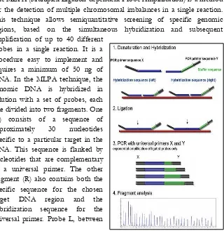
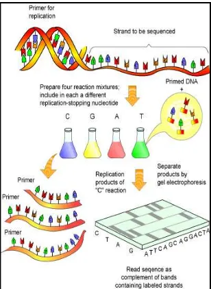
Figure 3. Overview of MLPA steps. In
this figure the main steps of MLPA are
shown (denaturation, hybridization,
ligation, PCR and fragment analysis) as
well as the structure of MPA probes
.
3 TECHNIQUES TO DETECT THE CAUSE OF THE
MALFORMATION
3.1 Techniques to study genetic rearrengements
There are multiple techniques that can be applied to the detection of possible
etiology of congenital malformation, either driven by the clinical suspicion or
as a screening approach. This review does not pretend to be exhaustive but
only to introduce the genetic and genomic technologies that have been
applied in the course of this project.
3.1.1 MLPA
The MLPA (Multiplex Ligation-dependent Probe Amplification) is a method
for the detection of multiple chromosomal imbalances in a single reaction.
This technique allows semiquantitative screening of specific genomic
regions, based on the simultaneous hybridization and subsequent
amplification of up to 40 different
probes in a single reaction. It is a
procedure easy to implement and
requires a minimum of 50 ng of
DNA. In the MLPA technique, the
genomic DNA is hybridized in
solution with a set of probes, each
one divided into two fragments. One
(L) consists of a sequence of
approximately
30
nucleotides
[image:42.499.96.416.234.563.2]INTRODUCTION
23
in the same reaction. This strategy allows electrophoretic resolution of the
amplified fragments (figure 3).
Both fragments of the MLPA probe complementary to the DNA sequence
are designed in order to hybridize with the target sequence one adjacent to
the other; then both are joined by ligase enzyme. The joining of both
fragments generates a single probe for each target region that can be
amplified by conventional PCR using universal primers; the fragments not
attached to another one cannot be amplified. The amount of bound probes
is proportional to the number of copies of the target sequence. After PCR
amplification the relative height of the peaks may indicate deletion or
duplication of the target sequence, namely the gene dose (104).
The MLPA technique is inexpensive, simple, fast and provides flexibility for
the study of regions of interest. Moreover, MLPA allows the study of a large
number of samples in the same experiment. Despite the advantages, the
main limitation of this technique is the inability to detect balanced
rearrangements, eg, translocations or inversions. MLPA might be useful to
screen easily several samples and loci of interest (aneuploidy screening in
prenatal diagnosis, regions associated to microdeletion and microduplication
syndromes in patients with mental retardation) or to validate results of other
techniques, such as the results obtained with CMA technologies.
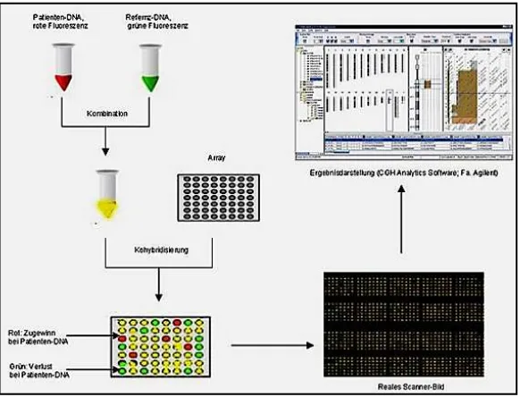
3.1.2 ACGH
The most recently developed technique for the detection of duplications and
deletions is based on comparative genomic hybridization, so called aCGH.
This technique is based on a competitive hybridization between two DNAs,
the sample of interest and a control sample. Each of these samples is labeled
with a different fluorochrome and it is hybridized on a support containing an
array of probes. Different types of probes might be used in aCGH such as
BACs (Bacterial Artificial Chromosome) or oligonucleotides. The resolution
of this technique depends on the size of the probes, the number of them and
their distribution through the genome (Figure 4).
INTRODUCTION
24
The aCGH permits the study of copy number variations in a large-scale
manner by the screening of regions throughout the genome, as well as a very
detailed analysis of some selected regions, depending on the design of the
array probes. The aCGH has the same limitation than the MLPA, as it does
not detect balanced alterations.
aCGH is useful for the screening of chromosomal imbalances for which
there is no suspicion, as it may have coverage throughout the genome with
high resolution. Using aCGH it is also possible to detect rearrangements in
mosaicism. Recent published articles describe a detection of mosaicism as
low as a 10% level.
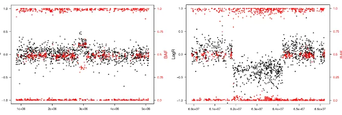
3.1.3 SNP arrays
[image:44.499.111.407.60.282.2]SNP arrays do not rely on the same system than comparative genomic
hybridization described in the previous section, but the same term is used
Figure 4.Schematic overview of aCGH. In the first step, genomic DNA
INTRODUCTION
25
when it is applied to study the copy number variants along the genome. SNP
arrays allow the study of the alleles present in each individual in a specific
number of SNPs, depending on the design of the experiment. This technique
quantifies the intensity of each of the alleles of the SNPs analyzed. The
information provided allows, in addition to deletions and duplications, the
analysis of heterozygosity and uniparental disomy. There are two relevant
parameters for the analysis of SNP array: the intensity of the fluorescent
signaling for each position and the BAF (B allele frequency), which
represents the proportion of signaling between both alleles, namely, the B
allele signal divided by the sum of the A and B signals.
The data for the detection of deletions and duplications is provided by the
intensities of the fluorescent signaling for each position and also by the
relative intensity between the two alleles in each nucleotide tested. For
example, in deleted regions the intensity is reduced and no heterozygous
positions are detected. In duplicated regions the intensity is higher and in
heterozygous positions one of the alleles is detected in a doubled intensity
compared to the other. Homozygous regions might be recognized because
normal intensities are detected but no heterozygous positions are identified
inside that fragment, namely, BAF is 0 (not B allele detected) or 1 (only B
allele is detected). Recently, some softwares have been developed to analyze
the presence of mosaic rearrangements by using data from SNP arrays. As in
the MLPA and aCGH, the main limitation of SNP arrays is the inability to
detect balanced rearrangements (108).
Figure 5. SNP array plots. A: Plot corresponding to a duplication. Within the
duplicated region the intensity (LogR) is higher and BAF shows a proportion of the alleles of 2:1. B: Plot showing a deletion. LogR of that region is reduced and there are none heterozygous positions within the region, as there are no BAF
[image:45.499.84.429.411.527.2]INTRODUCTION
26
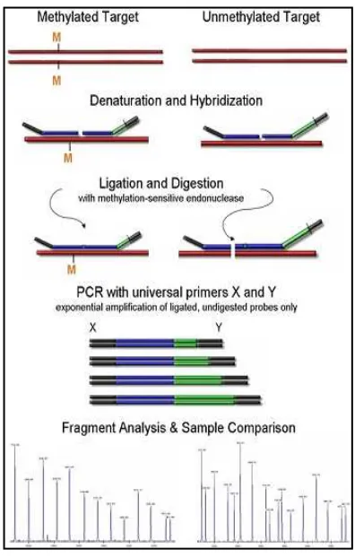
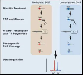
3.2 Techniques to study methylation alterations
3.2.1 Methylation arrays
Methylation arrays have been developed recently in order to interrogate
thousands of CpG sites per sample in the same experiment. Before the
genome-wide methylation array, a bisulfite treatment of DNA is required.
This procedure converts unmethylated citosine residues to uracile but does
not modify 5-methylcitosine residues. Therefore, bisulfite treatment
introduces modification in the DNA sequence depending on the methylation
status of citosine nucleotides. The microarray contains two different
bead-bound probes for each interrogated CpG. One of the bead-bead-bound probe is
complementary to the DNA sequence without the modification in citosine
residue, while the other one is complementary to the DNA sequence with
the conversion of the unmethylated citosine to uracile. The hybridization of
DNA with bead-bound probe is followed by a single-base extension with a
labeled nucleotide. The ratio of fluorescent intensity between the two
bead-bound probes for each CpG provides information about the level of
methylation in each CpG. Available softwares permit the comparison of
methylation patterns between single samples and between groups of samples
(109).
Figure 6.Principle of methylation array. A: as the CpG analyzed is unmethylated,
the cytosine residue is converted to uracile in bisulfate treatment. Due to the modification, the gDNA sequence is complementary to the unmethyalted bead. Therefore, the extension only occurs in the unmethylated probe. B: the CpG tested is methylated. For that reason no modification occurs during bisulfate treatment and the gDNA sequence hybridizes with the methyalted probe and extension takes place
[image:46.499.86.414.365.482.2]