Sociedad Cubana de Cardiología
______________________
Casos Clínicos
Síndrome de Marfan en edad adulta: a propósito de un caso
MSc. Dr. Geovedy Martínez García
a*
y MSc. Dr. Gerardo Rodríguez Lemus
b*
a Hospital Militar Central “Dr. Carlos J. Finlay”. La Habana, Cuba.b Hospital Provincial “Agustino Neto”. Guantánamo, Cuba.
* Colaboración Médica Cubana en la República Popular de Angola. Hospital do Prenda. Luanda, Angola.
Full English text of this article is also available
INFORMACIÓN DEL ARTÍCULO
Recibido: 09 de junio de 2014 Aceptado: 03 de julio de 2014
Conflictos de intereses
Los autores declaran que no existen conflictos de intereses
Versiones On-Line: Español - Inglés
G Martínez García
Anita 936 e/ Gertrudis y Lagueruela, 10 de octubre, La Habana, Cuba. Correo electrónico:
RESUMEN
El síndrome de Marfan es una enfermedad del tejido conectivo, autosómica dominan-te, causada por defecto en el gen fibrilina-1, el cual desempeña un papel importante en la formación de los tejidos elásticos del cuerpo. Se diagnostica según datos clíni-cos, algunos de los cuales dependen del crecimiento. Nuestro objetivo es describir un caso atípico de síndrome de Marfan en un hombre de 44 años de edad, con antece-dentes de salud, que acude al Servicio de Urgencias con disnea, dolores abdominales y edema intenso en miembros inferiores. En el examen físico se encontró soplo cardíaco, ingurgitación yugular y hepatomegalia congestiva. Se le realizó radiografía de tórax, ecocardiograma transtorácico y examen oftalmológico, y se llegó a la conclusión de estar en presencia de síndrome de Marfan, diagnóstico realizado en dependencia del cuadro clínico y de los criterios revisados de Ghent.
Palabras clave:Síndrome de Marfan, Insuficiencia aórtica, Alteraciones esqueléticas, Aneurisma aórtico, Subluxación del cristalino
Marfan syndrome in adulthood: a case report
ABSTRACT
Marfan syndrome is an autosomal dominant connective tissue disorder, caused by a defect in the fibrillin-1 gene, which plays an important role in the formation of elastic tissues. It is diagnosed on clinical grounds, some of which depend on growth. Our purpose is to describe an atypical case of Marfan syndrome in a 44 year-old-male patient, with no history of health problems, who arrives at the Emergency Depart-ment with shortness of breath, abdominal pain and great pedal edema. On physical examination, heart murmur, elevated jugular venous pressure and congestive hepato-megaly were found. Chest radiograph, transthoracic echocardiogram, and ophthal-mologic examination were performed, after which Marfan syndrome was diagnosed, according to the clinical picture and the revised Ghent criteria.
Key words:Marfan syndrome, Aortic regurgitation, Aortic aneurysm, skeletal
distur-bances, Lens subluxation
INTRODUCCIÓN
del tejido conectivo. Esta enfermedad, autosómica do-minante, tiene una incidencia de 2-3 por 10.000 indivi-duos, sin predilección de género, raza o etnia1. Se ha
demostrado que la causa de esta enfermedad es un cambio en el gen FBN1, el cual codifica la proteína fibrilina-1, componente de la red de microfibrillas que sirve de armazón para el depósito de elastina y el ensamblaje de las fibras elásticas. En este gen se han descrito más de 500 mutaciones y casi todas son únicas para un individuo o familia afectada, lo que da origen a un defecto hereditario de la fibrilina-1, y ocasiona una formación de fibras elásticas anormales, con la consiguiente disfunción de los tejidos que la poseen2-10. Además, se ha postulado que la fibrilina
normal inhibiría el crecimiento de los huesos largos y que las fibras elásticas a través de su tensión contro-larían el crecimiento de estos, por lo tanto, al existir una alteración en estas estructuras se produciría el crecimiento óseo exagerado propio de la enfermedad.
Sus complicaciones afectan a ojos, pulmones y sis-tema musculoesquelético; pero la alta mortalidad de casos no tratados resulta casi exclusivamente de las
complicaciones cardiovasculares, lo que incluye la di-sección aórtica y su ruptura5-6.
Los avances en el conocimiento de las causas del síndrome de Marfan, su diagnóstico temprano y la consecuente terapéutica médica o quirúrgica ha favo-recido la mejoría marcada en el pronóstico de la po-blación afectada, comparada con décadas anteriores. La identificación temprana de los pacientes asintomá-ticos es fundamental para reducir la frecuencia de los accidentes aórticos fatales. El presente trabajo tiene como objetivo presentar el caso de un paciente adulto con antecedentes de salud, que debutó con un cuadro clínico de insuficiencia cardíaca global, y que en el ingreso se le diagnosticó un síndrome de Marfan. Su importancia está dada por lo tardío del diagnóstico, que solo fue realizado en colaboración con la Brigada Cubana.
CASO CLÍNICO
Paciente masculino, de 44 años de edad, con antece-dentes personales de salud, que acude a Urgencias del Hospital do Prenda, en Luanda, con falta de aire, la cual había comenzado hacía dos semanas, primero de esfuerzo, y luego lo obligaba a levantarse por las noches cuando estaba dur-miendo, aliviándose al sentarse, con duración aproximada de una hora. También refería dolor abdo-minal en el hipocondrio derecho y aumento de volumen de miem-bros inferiores hasta la mitad de ambos muslos.
Datos positivos al examen físico
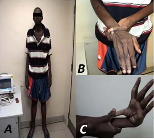
- Talla: 2,10 metros, brazos y de-dos alargade-dos. Aranodactilia. Signo muñeca-pulgar positivo. Laxitud articular, pie plano, y panículo adiposo disminuido (Figura 1).
- Pectus carinatum, con dismi-nución de la expansibilidad to-rácica, polipnea superficial, fre-cuencia respiratoria de 34 por minuto y estertores crepitantes
bibasales, más pronunciados
en el hemitórax izquierdo. - Pulsoirregularysaltón.Choque
Figura 1. Características esqueléticas del paciente. A. Talla alta, brazos
de la punta visible, palpable y desplazado fuera de la línea media clavicular. Primer ruido disminuido en la punta, soplo sisto-diastólico en borde esternal izquierdo con predominio del componente diastóli-co (grado IV/VI), diastóli-con irradiación al cuello. Ingurgi-tación yugular. Tensión arterial: 100/70 mmHg. - Abdomen blando y depresible, doloroso a la
palpa-ción en hipocondrio derecho, con hepatomegalia de 4 cm por debajo del reborde costal. Presencia de reflujo hepatoyugular.
- Edemas blandos, fríos y no dolorosos en miembros inferiores hasta la mitad de ambos muslos, con pre-sencia de fácil godet.
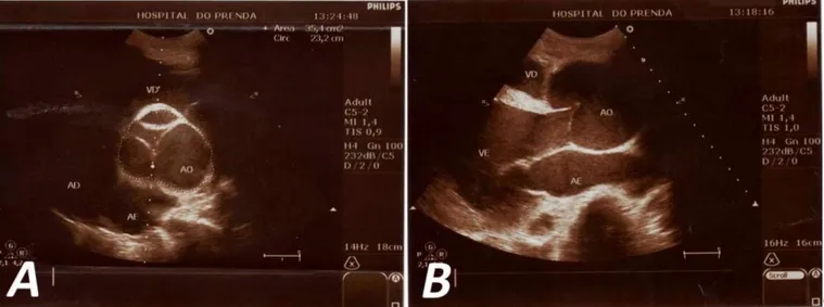
Se decide ingresar al paciente con el diagnóstico de insuficiencia cardíaca global y se inicia tratamiento farmacológico, con el que se logra mejorar el cuadro clínico. Teniendo en cuenta las características fenotí-picas de este paciente, así como los hallazgos ausculta-torios sugerentes de enfermedad valvular aórtica, se decide realizar otros exámenes complementarios: 1. Ecocardiograma (Figura 2): Se observa aorta
ascen-dente dilatada, de aspecto periforme, con diámetro de 77 mm, anillo aórtico no dilatado y cayado de la aorta normal (34 mm.). Regurgitación aórtica gra-ve, que ocupa totalmente el tracto de salida del ventrículo izquierdo, con volumen regurgitante de 80 ml y orificio regurgitante efectivo de 0,95 cm2.
Ventrículo izquierdo dilatado, con hipocinesia glo-bal leve y función sistólica disminuida (fracción de
eyección 40 %). Insuficiencias tricuspídea y mitral leves.
2. Examen oftalmológico: Lámpara de hendidura: en ambos ojos se observa córnea transparente, cáma-ra anterior con profundidad aumentada, signo del sol naciente hora uno (subluxación del cristalino) e iridodonesis.
Ante tales hallazgos se concluye que el paciente presenta un síndrome de Marfan. Se le plantea la posi-bilidad de tratamiento quirúrgico de la enfermedad valvular aórtica y acepta. Durante el ingreso en el hospital el paciente presenta parada cardiorrespira-toria y fallece.
COMENTARIO
El síndrome de Marfan se produce por la alteración en las microfibrillas, causada generalmente por la muta-ción del gen FBN1, que codifica la fibrilina-1, y se loca-liza en el cromosoma 15q21; todo lo cual fue descrito inicialmente por Dietz et al.1, en 1991. La fibrilina-1,
una glucoproteína extracelular esencial para la fibrino-génesis, es el principal componente de las microfibri-llas de 10-12 nm, que junto con la elastina forman las fibras elásticas encontradas en los tejidos. Mutaciones del FBN1 incrementan la susceptibilidad de la fibrilina-1 para la proteólisis in vitro, con la fragmentación de las microfibrillas como consecuencia11,12.
El diagnóstico se basa fundamentalmente en los hallazgos clínicos. Existen tres formas de presentación
Figura 2. Ecocardiografía transtorácica. A. Eje corto a nivel de grandes vasos. Se observa la dilatación en raíz de la aorta con
relacionadas estrechamente con la edad, con cuadros clínicos y pronósticos bien definidos: neonatal, infantil y clásica.Esta última es la más frecuente y reconocida, y se presenta en niños, adolescentes y adultos. Con-forme con los nuevos criterios diagnósticos para el síndrome de Marfan publicados en 2011, que surgen de la revisión de los clásicos criterios de Ghent, el fenotipo del paciente caracteriza el diagnóstico de este síndrome de otras enfermedades con fenotipos similares2.
Las alteraciones esqueléticas son las más frecuen-tes y precoces de detectar, y por ello son las que primero establecen la sospecha de la enfermedad. Son progresivas con la edad y se completan en la adoles-cencia. Destacan por su frecuencia la talla alta con mayor envergadura, pectum excavatum o carinatum,
aracnodactilia, escoliosis, hipermotilidad articular y paladar ojival2,4,6-11. Las lesiones cardiovasculares
de-terminan el pronóstico del síndrome de Marfan, ya que son las que ocasionan la mayor mortalidad, entre el 70-95 % de los casos, la más específica y frecuente ha sido la dilatación aórtica. Su incidencia depende de la edad, 40-80 % en niños y 80-100 %, en adultos13.
El compromiso ocular es frecuente (70 %) y pro-gresivo. La lesión más específica para el diagnóstico es la subluxación del cristalino; sin embargo, es necesario también identificar vicios de refracción para preservar la máxima función visual14.
Al considerar los criterios diagnósticos antes descri-tos, en este paciente se encuentra:
1. Historia familiar y genética: se realizó examen físico a familiares de primer grado sin encontrar eviden-cia de la enfermedad.
2. Sistema esquelético:
• Pectus carinatum: 2 puntos. • Pie plano: 1 punto.
• Signo muñeca-pulgar: 3 puntos.
3. Sistema cardiovascular: dilatación de aorta ascen-dente con insuficiencia aórtica grave (presencia de criterio mayor).
4. Sistema ocular: subluxación del cristalino (presen-cia de criterio mayor).
Al excluir clínicamente los síndromes de Ehlers-Danlos y Loeys-Dietz, y teniendo en cuenta que no existían antecedentes familiares de la enfermedad, la presencia de dilatación de la raíz aórtica y de subluxa-ción del cristalino permite el diagnóstico del síndrome de Marfan.
En la literatura revisada2-4,6 se mencionan todos los
tipos clínicos de este síndrome, pero el diagnóstico de la enfermedad generalmente se realiza en la etapa infantil y en la adolescencia. Lo interesante del caso presentado es lo tardío del diagnóstico, en un paciente que tuvo varios ingresos por insuficiencia cardíaca y no contó con una evaluación completa y certera. En el momento actual se brinda asesoramiento genético a los otros miembros de la familia.
REFERENCIAS BIBLIOGRÁFICAS
1. Dietz H, Cutting G, Pyeritz RE, Maslen CL, Sakai LY, Corson GM, et al. Marfan syndrome caused by a recurrent do novo missense mutation in the fibrillin gene. Nature. 1991;352(6333):337-9.
2. Loeys BL, Dietz HC, Braverman AC, Callewaert BL, De Backer J, Devereux RB, et al. The revised Ghent nosology for the Marfan syndrome. J Med Genet. 2010;47(7):476-85.
3. von Kodolitsch Y, Robinson PN. Marfan syndrome: anupdateofgenetics,medicalandsurgicalmanage- ment. Heart. 2007;93(6):755-60.
4. Lebreiro A, Martins E, Cruz C, Almeida J, Maciel MJ, Cardoso JC, et al. Marfan syndrome: clinical mani-festations, pathophysiology and new outlook on drug therapy. Rev Port Cardiol. 2010;29(6):1021-36. 5. Sohn GH, Jang SY, Moon JR, Yang JH, Sung K, Ki CS,
et al. The usefulness of multidetector computed to-mographic angiography for the diagnosis of Marfan syndrome by Ghent criteria. Int J Cardiovasc Ima-ging. 2011;27(5):679-88.
6. Ammash NM, Sundt TM, Connolly HM. Marfan
syn-drome – diagnosis and management. Curr Probl Cardiol. 2008;33(1):7-39.
7. Jondeau G, Michel JB, Boileau C. The translational science of Marfan syndrome. Heart. 2011;97(15): 1206-14.
8. Iams HD. Diagnosis and management of Marfan
syndrome. Curr Sports Med Rep. 2010;9(2):93-8.
9. Chan YC, Ting CW, Ho P, Poon JT, Cheung GC,
Cheng SW. Ten-year epidemiological review of in-hospital patients with Marfan syndrome. Ann Vasc Surg. 2008;22(5):608-12.
10.Ozdemir O, Olgunturk R, Kula S, Tunaoglu FS. Echo-cardiographic findings in children with Marfan syn-drome. Cardiovasc J Afr. 2011;22(5):245-8.
2010;92(9):1868-75.
12.Gao LG, Yao XP, Zhang L, Hui RT, Zhou XL. Mole-cular analysis for diagnosis of Marfan syndrome and Marfan-associated disorders. Chin Med J. 2011;124(6):930-4.
13.Pearson GD, Devereux R, Loeys B, Maslen C,
Milewicz D, Pyeritz R, et al. Report of the National
Heart, Lung, and Blood Institute and National fan Foundation Working Group on research in Mar-fan syndrome and related disorders. Circulation. 2008;118(7):785-91.
______________________
Case Report
Marfan syndrome in adulthood: a case report
Geovedy Martínez García
a*
, MD, MSc, and Gerardo Rodríguez Lemus
b*, MD, MSc
a Dr. Carlos J. Finlay Central Military Hospital. Havana, Cuba.b Agustino Neto Provincial Hospital. Guantanamo, Cuba.
* Cuban Medical Collaboration in the People's Republic of Angola. Hospital do Prenda. Luanda, Angola.
Full English text of this article is also available
ARTICLE INFORMATION
Received: June 9, 2014 Accepted: July 3, 2014
Competing interests
The authors declare no competing interests
On-Line Versions: Spanish - English
G Martínez García
Anita 936 e/ Gertrudis y Lagueruela, 10 de octubre, La Habana, Cuba. E-mail address:
ABSTRACT
Marfan syndrome is an autosomal dominant connective tissue disorder, caused by a defect in the fibrillin-1 gene, which plays an important role in the formation of elastic tissues. It is diagnosed on clinical grounds, some of which depend on growth. Our purpose is to describe an atypical case of Marfan syndrome in a 44 year-old-male patient, with no history of health problems, who arrives at the Emergency Depart-ment with shortness of breath, abdominal pain and great pedal edema. On physical examination, heart murmur, elevated jugular venous pressure and congestive hepato-megaly were found. Chest radiograph, transthoracic echocardiogram, and ophthal-mologic examination were performed, after which Marfan syndrome was diagnosed, according to the clinical picture and the revised Ghent criteria.
Key words:Marfan syndrome, Aortic regurgitation, Aortic aneurysm, skeletal
distur-bances, Lens subluxation
Síndrome de Marfan en edad adulta: a propósito de un caso
RESUMEN
El síndrome de Marfan es una enfermedad del tejido conectivo, autosómica dominan-te, causada por defecto en el gen fibrilina-1, el cual desempeña un papel importante en la formación de los tejidos elásticos del cuerpo. Se diagnostica según datos clíni-cos, algunos de los cuales dependen del crecimiento. Nuestro objetivo es describir un caso atípico de síndrome de Marfan en un hombre de 44 años de edad, con antece-dentes de salud, que acude al Servicio de Urgencias con disnea, dolores abdominales y edema intenso en miembros inferiores. En el examen físico se encontró soplo cardíaco, ingurgitación yugular y hepatomegalia congestiva. Se le realizó radiografía de tórax, ecocardiograma transtorácico y examen oftalmológico, y se llegó a la conclusión de estar en presencia de síndrome de Marfan, diagnóstico realizado en dependencia del cuadro clínico y de los criterios revisados de Ghent.
Palabras clave:Síndrome de Marfan, Insuficiencia aórtica, Alteraciones esqueléticas, Aneurisma aórtico, Subluxación del cristalino
INTRODUCTION
sue disorder. This autosomal dominant disease has an incidence of 2-3 per 10,000 individuals, without predi-lection of gender, race or ethnic group1. It has been
demonstrated that the cause of this disease is a change in the FBN1 gene, which encodes the fibrillin-1 protein, a component of a microfibrils network serving as a frame to elastin deposition and assembly of elastic fibers. In this gene, over 500 mutations have been described and almost all are unique for an individual or family affected, giving rise to a hereditary defect in the fibrillin-1 and causing a formation of abnormal elastic fibers, with the consequent dysfunc-tion of the tissues that present it2-10. Furthermore, it
has been postulated that normal fibrillin would inhibit growth of the long bones and elastic fibers through its tension would control the growth of these; therefore, since there is an alteration in these structures an exaggerated bone growth would occur, typical of the disease.
Its complications affect the eyes, lungs and muscu-loskeletal system; but the high mortality of untreated cases results almost exclusively from cardiovascular
complications, including aortic dissection and its rup-ture5-6.
Advances in the understanding of the causes of Marfan syndrome, its early diagnosis and the sub-sequent medical or surgical therapy has led to the marked improvement in the prognosis of the affected population, compared to previous decades. Early iden-tification of asymptomatic patients is essential to reduce the frequency of fatal aortic accidents. This paper aims to present the case of an adult patient with no history of health problems that started with a cli-nical picture of global heart failure and, at admission, a Marfan syndrome was diagnosed. Its importance is given by the lateness of diagnosis, which was only achieved in collaboration with the Cuban Brigade.
CASE REPORT
44 year-old-male patient, with no history of health problems, who arrives at the Emergency Department of Hospital do Prenda with shortness of breath, which began two weeks ago, first on ex-ertion, and then it forced him to rise at night when he was sleep-ing, relieved by sittsleep-ing, with ap-proximately one hour duration. He alsocomplainedofabdominalpain in the right upper quadrant and in-creased volume of lower limbs up to half of both thighs.
Positive data on physical examin-ation
- Height: 2.10 meters, long arms and fingers. Arachnodactyly. Positive wrist/thumb sign. Joint laxity, flat feet, and decreased adipose tissue (Figure 1). - Pectus carinatum, with
de-creased thorax expansion, su-perficial polypnea, respiratory rate of 34 beats per minute and crackling rales in both lung
bases, more pronounced on
the left hemithorax.
- Irregular and bounding pulse.
Figure 1. Skeletal characteristics of the patient. A. Above-average height,
Apex beat was visible, palpable and displaced out-side the midclavicular line. First noise decreased in the apex, sisto-diastolic murmur in the left sternal border with prevalence of the diastolic component (grade IV/VI) with neck irradiation. Elevated jugular venous pressure. Blood pressure: 100/70 mmHg. - Soft and palpable abdomen, painful on palpation in
the right upper quadrant, with hepatomegaly of 4 cm below the costal margin. Presence of hepato-jugular reflux.
- Soft, cold and painless edema in the lower extre-mities to the middle of both thighs, with presence of pitting edema.
It was decided to admit the patient with the diag-nosis of overall heart failure, and drug treatment was started, with which the clinical picture improved. Gi-ven the phenotypic characteristics of this patient and auscultatory findings suggestive of aortic valve dis-ease, it was decided to perform other complementary tests:
1. Echocardiography (Figure 2): Dilated ascending aorta, with pearly appearance, 77 mm diameter, non-dilated aortic annulus and normal aortic arch (34 mm) were observed. Severe aortic regurgita-tion which fully occupied the outflow tract of the left ventricle with 80 ml regurgitant volume and ef-fective regurgitant orifice area of 0.95 cm2. Dilated
left ventricle with mild global hypokinesia and de-creased systolic function (ejection fraction 40%).
Mild tricuspid and mitral insufficiencies.
2. Eye exam: Slit lamp: clear cornea is observed in both eyes, anterior chamber with increased depth, lens subluxation and iridodonesis.
Given these findings it was concluded that the pa-tient presented Marfan syndrome. He was told about the possibility of surgical treatment of aortic valve disease and he agreed. During hospital stay the patient had a cardiac arrest and died.
COMMENT
Marfan syndrome is caused by the alteration in the microfibrils, usually caused by mutation of FBN1 gene encoding fibrillin-1, and is located on chromosome 15q21; all of which was first described by Dietz et al.1
in 1991. Fibrillin-1, an extracellular glycoprotein essen-tial for fibrogenesis is the major component of the microfibrils of 10-12 nm, which forms, together with elastin, the elastic fibers found in tissues. FBN1 muta-tions increase susceptibility of fibrillin-1 for in vitro
proteolysis, with the fragmentation of microfibrils as a result11,12.
Diagnosis is mainly based on clinical findings. There are three forms of presentation closely related to age, and to well-defined clinical conditions and prognosis: neonatal, child and classic. The latter is the most com-mon and recognized, and occurs in children,
adoles-Figure 2. Transthoracic echocardiography. A. Short axis at the level of large vessels. Dilation is observed in aortic root with
poorly closing of its sigmoids. B. Parasternal long axis.
cents and adults. In accordance with the new diagnos-tic criteria for Marfan syndrome published in 2011, arising from the review of the classical Ghent criteria, the patient phenotype characterizes the diagnosis of this syndrome from other diseases with similar pheno-types2.
Skeletal disorders are the most common and ear-liest to detect, and therefore are the ones to first establish the suspicion of the disease. They are pro-gressive with age and are completed in adolescence. The most frequent are: extreme height, pectus excava-tum or carinatum, arachnodactyly, scoliosis, joint hy-permobility and high arched palate2,4,6-11.
Cardiovas-cular injuries determine the prognosis of Marfan syndrome, since they cause the highest mortality, between 70-95% of cases, aortic dilatation has been the most specific and frequent problem. Its incidence depends on the age, 40-80% in children and 80-100% in adults13.
Ocular involvement is common (70%) and progres-sive. The more specific lesion for diagnosis is lens subluxation; however, you must also identify refrac-tion errors to preserve the maximum visual funcrefrac-tion14.
When considering the diagnostic criteria described above, this patient presented:
1. Family and genetic history: Physical examination on first-degree relatives was performed without find-ing evidence of the disease.
2. Skeletal System:
• Pectus carinatum: 2 points. • Flat foot: 1 point.
• Wrist/thumb sign: 3 points.
3. Cardiovascular system: dilated ascending aorta
with severe aortic regurgitation (presence of major criterion).
4. Ocular system: lens subluxation (presence of major criterion.
By clinically excluding Ehlers-Danlos and Loeys-Dietz syndromes, and considering that there was no family history of the disease, the presence of dilated aortic root and lens subluxation allows the diagnosis of Marfan syndrome.
In the literature revised2-4,6 all clinical types of this
syndrome are mentioned, but diagnosis of the disease is usually performed during childhood and adoles-cence. The interesting thing in this case is its late diag-nosis in a patient who had several admissions for heart failure and did not have a complete and accurate
assessment. At present genetic counseling is provided to other family members.
REFERENCES
1. Dietz H, Cutting G, Pyeritz RE, Maslen CL, Sakai LY, Corson GM, et al. Marfan syndrome caused by a recurrent do novo missense mutation in the fibrillin gene. Nature. 1991;352(6333):337-9.
2. Loeys BL, Dietz HC, Braverman AC, Callewaert BL, De Backer J, Devereux RB, et al. The revised Ghent nosology for the Marfan syndrome. J Med Genet. 2010;47(7):476-85.
3. von Kodolitsch Y, Robinson PN. Marfan syndrome: anupdateofgenetics,medicalandsurgicalmanage- ment. Heart. 2007;93(6):755-60.
4. Lebreiro A, Martins E, Cruz C, Almeida J, Maciel MJ, Cardoso JC, et al. Marfan syndrome: clinical mani-festations, pathophysiology and new outlook on drug therapy. Rev Port Cardiol. 2010;29(6):1021-36. 5. Sohn GH, Jang SY, Moon JR, Yang JH, Sung K, Ki CS,
et al. The usefulness of multidetector computed to-mographic angiography for the diagnosis of Marfan syndrome by Ghent criteria. Int J Cardiovasc Ima-ging. 2011;27(5):679-88.
6. Ammash NM, Sundt TM, Connolly HM. Marfan
syn-drome – diagnosis and management. Curr Probl Cardiol. 2008;33(1):7-39.
7. Jondeau G, Michel JB, Boileau C. The translational science of Marfan syndrome. Heart. 2011;97(15): 1206-14.
8. Iams HD. Diagnosis and management of Marfan
syndrome. Curr Sports Med Rep. 2010;9(2):93-8.
9. Chan YC, Ting CW, Ho P, Poon JT, Cheung GC,
Cheng SW. Ten-year epidemiological review of in-hospital patients with Marfan syndrome. Ann Vasc Surg. 2008;22(5):608-12.
10.Ozdemir O, Olgunturk R, Kula S, Tunaoglu FS. Echo-cardiographic findings in children with Marfan syn-drome. Cardiovasc J Afr. 2011;22(5):245-8.
11.Sponseller PD, Erkula G, Skolasky RL, Venuti KD, Dietz HC. Improving clinical recognition of Marfan syndrome. J Bone Joint Surg Am. 2010;92(9):1868-75.
13.Pearson GD, Devereux R, Loeys B, Maslen C, Milewicz D, Pyeritz R, et al. Report of the National Heart, Lung, and Blood Institute and National fan Foundation Working Group on research in Mar-fan syndrome and related disorders. Circulation.
2008;118(7):785-91.