Otras secciones de este sitio: ☞ ☞ ☞ ☞
☞ Índice de este número ☞
☞ ☞ ☞
☞ Más revistas ☞
☞ ☞ ☞
☞ Búsqueda
Others sections in this web site: ☞
☞ ☞ ☞
☞ Contents of this number ☞
☞ ☞ ☞
☞ More journals ☞
☞ ☞ ☞ ☞ Search Artículo:
Assessment of a predictive index for coronary artery disease in patients with rheumatic valvular disease
Derechos reservados, Copyright © 2005: Academia Mexicana de Cirugía Cirugía y Cirujanos
Número
Number 2
Marzo-Abril
March-April 2 0 0 5
Volumen
edigraphic.com
Assessment of a predictive index for coronary artery
disease in patients with rheumatic valvular disease
Dr. Víctor Manuel Salas-Lara,* Acad. Dr. Alberto Rangel-Abundis,*
Dr. Sergio Solorio-Meza,* Dr. Héctor Albarrán-López*
* Servicio de Hemodinámica, Centro Médico Nacional La Raza, Ins-tituto Mexicano del Seguro Social.
Solicitud de sobretiros:
Acad. Dr. Alberto Rangel-Abundis, Pajarera 1,
Unidad Independencia, 10100 México, D. F.,
Recibido para publicación: 19-05-2004 Aceptado para publicación: 13-10-2004
Resumen
Objetivo: se estudió la sensibilidad, especificidad y valores predictivos positivo y negativo, de un índice pronóstico para diagnosticar enfermedad arterial coronaria significativa en pa-cientes con valvulopatía cardiaca reumática. Dicho índice fue obtenido a partir de la siguiente información: presión arterial diastólica, número total de cigarrillos consumidos a lo largo de la vida, severidad de la angina de pecho, antecedentes de historia familiar de cardiopatía isquémica, edad en años, tabaquismo actual y, finalmente, razón entre la concentración total de lipoproteínas HDL y colesterol séricos.
Material y métodos: se llevó a cabo estudio prospectivo, observacional, transversal, consecutivo, comparativo, sin estar sujeto al azar, en hombres y mujeres entre los 30 y 78 años de edad, con valvulopatía cardiaca reumática, sometidos a cateterismo cardiaco y coronariografía.
Resultados: 102 pacientes (61 mujeres y 41 hombres), 55.63 ± 9.88 años de edad, con intervalo de 30 a 78 años (mujeres 56.09 ± 11.48 y hombres 54.6 ± 11.35 años). Los pacientes con enfer-medad mitral fueron 30 (29.41 %), en 49 (48.05 %) estaba aso-ciada enfermedad de la válvula aórtica y la válvula mitral y en 23 (22.55 %) con enfermedad de la válvula aórtica. La coronariografía reveló que sólo ocho (7.84 %) presentaron aterosclerosis signi-ficativa de las arterias coronarias. La sensibilidad de la prueba fue de 50 % y la especificidad de 80.85 %. El valor predictivo positivo fue de 0.18 y el negativo de 0.95.
Conclusiones: el índice analizado es útil para predecir la au-sencia de enfermedad coronaria significativa en pacientes con valvulopatía cardiaca reumática. Por otro lado, dicho índice no es útil para identificar enfermedad arterial coronaria significativa en esos pacientes.
Palabras clave: valvulopatía cardiaca reumática, cardiopatía isquémica, enfermedad arterial coronaria, coronariografía.
Summary
Objective: the authors studied the sensitivity and specificity, as well the positive and negative predictive values, of a prognostic index conformed by diastolic blood pressure, total number of cigarettes smoked during the lifetime, severity of angina pectoris, positive family history of ischemic heart diseases, age (years), current cigarette smoking, and total to HDL-cholesterol ratio in order to anticipate the presence of significant coronary artery disease in patients with rheumatic cardiac valvulopathy. Material and methods: a prospective, observational, non-randomized, cross-sectional and comparative study was performed in men and women ≥ 30 and ≤ 78 years of age, with rheumatic valve cardiopathy and who were submitted to catheterization and coronary angiography.
Results: we studied 102 patients (61 women and 41 men) 55.63 ± 9.88 years of age, range: 30-78 years (women 56.09 ± 11.48, and men 54.6 ± 11.35 years of age, respectively). The patients had mitral valve disease 30 (29.41 %), 49 (48.03 %) had mitral valve disease associated with aortic valve disease and 23 (22.55%) had aortic valvular disease. Significant coronary artery athero-sclerosis was present in eight patients (7.84 %). Sensitivity and specificity analysis resulted as follows: sensitivity, 50 % and specificity, 80.85 %. Positive predictive value was 0.18 and negative predictive value 0.95.
Conclusions: the index analyzed here is useful to predict cases without significant coronary artery disease in patients with rheumatic heart valvulopathy, but this index is not useful to identify significant coronary artery disease in such patients.
Key words: rheumatic cardiac valvulopathy, coronary artery disease, coronary angiography, coronary heart disease.
Introduction
To recommend the correct surgical treatment in male patients
≥ 35 year old (for some authors) with rheumatic valvular heart disease, it is mandatory to assess the presence of significant coronary artery disease by means of coronary angiography.
Rheumatic valve heart disease associated with coronary heart disease varies between 8.00 % and 28.9 %.1-6 Normal
MG Salas-Lara VM et al.
edigraphic.com
When coronary arteriography is indiscriminately indicated in patients with heart valvulopathy, it increases the catheteriza-tion endurance, the radiacatheteriza-tion exposure (for the patient and for the health staff), the wear and tear on equipment and material, and the patient morbi-mortality. The frequency of coronary artery disease is low (3 %) when ischemic cardiopathy risk factors are absent. Under these conditions, the cost-benefit ratio increases. To avoid such abnormalities, several investigations have been attempted to identify, prior to coronary angiography, true positive patients with significant coronary artery disease. It is well known that age and gender are poor indices for indicating coronary angiography in valvular heart disease patients.1 Uncertain results have been
obtained when isolated coronary artery disease risk factors are related with the presence of significant coronary artery atherosclerosis, but good correlation is obtained when the influences of these risk factors are studied together. Ramsdale and colleagues found that isolated angina pectoris is an irrelevant predictor of significant coronary artery disease, but correlation between risk factors and coronary artery disease increases with the addition of risk factors considered in the pool.2,7,8 Based on the figures of merit found by Ramsdale and
colleagues,2 we studied the sensitivity and specificity of such
figures to anticipate the presence of significant coronary artery disease in patients with rheumatic cardiac valvulopathy.
Materials and methods
A prospective, observational, non-randomized, cross-sectional, comparative study was performed in men and women ≥ 30 and
≤ 78 years of age, with cardiac rheumatic valve cardiopathy
submitted to catheterization and coronary angiography. Patients with previous myocardial infarction or non-rheumatic-valve disease were excluded, as well as patients who refused participation in the study.
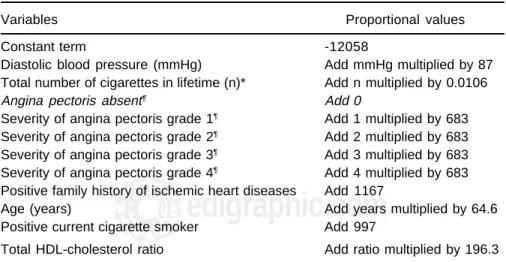
Table I shows the risk factors of ischemic cardiopathy studied and the proportional numeric values found from the covariate study of Ramsdale and colleagues.2 According to
the Ramsdale et al. multivariate regression study, the sum of the arithmetic operator shown in Table I was added to a basal number (-12058) to obtain a critical value of 600.2 We chose a
critical score of 600, instead of 500.8 (value elected by Ramsdale) because we wanted to improve the specificity of the analysis. Once the score was calculated, the patients were catheterized to obtain the hemodynamic diagnosis related to the valvulo-pathy, as well as the coronary artery disease.
We considered significant coronary artery disease when at least one principal coronary artery was stenosed ≥ 75 %. Patients with a score ≥ 600 and significant coronary artery disease were considered true positives (Group A). Patients with a score ≥ 600 and no coronary artery disease were false positives (Group B). Patients with a score < 600 and coronary artery disease were false negatives (Group C). Finally, patients with a score < 600 and no significant coronary artery disease were true negatives (Group D).
Statistical analysis
We calculated the power of the sample in > 30 patients. Descriptive statistical analysis was applied to numerical va-riables. Sensitivity and specificity analysis was applied to evaluate the prediction of the presence of significant coronary artery disease in patients with rheumatic cardiac valvulopathy.
Table I. Coronary artery disease: risk factors and proportional numeric values
Variables Proportional values
Constant term -12058
Diastolic blood pressure (mmHg) Add mmHg multiplied by 87 Total number of cigarettes in lifetime (n)* Add n multiplied by 0.0106
Angina pectoris absent¶ Add 0
Severity of angina pectoris grade 1¶ Add 1 multiplied by 683 Severity of angina pectoris grade 2¶ Add 2 multiplied by 683 Severity of angina pectoris grade 3¶ Add 3 multiplied by 683
Severity of angina pectoris grade 4¶ Add 4 multiplied by 683
Positive family history of ischemic heart diseases Add 1167
Age (years) Add years multiplied by 64.6
Positive current cigarette smoker Add 997
Total HDL-cholesterol ratio Add ratio multiplied by 196.3
* Average of cigarettes smoked annually multiplied by number of years (n).
¶ Grading angina severity: 0 = no angina; 1 = on strenuous exertion; 2 = on moderate exertion; 3 = on slight
edigraphic.com
Results
We studied 102 patients (61 women and 41 men) 55.63 ± 9.88 years of age, range: 30-78 years (women 56.09 ± 11.48, and men 54.6 ± 11.35 years, respectively). The ratio of women vs. men was 1.48. Figure 1 shows the cumulative frequency of the age of patients with normal coronary arteries and with coronary arteries lesions, respectively. The percentile 50 for patients with normal coronary arteries was 52 years old and 59 years old for patients with coronary artery lesions. We excluded four patients with coronary artery lesions other than atherosclerotic: two men and a woman with coronary artery ectasia and a man with an arteriovenous coronary fistula. None of these patients showed symptoms or signs of coronary artery disease.
Diastolic blood pressure was 68.09 ± 13.37 mmHg for the entire group (range: 40-129 mmHg); number of cigarettes smoked during their lifetime 29,169.56 ± 71,657.84; total cholesterol 184.34 ± 51.74 mg dL-1 (range: 103-254 mg dL-1),
and HDL cholesterol 35.08 ± 10.56 mg dL-1 (range: 23-69 mg
dL-1). Family history of ischemic heart disease was present in
17 patients (16.66 %), angina pectoris in 23 (22.55 %), and 4 patients were current cigarette smokers (3.92 %). Thirty patients had mitral valve disease (29.41 %), 49 (48.05 %) aortic and mitral valve disease, and 23 (22.55 %) presented isolated aortic valve disease. Significant coronary artery atherosclero-sis was present in only eight patients (7.84 %): five men and three women (62 ± 9 years of age, range: 41–70 years). True positive patients were 4, false positive 18, true negative 76 and false negative 4. Index sensitivity was 50 % and specificity 80.85 %. The results of the analysis are expressed in Table II. In this study we found a relevant difference between the age of the patients with and without coronary artery disease (Figure 1). Fifty percent of the patients without coronary artery disease were ≤ 52 years old. On the other hand, 50 % of the patients with coronary artery lesions were ≤ 60 years old. In the present study it was not possible to separate the age of men and women with coronary artery lesions.
Discussion
Rahimtoola recommended coronary angiography in ≥ 35-year-old patients with valvular cardiopathy or in < 35-year-35-year-old patients with two or more risk factors.9 Muñoz concluded that
coronary angiography in valvular patients is indicated in men > 60 years of age, and in women > 65 years of age.10 Historically,
each period of 5 or 10 years was taken as a limit to indicate coronary angiography for patients with valvular cardiopathy, from the age of 35 to 65 years. According our results shown in Figure 1, if we indicate coronary artery angiography in patients in their 40s, 88 % of the patients will show normal coronary angiography. It is well known that women with coronary artery disease are older than men. Previously, we reported the following results for patients with cardiac valve disease and coronary artery lesions: percentile 50, ≤ 60 years old for women, and ≤ 53 years old for men.1
Many authors consider the presence of angina pectoris as the keystone to indicate coronary angiography; for instance,
Table II. Statistical analysis of the results
Coronary disease
Index Positive Negative Total Predictive values
Positive (a) 4 (b) 18 22 Positive 0.18 Negative (c) 4 (d) 76 80 Negative 0.95
Total 8 94 102
Sensitivity Specificity 50 % 80.85 %
(a) true positive patients; (b) false positives patients; (c) false negative patients; (d) true negative patients.
Figure 1. Cumulative frequency of age in patients with cardiac valve disease and with normal coronary arteries, and coro-nary artery lesions shown during corocoro-nary artery angiography.
Age in years
CUMULATIVE FRECUENCY
Patients with and without coronary artery lesions
MG Salas-Lara VM et al.
edigraphic.com
sustraídode-m.e.d.i.g.r.a.p.h.i.c cihpargidemedodabor
:rop odarobale FDP
VC ed AS, cidemihparG
arap
acidémoiB arutaretiL :cihpargideM
sustraídode-m.e.d.i.g.r.a.p.h.i.c
young patients with valve cardiopathy and angina pectoris.11,12
Ramsdale and colleagues studied a great number of variables to find a predictive index for significant coronary artery disease. The method of these authors showed a sensitivity of 95.9 % and a specificity of 55.2 %.2 These data suggest that the index
was useful to recognize patients with significant coronary artery atherosclerosis. Using the same index, we found a sensitivity of 50 % and a specificity of 80.85 %, suggesting that we can predict cases without coronary artery disease.
According to the medical literature, in western Europe and in the U.S., a high prevalence of degenerative cardiac valvulopathy is observed. On the contrary, in Mexico, a high prevalence of rheumatic cardiac valvulopathy is observed. Although Ramsdale and colleagues did not mention the nature of the valvular disease in their patients, we are prone to believe that degenerative and rheumatic were included. On the contrary, we excluded patients with non-rheumatic cardiac valvulopathy. For that reason, we do not expect to find a coincidence of our results with those of Ramsdale et al.,2
especially about the specificity and sensibility; however, the mean age coincided in both studies. The ratio of women to men was 1.48 in our study, and 1.35 in the Ramsdale investigation.2 This might mean that patients with degenerative
cardiac valvulopathy were include in the Ramsdale study. Although the number of patients studied by Ramsdale and colleagues was higher than the number of patients collected in the present study, the calculated power of our sample showed an acceptable number of patients included in our study. As mentioned, sensitivity and specificity found by Ramsdale et al. were 95.9 and 52.2 %, respectively. Our results were sensitivity 50 % and specificity 80.85 %. In the Ramsdale group, true positive cases were clearly identified; in our study true negative cases were well identified. According to the results of Ramsdale and colleagues, it is possible to diagnose patients with coronary artery disease with an error of 5 %, but not possible to recognize patients without coronary artery lesions. According to our results, we can predict absence of coronary artery lesions, with an error of 5 %, but are unable to predict patients with coronary artery lesions (Table II).
According to our results, the sensitivity of the test was 50 %: almost half of the patients with an index ≥ 600 had coronary artery lesions, and the specifity was 80.85 %: 4/5 of the cases with an index < 600 had no coronary artery lesions. The positive predictive value was 0.18, indicating that patients with an index ≥ 600 have a probability, a posteriori, of 18 % to present coronary artery lesions. An index < 600 predicts, a posteriori, the absence of coronary artery lesions with a probability of 95 % (negative predictive value, 0.95).
Generally, it is desirable to account with a diagnostic index with high specificity and sensitivity. But in some cases this is not possible to obtain this degree of confidence. According to the pathology, it is possible to renounce to one of these
statistical constants. In the case of patients with cardiac valve disease, an index with high specificity is required because we cannot obviate the coronary angiography in patients with false negatives. In false-positive patients, coronary artery angiography could correct the error without consequences.
The presence of significant coronary artery disease worsens the prognosis of patients with heart valve disease. Scanlon and colleagues recommend performing coronary artery angiography in adult patients, before valve surgery or intraluminal valvotomy, with chest discomfort or with myocar-dial ischemia demonstrated by means of non-invasive imaging studies, or in patients without angor pectoris, but with multiple factor risks for coronary artery disease.13 As we can see at first
glance, these recommendations are not useful to differentiate the true from the false positives in the group of patients previously studied by us and other authors.2 In the present
study we found only eight patients (7.84 %) with significant coronary artery atherosclerosis associated with cardiac valve rheumatic disease. Previously, we have reported 407 patients with rheumatic heart disease and found only 8.3% with coronary atherosclerosis.1
Although the frequency of the coronary arteriovenous fistulae is very low (< 1 of all congenital cardiopathies), the surprisingly discovery of these fistula during coronary arterio-graphy in patients with cardiac valve disease or ischemic car-diopathy is not infrequent.14-16
In previous decades, coronary angiography was cautiously indicated in patients with clinical diagnosis of ischemic cardiopathy. At that time, few cases of normal coronary arteries were detected. Since that time, coronary angiography became an easy, quick, and safe procedure, and indications for coronary angiography were more numerous. For that reason, an increase in the number of patients with normal coronary arteries was observed. However, such an increase is less (almost 20 %) than the rate of normal coronary arteries found in patients with cardiac valvulopathy. Many investigations have been carried out to discriminate patients with cardiac valvulopathy isolated or associated with coronary artery atherosclerosis. Although none of these investigations has obtained optimal results, it is at least a good approach to know that age or presence of the isolated risk factor of ischemic cardiopathy are poor prognostic indices for significant coronary artery atherosclerosis in these patients.
edigraphic.com
Because age, gender or isolated signs and symptoms are poor predictors of significant coronary artery lesions in these patients, it is mandatory to find an index that impeaches unnecessary coronary artery angiography.
References
1 . Rangel A, Hernández J, Iris J, Baduí E, Chávez E. Indicaciones de la coronariografía en las valvulopatías cardiacas. Arch Inst Cardiol Mex 1996;66:60-69.
2 . Ramsdale DR, Fargher EB, Bennett DH, Bray CL, Ward C, Beton DC. Preoperative prediction of significant coronary artery disease in pa-tients with valvular heart disease. Br Med J 1982;284:223-226. 3 . Morrison GW, Thomas RD, Grimmer SF, Silverton PN, Smith DR.
Incidence of coronary artery disease in patients with valvular heart disease. Br Heart J 1980;44:630-637.
4 . Kay PH, Nunley DL, Grunkemeier GL, Pinson CW, Starr A. Late results of combined mitral valve replacement and coronary bypass surgery. J Am Coll Cardiol 1985;5:29-33.
5 . Marchant E, Pichard A, Casanegra P. Association of coronary ar-tery disease and valvular heart disease in Chile. Clin Cardiol 1983; 6:352-356.
6 . Lacy J, Goodin R, McMartin D, Masden R, Flowers N. Coronary atherosclerosis in valvular heart disease. Ann Thorac Surg 1977;23: 429-435.
7 . Ramsdale DR, Bennett DH, Bray CL, Faragher EB. Incidence of coronary artery disease in patients with valvular heart disease. Br Heart J 1981;46:222-223.
8 . Ramsdale DR, Bennett DH, Bray CL, Ward C, Beton DC, Faragher EB. Angina, coronary risk factors and coronary artery disease in patients with valvular disease. A prospective study. Eur Heart J 1984;5:716-726.
9 . Rahimtoola SH. Aortic valve disease. In: Hurst´s: The Heart. 9th ed. New York: McGraw-Hill;1998. pp. 1753-1787.
10.Muñoz JC, de la Fuente L, Garcimartin I, de la Torre M, Bermejo J, et al. Preoperative coronary angiography in valve patients. Criteria for indication in a determined population. Rev Esp Cardiol 1997;50:467-473
11.Schaefer A, Jehle J, Loogen F. Indications for coronary angiography in patients with acquired heart valve disease with reference to risk factors. Kardiol 1987;76:276-283.
12.Olofsson BO, Bjerie P, Aberg T, Osteram G, Jacobsson KA. Preva-lence of coronary artery disease in patients with valvular heart disease. Acta Med Scand 1985;218:365-371.
13.Scanlon PJ, Faxon DP, Audet AM, Carabello B, Dehmer GJ, Eagle KA, et al. ACC/AHA Guidelines for coronary angiography. J Am Coll Cardiol 1999;33:1756-1824.
14.Rangel-Abundis A, Muñoz-Castellanos, Marín G, Baduí E. Correlación morfofuncional en las anomalías congénitas de las arterias coronarias. I. Fístulas arteriales coronarias. Arch Inst Cardiol Méx 1994;64:48-62.
15.Rangel A, Muñoz-Castellanos L, Solorio S. Fístulas arteriovenosas coronarias múltiples. ¿Azar o predeterminación? Arch Cardiol Mex 2003;73:31-37.