Decreased serum total T3 level in hepatitis B and C
related cirrhosis by severity of liver damage
Fariborz Mansour-Ghanaei,* Mojtaba Mehrdad,** Sahereh Mortazavi,** Farahnaz Joukar,** Mohammad Khak***,**** Zahra Atrkar-Roushan*
* Gastrointestinal & Liver Diseases Research Center, Guilan University of Medical Sciences, Rasht, Guilan, Iran. ** Department of Endocrinology, Guilan University of Medical Sciences, Rasht, Guilan, Iran.
*** Department of Internal Medicine, Tehran University of Medical Sciences, Tehran, Iran. **** Farzan Clinical Research Institute, Tehran, Iran.
ABSTRACT
Objective. Thyroid hormones profile in patients with hepatic cirrhosis due to chronic HBV and HCV infec-tions was evaluated in order to find any relainfec-tionship between thyroid hormones and severity of liver dama-ge. Material and methods. Patients with the diagnosis of hepatic cirrhosis due to hepatitis B or C were screened for thyroid function status. Child-Pugh and model for end-stage liver disease (MELD) scores were calculated. Considering each thyroid function test, patients were divided into two groups with lower than normal and normal range of thyroid hormones, separately for each (for TSH, normal and upper than nor-mal). The correlation between thyroid function tests and severity of liver disease was taken into account.
Results. Number of patients with a T3 level lower than normal range (70-110 ng/dL) significantly increased along with Child-Pugh scores A, B and C. A negative correlation was found between Child-Pugh scores and total serum T3 level (r = -0.453, P < 0.001). Also a reverse correlation was observed between MELD score and T3 levels (r = -0.305, P = 0.14). Conclusion. In conclusion serum T3 concentration is a good index of hepatic function, decreasing by the severity of liver damage.
Key words. Cirrhosis. Thyroid hormones. Thyroid dysfunction. Hypothyroidism.
Correspondence and reprint request: Fariborz Mansour-Ghanaei, M.D. AGAF Division of Gastroenterology, Gastrointestinal & Liver Diseases Research Center, Guilan University of Medical Sciences.
Rasht, Iran. P.O. Box: 41635-3677
Tel.: +98 131 553-5116. Fax: +98 131 553-4951 E-mail: [email protected]
Manuscript received: June 22, 2012. Manuscript accepted: June 26, 2012. INTRODUCTION
Liver plays an important role in the metabolism of thyroid hormones, as it is the most important or-gan in the peripheral conversion of tetraiodothyro-nine (T4) to triiodothyrotetraiodothyro-nine (T3) by type I iodinase resulting to 5' deiodination of T4.1 Moreover, it is involved in conjugation and circulation of thyroid hormones by synthesis of thyroid binding pro-teins.2,3
There are evidences showing an association bet-ween chronic liver diseases and changes in thyroid gland.2-6 A 17% increase in thyroid glandular volu-me in cirrhotic patients is reported when compared with controls.4 Furthermore, it is demonstrated that levels of thyroid hormones and their binding
pro-teins are altered in patients with hepatic disorders, especially cirrhosis;5 however, almost all are clinica-lly euthyroid.3 The most frequent change in plasma levels of thyroid hormones is decreased total T3 and free T3 concentration which is reported to be asso-ciated with the severity of hepatic dysfunction.2,3,6
The relative high prevalence of hepatitis B and C viruses (HBV, HCV) infection in Iran7,8 and their major role in development of hepatic cirrhosis led us to evaluate thyroid hormones profile in patients with hepatic cirrhosis due to chronic HBV and HCV infections to find any relationship between thyroid hormones and severity of liver damage.
MATERIAL AND METHODS
than 6 months of abnormal serum aminoteransfera-se; if associated with positive serum HBS antigen, was considered due to HBV, and if accompanied by positive serum anti-HCV antibody and positive HCV RNA PCR, was considered due to HCV. Patients su-ffering from previously known thyroid dysfunction or with clinical findings of thyroid disease and those positive for both HBV and HCV infections, were not considered in the study. Patients on medications that could affect the study outcome, including Car-bamazepine, Phenytoin, Phenobarbitone, Salicylates and Nonsteroidal anti-inflamatory druges (NSAIDs) were also excluded; none of the patients had been under treatment by thyroid stimulating/inhibiting agents. As we wanted to study the changes in thyroid hormones in patients suffering from HBV or HCV related cirrhosis, subjects with a history of alcohol consumption in the last 6 months were omitted.
Gastrointestinal and Liver Diseases Research Center of Guilan University of Medical Sciences ethics committee approved the study protocol. Pa-tients fulfilling the inclusion criteria were recruited in the study after taking informed written consent. Thyroid function, liver function and clinical compli-cations including ascites, encephalopathy, bleeding varices, and spontaneous bacterial peritonitis (SBP) were evaluated. Child-Pugh score was recorded9 and MELD risk score (RS) was calculated according to the following formula:10
RS = 0.957 x loge (creatinine mg/dL) + 0.378 x loge (bilirubin mg/dL) + 1.120 x loge (INR) + 0.643 x (cause of cirrhosis).
For which the value for cause of cirrhosis, as it was viral in all the patients, was considered 1.
Circulating free T3 (reference, 1.5 to 4.1 ng/dL), free T4 (reference, 0.9 to 1.7 mcg/dL), total T3 (refe-rence, 70 to 190 ng/dL), total T4 (refe(refe-rence, 4.8 to 12 mcg/dL) and TSH (reference, 0.3 to 5.5 mcIU/ mL), total albumin and thyroxine-binding globulin (TBG) levels were measured with radioimmunologi-cal assay kits (Monobind, USA). Liver tests were examined using Pars Azmoon-co kits (Tehran, Iran).
Statistical analysis
During analysis, in order to figure out significant differences, patients were divided into different groups according to type of hepatitis, comorbidities, Child-Pugh classes and MELD score more vs. less
than 20. Statistical analysis was performed using SPSS 14.0 (SPSS Inc., Chicago, IL). Chi-square test, Fisher exact test and univariate logistic regres-sion model were used as appropriate. As child-Pugh and MELD scores were not normally distributed, Spearman’s rho was used to report the correlations. A P-value < 0.05 was considered sta-tistically significant.
RESULTS
Seventy two cirrhotic patients attended our liver clinic during the study period; 3 were excluded due to chronic alcohol consumption and 5 due to history of hypothyroidism. A total of 64 patients (42 male and 22 female) with a mean age of 55.03 ± 12.05 were enrolled in the study.
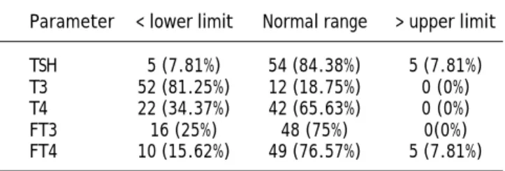
The mean duration of cirrhosis before enrollment was 25 months (range = 1 month to 6 years). HBV was specified in 34 patients as the etiology of cirrho-sis while in 30 patients HCV was responsible. Asso-ciated comorbidities included ascites (n = 59), encephalopathy (n = 31), bleeding varices (n = 21), and SBP (n = 10). The results of thyroid function tests are expressed in table 1.The total albumin and the TBG levels were measured as 3.04 ± 0.6 (NL: 4-5.3 gr/dL) and 1.02 ± 0.9 (NL: 1.3-3.0 mgr/dL), respectively.
When patients were divided into two groups with lower than normal and normal range of thyroid hor-mones, separately for each thyroid hormone test (for TSH, normal and upper than normal), only for total T3, significant differences were found in number of patients suffering from ascites, bleeding varices, and Child-Pugh scores. Among the patients presented with ascites, 16 had severe (all with T3 < 70 ng/dL), 31 moderate (26 with T3 < 70 ng/dL), and 12 mild (2 with T3 < 70 ng/dL) ascites. In a logistic regres-sion model adjusted only for ascites, patients with a higher grade ascites were more probable to have a serum T3 level below the normal range (OR = 3.56, 95% CI = 1.54 to 8.23); hence, the severity of ascites
Table 1. Thyroid function tests in the study population cate-gorized according to normal ranges (n; %).
was correlated with the number of patients with a serum T3 level of less than the normal range (P = 0.011). For all 20 patients with bleeding varices, serum T3 level was < 70; compared with the rest (32 with serum T3 lower than normal range out of 44), the difference was significant (P = 0.012). However, TSH, T4, FT3 and FT4 were not different in patients with and without comorbidities.
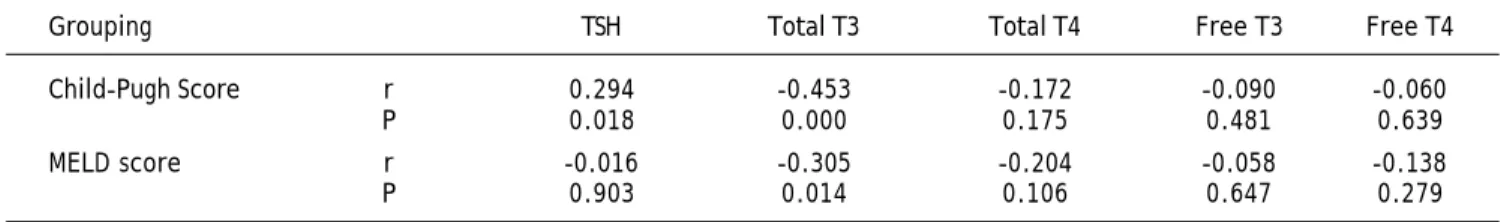
Patients were categorized into Child-Pugh scores A, B and C. Dividing patients into subgroups with thyroid function test variables lower than normal and normal (for TSH, normal and upper than nor-mal), the difference was again significant only for T3; number of patients with T3 < 70 ng/dL was 2 (out of 5), 16 (out of 22) and 34 (out of 35) for Child-Pugh scores A, B and C, respectively (P < 0.001). Also a negative correlation was found bet-ween Child-Pugh scores and total serum T3 level (r = -0.453, P < 0.001). The same analysis for MELD score over 20 versus less than 20 showed no significant difference for any of the thyroid function test variables. However, a reverse correlation was observed between MELD score and T3 levels (r = -0.305, P = 0.014). Also there was a positive correlation between TSH and Child-Pugh score (r = 0.294, P = 0.018) (Table 2). Patients with HBV/HCV related cirrhosis were classified by thyroid test variables lower than normal/within normal range, separately for each thyroid hormone test (for TSH, normal and upper than normal). No significant difference was recorded for any thyroid test variables (for TSH, P = 0.540; T3, P = 0.810; T4, P = 0.081; free T3, P = 0.583; free T4, P = 0.810).
DISCUSSION
In the present study, it was confirmed that a more severe liver status was inversely associated with serum total T3 levels; using Child-Pugh score and MELD risk score such a negative correlation was observed. Categorizing patients according to Child-Pugh score to A, B and C, we found that the number of patients with a serum T3 below the
normal range significantly increased with Child-Pugh score; such relationship was not present when setting a cutoff of 20 as the indicator of severity of hepatic status in MELD risk score. Severity of asci-tes and presence of bleeding varices, as indicators of severity of cirrhosis, also showed negative correla-tion with serum T3 levels. However, no significant difference was found according to the viral etiology of cirrhosis (HBV/HCV).
It is proved that hypothyroidism is associated with conditions mimicking liver diseases, including eleva-ted aspartate amino transferase and myxoedema asci-tes.11,12 As a result, patients with a history of treated or under treatment hypothyroidism were excluded in this study. Alcohol is stated to be toxic for thyroid gland parenchyma;13 hence, patients with a history of alcohol consumption were also omitted. In this study, in addition to Child-Pugh score, the correlation bet-ween MELD score and thyroid function tests was also examined. We also tried to investigate the de-crease in serum T3 level in cirrhotic patients accor-ding to severity of liver damage by classifying the patients into under normal/normal range T3 groups along with finding the correlation between T3 and se-verity of the liver disease.
Thyroxine and tri-iodothyronine modulate hepatic function by regulating hepatocyte basal metabolic rate, besides all other body cells. Liver also regula-tes circulating T3 and T4 by hormone metabolizing, resulting in a supervised endocrine effect.6 Two main enzymes acting in the liver as part of the iodo-thyronine seleno-deiodinase enzyme system are type 1 and type 3 deiodinases, responsible for extra-thyroidal production of T3 and inactivation of thyroid hormones, respectively.14-16 The decrease in total T3 probably reflects a decrease in deiodinase 1 activity in the liver of cirrhotic patients.2,6,17 Howe-ver, a contemporary increase in rT3 values18,19 asso-ciated with sick euthyroid syndrome is attributed to factors inhibiting conversion of rT3 to rT2,6,20 which was not observed in our study.
Lipophilic thyroid hormones in plasma are more than 99% bound to thyroxine-binding globulin, Table 2. The correlation between thyroid profile and Child-Pugh score and MELD score.
Grouping TSH Total T3 Total T4 Free T3 Free T4 Child-Pugh Score r 0.294 -0.453 -0.172 -0.090 -0.060
thyroxine-binding prealbumin and albumin. The li-ver role in synthesis of these proteins justifies a lo-wer serum total T3 after hepatocyte damage; negative correlation between thyroxine-binding glo-bulin and Child-Pugh score is reported. Decreased serum T3 is, as a result, associated with severity of Child-Pugh score.21,22 However, serum TSH and T4 are reported to be at steady levels2,3,6,17,22 despite al-teration in serum T3, indicating adaptive mechanisms by which the body reduces basal metabolic rate to decrease caloric requirements, however, preserving the patient euthyroid. Moreover, lower total serum T3 dedicates to a lower basal metabolic rate within hepatocytes and the resultant preservation of liver function and total body protein storage.6
Experimental studies on rats showed subclinical hypothyroidism to be beneficial both in protecting the liver from further damage and in regression of established fibrosis in induced liver fibrosis.23 It is also suggested that liver function in hypothyroid pa-tients tend to be better compared with euthyroid controls.24 Besides, Oren et al also showed that a controlled hypothyroidism might be beneficial for euthyroid cirrhotic patients.25 These studies, howe-ver, could be suggestive of a protective mechanism in the body in which lower circulating T3 in the body contributes to protection of liver from further fibrosis and helps the liver reverse the damages.
Hepatitis C is reported to be more associated with hypothyroidism rather than hepatitis B,22,25 however, we did not find any significant relation between types of hepatitis and serum level of thyroid hormones which was in accordance with the results of Zietz, et al.21
CONCLUSION
Our results confirm other studies in which serum T3 concentration is reported to be a good indicator of hepatic function. No difference was observed in thyroid hormones regarding etiology of viral hepatitis.
ABBREVIATIONS
• MELD: model for end-stage liver disease.
• SBP: spontaneous bacterial peritonitis. • RS: risk score.
• HBV, HCV: hepatitis B and C viruses.
ACKNOWLEDGEMENT
The authors would like to thank members of Gas-troenterology ward, Laboratory personnel’s of Razi
Hospital for their priceless assistance in performing this study. We also thank Farzan Institute of Science Research and Technology for technical assistance.
REFERENCES
1. Sorvillo F, Mazziotti G, Carbone A, Morisco F, Cioffi M, Ro-tondi M, et al. Increased serum reverse triiodothyronine levels at diagnosis of hepatocellular carcinoma in patients with compensated HCV-related liver cirrhosis. Clin Endo-crinol (Oxf) 2003; 58: 207-12.
2. Kayacetin E, Kisakol G, Kaya A. Low serum total thyroxine and free triiodothyronine in patients with hepatic ence-phalopathy due to non-alcoholic cirrhosis. Swiss Med Wkly
2003; 5: 210-3.
3. Borzio M, Caldara R, Borzio F, Piepoli V, Rampini P, Ferrari C. Thyroid function tests in chronic liver disease: eviden-ce for multiple abnormalities despite clinical euthyroidism.
Gut 1983; 24: 631-6.
4. Bianchi GP, Zoli M, Marchesini G, Volta U, Vecchi F, Ierve-se T, et al. Thyroid gland size and function in patients with cirrhosis of the liver. Liver 1991; 11: 71-7.
5. Yamanaka T, Ido K, Kimura K, Saito T. Serum levels of thyroid hormones in liver diseases. Clin Chim Acta 1980; 14: 45-55.
6. Malik R, Hodgson H. The relationship between the thyroid gland and the liver. QJM 2002; 95: 559-69.
7. Merat S, Rezvan H, Nouraie M, Jafari E, Abolghasemi H, Radmard AR, Zaer-Rezaii H, et al. Seroprevalence of hepa-titis C virus: the first population-based study from Iran.
Int J Infect Dis 2010; Suppl. 3: e113-6.
8. Poorolajal J, Majdzadeh R. Prevalence of chronic hepatitis B infection in Iran: A review article. J Res Med Sci 2009; 14: 249-58.
9. Pugh RN, Murray-Lyon IM, Dawson JL, Pietroni MC, Williams R. Transection of the oesophagus for bleeding oesophageal varices. Br J Surg 1973; 60: 646-9.
10. Malinchoc M, Kamath PS, Gordon FD, Peine CJ, Rank J, ter Borg PCJ. A model to predict poor survival in patients un-dergoing transjugular intrahepatic portosystemic shunts.
Hepatology 2000; 31: 864-71.
11. Laycock MA, Pascuzzi RM: The neuromuscular effects of hypothyroidism. Semin Neurol 1991; 11: 288-94.
12. Thobe N, Pilger P, Jones MP. Primary hypothyroidism mas-querading as hepatic encephalopathy: case report and re-view of the literature. Postgrad Med J 2000; 76: 424-6. 13. Hegedus L. Decreased thyroid gland volume in alcoholic
ci-rrhosis of the liver. J Clin Endocrinal Metab 1984; 106: 203-8.
14. Sanders JP, Van Der GS, Kaptein E, Darras VM, Kuhn ER, Leonard JL, et al. Characterization of a propylthiouracil-insensitive type I iodothyronine deiodinase. Endocrinolo-gy 1997; 138: 5153-60.
15. Bianco AC, Salvatore D, Gereben B, Berry MJ, Larsen PR. Biochemistry, cellular and molecular biology, and physiolo-gical roles of the iodothyronine selenodeiodinases. Endocr Rev 2002; 23: 38-89.
16. Tu HM, Legradi G, Bartha T, Salvatore D, Lechan RM, Lar-sen PR. Regional expression of the type 3 iodothyronine deiodinase messenger ribonucleic acid in the rat central nervous system and its regulation by thyroid hormone. En-docrinol 1999; 140: 784-90.
18. De Groot LJ. Dangerous dogmas in medicine: the non-thyroidal illness syndrome. J Clin Endocrinol Metab 1999; 84: 151-64.
19. Kabadi UM, Premachandra BN. Serum T3 and reverse T3 le-vels in hepatic cirrhosis: relation to hepatocellular dama-ge and normalization on improvement in liver dysfunction.
Am J Gastroenterol 1983; 78: 750-5.
20. Caregaro L, Alberino F, Amodio P, Merkel C, Angeli P, Ple-bani M, et al. Nutritional and prognostic significance of serum hypothyroxinemia in hospitalized patients with liver cirrhosis. J Hepatol 1998; 28: 115-21.
21. Zietz B, Lock G, Plach B, Drobnik W, Grossmann J, Schölmerich J, et al. Dysfunction of the hypothalamic-pi-tuitary-glandular axes and relation to Child-Pugh classi-fication in male patients with alcoholic and
virus-related cirrhosis. Eur J Gastroenterol Hepatol
2003; 15: 495-501.
22. Huang MJ, Liaw YF. Clinical associations between thyroid and liver diseases. J Gastroenterol Hepatol 1995; 10: 344-50.
23. Bruck R, Weiss S, Traister A, Zvibel I, Aeed H, Halpern Z, et al. Induced hypothyroidism accelerates the regression of liver fibrosis in rats. J Gastroenterol Hepatol 2007; 22: 2189-94.
24. Oren R, Sikuler E, Wong F, Blendis LM, Halpern Z. The effects of hypothyroidism on liver status of cirrhotic pa-tients. J Clin Gastroenterol 2000; 31: 162-3.