Intrapulmonary vascular dilation
in children with chronic liver diseases:
pre- and post-liver transplantation
Anant Khositseth,** Suporn Treepongkaruna,* Khemika Khemakanok,** Sumate Teeraratkul,** Vichai Pansrimangkorn,** Chollasak Thirapattaraphan,** Surasak Leelaudomlipi*
* Department of Pediatrics. ** Department of Surgery. Faculty of Medicine Ramathibodi Hospital, Mahidol University, Bangkok, Thailand.
A B S T R A C T A B S T R A C T A B S T R A C T A B S T R A C T A B S T R A C T
Background and study aims. Background and study aims.Background and study aims. Background and study aims.
Background and study aims. Chronic liver disease (CLD) can cause hepatopulmonary syndrome (HPS), defined as triad of liv-er disease, hypoxemia, and intrapulmonary vascular dilation (IPVD). The aim of this study was to detliv-ermine the evidence of IPVD in a cohort of pediatric patients with CLD pre- and post-liver transplantation (LT). Material and methods.Material and methods.Material and methods.Material and methods. All pediatric patients withMaterial and methods. CLD listed for LT were studied. Pulse oxygen saturation (SpO2), technetium-99m–labeled macroaggregated albumin (99mTc- MAA) perfusion scan (positive test: uptake of the isotope ≥ 6% in the brain), and echocardiography with saline bubble test (SBT) were per-formed. SBT was re-evaluated at 3-6 months after LT. Grading of SBT included grade 0 (no bubble), I (1-9 bubbles), grade II (10-20 bubbles), and grade III (> 20 bubbles). Results.Results.Results.Results.Results. Eighteen patients, median age 22.5 months (8-108), were enrolled. Most had bil-iary atresia (77.8%). Pre-LT, all patients had SpO2 of 100% and none had positive 99mTc- MAA perfusion scan. Two patients (11%) had negative SBT (grade 0), 1 (5.5%) had grade I, 3 (16.5%) had grade II, and 12 (67%) had grade III, respectively. Post-LT SBT became negative in all survivors (n = 16), (p = 0.0001). Conclusions.Conclusions.Conclusions.Conclusions.Conclusions. Most cirrhotic children in this cohort study had evidence of IPVD by positive SBT. However, none of these met the criteria for diagnosis of HPS. This evidence of IPVD subsided after LT.
Key words. Key words.Key words. Key words.
Key words. Echocardiography. Cirrhosis. Cyanosis.
January-February, Vol. 15 No. 1, 2016: 47-52
BACKGROUND
Portopulmonary hypertension and hepatopulmonary syndrome (HPS) are two common complications causing hypoxemia in patients with advanced chronic liver disease (CLD).1 Portopulmonary hypertension, defined as an
in-creased pulmonary arterial pressure in patients with liver disease caused by pulmonary arterial constriction, is found less common than HPS.2 HPS is characterized by triad of
liver disease, arterial hypoxemia, and intrapulmonary vascular dilation (IPVD).3 The prevalence of HPS in patients
with liver disease has been reported to be between 4% and 47%.4,5 The data on prevalence of HPS in children with
CLD have been limited, however, it is estimated to be less than that found in adult patients.6 IPVD plays an important
role in the HPS.3 Liver transplantation (LT) is the only
definitive treatment for HPS.7 The aim of this study was
to determine the evidence of IPVD in a cohort of pediat-ric patients with CLD pre- and post-LT.
MATERIAL AND METHODS
Pediatric patients with chronic liver disease listed for liver transplantation were included. Chronic liver diseases in this study meant cirrhosis which defined as progressive chronic liver diseases with poor synthetic functions and/ or an evidence of portal hypertension. The etiologies of cirrhosis were determined according to clinical findings. Standard investigations included liver function tests, hepa-tobiliary ultrasonography, hepahepa-tobiliary scintigraphy and percutaneous liver biopsy. Intraoperative cholangiography was done if biliary atresia was suspected. Investigations for metabolic liver diseases and genetic studies were per-formed in selected cases. If the causes could not be
identi-The Official Journal of the Mexican Association of Hepatology, the Latin-American Association for Study of the Liver and
the Canadian Association for the Study of the Liver
Manuscript received: Manuscript received:Manuscript received:
fied the diagnosis was categorized as unknown cause. The diagnosis of liver disease was based on the standard crite-ria for each disease.
The informed consent in writing was obtained from each patient’s parents. This study which the study protocol was conformed was approved by the Institute Research Board Committee. The study protocol conformed to the ethical guide lines of the Declaration of Helsinki (1975). Demographic data including laboratory findings and pedi-atric end-stage liver disease (PELD) score8 were
record-ed. Pre-LT studies included oxygen saturation measurement, contrast-enhanced transthoracic echocardi-ography (CEE) and Technetium-99m (99mTc)-labeled
macroaggregated albumin (MAA) perfusion scan. CEE was re-evaluated in all survivors at 3-6 months after LT. MAA perfusion scan was be re-evaluated at 3-6 months af-ter LT if pre-LT results were abnormal.
Measure of oxygen saturation
A pulse oximetry was used to measure pulse oxygen saturation (SpO2) in all patients while breathing in room-air at rest.
Methods for the identification of intrapulmonary vascular dilation (IVPD)
• Contrast-enhanced transthoracic echocardiography (CEE), so called saline bubble test (SBT) was per-formed as previously described.9 A standard real-time
two-dimensional (2-D) transthoracic echocardiogra-phy was performed in an apical four-chamber view. A 10 mL NSS in a luerlock syringe was injected into an-other syringe connecting by 3-way stopcock back and forth to create agitated (shaken) NSS. Then, this agi-tated NSS was injected via a peripheral vein into supe-rior/inferior vena cava, right atrium (RA), and right ventricle (RV). The agitated NSS, acting like a contrast medium, created a stream of micro-bubbles (diameter >10 μm)9,10 seen as increased echogenicity in RA and
RV on 2-D echocardiography.9,11
A positive SBT is defined as micro-bubble appearance in the LA after its appearance in RA and RV at the time of more than three cardiac cycles. The immediate ap-pearance of micro-bubble in LA following its appear-ance in RA within 3 cardiac cycles suggested that the right to left shunt occurred across the patent foramen ovale or the atrial septal defect. SBT was grading as:
° Grade 0 (no micro-bubble in the left atrium). ° Grade I (1-9 bubbles).
° Grade II (10-20 bubbles). ° Grade III (> 20 bubbles).12
• Technetium-99m (99mTc)-labeled macroaggregated
al-bumin (MAA) perfusion scan was performed to detect uptake over the kidneys and brain, indicating that the MAA particles which were larger than 20 microns passed through abnormally dilated pulmonary vascular channels in case of normal cardiac chambers. Patient was in the standing position for 10 min and room air breathing, 2 mCi of 99mTc-MAA (Dupont Pulmolite;
Billerica, MA; 90% of the MAA particle size between 10 and 90 mm) was injected via a peripheral IV site. At 20 min after injection, quantitative brain imaging was conducted in the supine position, and a brain uptake percent (assuming a constant 13% blood flow to the brain)13 was obtained via the following calculation,
ge-ometric mean of technetium (GMT) counts around the brain and lung: (GMTbrain)/GMTbrain + GMTlung).9
In normal healthy children, less than 5% of isotope can be quantified in the brain. IPVD was defined as either positive SBT or abnormal uptake in the brain (≥ 6%) with 99mTc-MAA perfusion scanning.9
Statistical analysis
Data were expressed in median and range for continu-ous variables, number, percentage for categorical varia-bles. Data of the numbers of patients with positive and negative saline bubble test pre- and post-liver transplanta-tion were compared using McNemar’ s test. A p value of < 0.05 was considered statistical significant. Data were an-alyzed using the Stata 13 software (StataCorp. 2013. Stata Statistical Software: Release 13. College Station, TX: Stata-Corp LP).
RESULTS
All cirrhotic children listed for liver transplantation and undergoing liver transplantation at our center were consecutively enrolled in 18-month period resulting in 18 patients. Median age of the study children was 22.5 months (range 8-108) and median weight was 9 kg (range 5.6-23.8). Most patients were less than 24 months of age (55.6%) and had body weight less than the 3rd percentile (66.7%) (Table 1). The most common etiology was biliary atresia (77.8%). The median PELD score was 19 (range 12-25) (Table 2). All patients in this study had at least one of the clinical evidences of portal hypertension including variceal bleeding, ascites and splenomegaly. All patients underwent LT after performing all tests with median du-ration of 37 days (range 2-104).
Pre-LT, there was no saline bubble detected in the left sided heart within the first three cardiac cycles, but saline bubbles were detected in the left sided heart after 3 cardi-ac cycles in 16 of 18 children (89%) (Figure 1). Severity of positive SBT was as following; grade I (n = 1, 5.5%), grade II (n = 3, 16.5%), and grade III 12 (67%). However, none of them had positive 99mTc- MAA perfusion scan.
There was no correlation between PELD score and SBT grade since most patients had high PELD score. Two chil-dren died after LT, resulting in 16 chilchil-dren with complete studies. Importantly, post-LT, SBT became negative in all 16 survived children including 14 children with positive pre-LT SBT (p = 0.0001).
DISCUSSION
Normally, the aorta and its branches (arteries) bring the oxygenated blood thru the tissues, their lumens being smaller until they connect to the capillary plexuses. After delivery of oxygen, the deoxygenated blood circulates into veins and back to the right-sided heart. The pulmonary ar-teries bring deoxygenated blood thru the lungs, their lu-mens being smaller until they connect to pulmonary capillary plexuses, receiving oxygen into the circulation of the pulmonary veins and back to the left-sided heart. The pulmonary capillary plexuses act as filters for small parti-cles such as small blood clots coming back from the body
Table 1 Demographic data of the study children (N = 18).
Demographic data Results
Age (months)* 22.5 (8-108)
Sex (M/F) 8/10
Weight (kg)* 9 (5.6-23.8)
Weight < P3, n (%) 10 (55.6%)
Weight P3-P50, n (%) 8 (44.4%)
Height (cm)* 73 (60-121)
Height < P3, n (%) 12 (66.7%)
BSA* 0.42 (0.31-0.89)
BSA: body surface area. F: female. M: male. P: percentile. * Data shown as median (range).
Figure 1 Figure 1Figure 1 Figure 1
Figure 1 AAAAA... Echocardiography in 4-chamber view demonstrates dense agi-tated saline bubbles in right atrium and right ventricle. B. B. B. B. B. After 3 cardiac cycles, these saline bubbles appeared in left atrium and left ventricle (arrow)...
A A A A
A... Saline bubbles in RA & RV.
B. B. B. B.
B. Saline bubbles in LA & LV.
Table 2 Characteristics of variables in the study patients
Variables Results
Clubbing of finger, n (%) 12 (66.7%)
Spleen size (cm) * 8 (2-13)
Liver size (cm) * 5.5 (2-9)
Ascites, n (%) 8 (44.4%)
SpO2 100%
PELD score*† 19 (12-25)
Causes of cirrhosis, n (%)
Biliary atresia 14 (77.8%)
Alagille syndrome 2 (11.1%)
PFIC 1 (5.6%)
Unknown 1 (5.6%)
Complications, n (%)
GI bleeding 13 (72.2%)
Infection 12 (66.7%)
Hepatic encephalopathy 1 (5.6%)
to prevent them from circulating to the left sided heart. The intrapulmonary vascular dilation is the main patho-logical feature of HPS in which many potential mecha-nisms explained this vascular dilation including endothelins, nitric oxide, and tumor necrotic factor-al-pha.14-16 The more degree of this intrapulmonary vascular
dilation, the more arteriovenous shunting which leading to HPS.14,17 The capillaries (parts of intrapulmonary
vascular beds) where gas exchange occurs, dilates to 15-50 mm in HPS.18 Intrapulmonary right to left shunt can be
caused by pulmonary arteriovenous malformation,
pulmo-nary arteriovenous anastomoses (fistulas), or intrapul-monary vascular dilation. Consequently, some of the deoxygenated blood could circulate into pulmonary veins, and when the degree of intrapulmonary shunt is much enough, hypoxemia and cyanosis can occur.
In this cohort study, among 18 children with cirrho-sis, 16 children (89%) had evidence of intrapulmonary shunting according to positive saline bubble test. Most (67%) had positive SBT grade III (> 20 bubbles). Nor-mally, these microbubbles which larger than 10 μm9,10
are trapped and absorbed in the normal pulmonary capillary
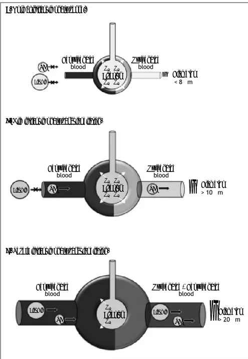
Figure 2. Figure 2. Figure 2. Figure 2.
Figure 2. Drawing demonstrates normal intrapulmonary vessel (A)(A)(A)(A)(A) which saline bubble and technetium-99m–labeled macroaggrega-ted albumin could not pass thru, mild intrapul-monary vascular dilation (B)(B)(B)(B)(B) which only saline bubble could pass thru but technetium-99m–labeled macroaggregated albumin could not pass thru, and severe intrapulmonary vascular dilation (C)(C)(C)(C)(C) which boths could pass thru. Albumin: techne-tium-99m–labeled macroaggregated albumin. O2: Oxygen. SB: saline bubble.
A. A. A. A.
A. Normal intrapulmonary vessel.
B. B. B. B.
B. Mild intrapulmonary vascular dilation.
C. C. C. C.
C. Severe intrapulmonary vascular dilation.
Diameter > 10 μm
Diameter > 20 μm Diameter
< 8 μm Oxygenated
blood Deoxygenated
blood
Alveolus O2 O2
O2 O2
Alveolus O2 O2
O2 O2
Alveolus O2
O2
Oxygenated blood Deoxygenated
blood
Oxygenated + Deoxygenated blood
Deoxygenated blood
SB SB
SB SB
Albumin Albumin
Albumin Albumin
SB
}
}
bed during the first pass, but they could pass through if the pulmonary vessels are dilated. When the agitated sa-line are seen in the left heart after 3 cardiac cycles, the intracardiac right to left shunt could be excluded,19
how-ever, we could not differentiate pulmonary arteriov-enous malformation, pulmonary arteriovarteriov-enous fistula, and intrapulmonary vascular dilation from each other. Nevertheless, in our study, all positive saline bubble tests disappeared post LT. This indirectly indicated that the positive SBT in all patients was due to intrapulmo-nary vascular dilation rather than the two formers. Since all pre-LT children did not have desaturation at room air, the definite diagnosis of HPS could not be made, al-though mild or early HPS was not excluded. In addition, the 99mTc- MAA perfusion scan was negative in all
pre-LT children. This indicated that the degree of IPVD was not much enough to let the molecule of MAA which had diameter larger than the intrapulmonary vessel to pass through the intrapulmonary vascular beds. Although the Tc-99m MAA perfusion scan is the standard method for the diagnosis of HPS, the major disadvantage of Tc-99m MAA perfusion scan is, first, it could not differentiate in-tracardiac shunt from intrapulmonary shunt.20 Secondly,
the MAA perfusion scan is less sensitive for the detection mild IPVD when compared to saline bubble as shown in this study. This could be explained by the size of Tc-99m-albumin (approximately more than 20 μm)21,22 will
be trapped in pulmonary vascular bed whereas the size of saline bubble (approximately more than 10 μm)9,10 could
pass thru the pulmonary vascular bed if there is mild IPVD (Figure 2). Previous studies demonstrated that sa-line bubble test or contrast enhanced echocardiography was more sensitive than MAA perfusion scan in detec-tion of intrapulmonary vascular diladetec-tion.9,23 European
Respiratory Society (ERS) Task Force on Pulmonary-Hepatic Vascular Disorders (PVD) has recommended contrast-enhanced echocardiography as the first line screening test for HPS.24 From our study, we agree with
this recommendation in performing echocardiography with saline bubble test as part of investigations in all children with chronic liver disease. Studies demonstrat-ed that most patients (> 85%) with HPS had improve-ment or resolution in hypoxemia after LT.7,25 Moreover,
the mortality after LT was significantly increased in pa-tients who developed HPS before performing liver transplantation.26 Our findings indicate that IPVD or
probable early stage of HPS can be identified in most children with CLD, since 89% of these children had evi-dence of IPVD by SBT. HPS has also been reported in patients with non-hepatic portal hypertension.27 In our
study, all cirrhotic patients had clinical evidences of por-tal hypertension, we speculated that porpor-tal hypertension may play role in the findings of IPVD. Early LT in
pa-tients with evidence of IPVD might prevent the progres-sion to definite HPS and thereby could improve the sur-vival rate after LT.
CONCLUSIONS
Most cirrhotic children in this cohort study had evi-dence of IPVD by positive SBT. However, none of these met the criteria for diagnosis of HPS. This might indicate mild degree of IPVD and might represent early stage of HPS. IPVD resolved after LT in all patients.
ABBREVIATIONS
• 2-D: 2 dimension. • 99mTc: Technetium-99m.
• CEE: contrast-enhanced transthoracic echocardiogra-phy.
• CLD: chronic liver disease.
• GMT: geometric mean of technetium. • HPS: hepatopulmonary syndrome. • IPVD: intrapulmonary vascular dilation. • LA: left atrium.
• LT: liver transplantation. • LV: left ventricle.
• MAA: macroaggregated albumin. • NSS: normal saline solution.
• PELD: pediatric end-stage liver disease. • RA: right atrium.
• RV: right ventricle. • SBT: saline bubble test.
FINANTIAL SUPPORT
This study was financially supported by Research Grant of Faculty of Medicine Ramathibodi Hospital, Mahidol University.
ACKNOWLEDGEMENTS
We acknowledge Mr. Dittapol Muntham for his statis-tical analysis, liver transplantation team at Faculty of Med-icine Ramathibodi Hospital, Mr. Uthen Pandee for performing echocardiography, and all patients participat-ing in this study.
REFERENCES
1. Nusrat S, Khan MS, Fazili J, Madhoun MF. Cirrhosis and its complications: Evidence based treatment. World J Gastro-enterol 2014; 20: 5442-60.
3. Krowka MJ, Cortese DA. Hepatopulmonary syndrome. Chest
1990; 98: 1053-4.
4. Martinez GP, Barbera JA, Visa J, Rimola A, Pare JC, Roca J, Navasa M, et al. Hepatopulmonary syndrome in candidates for liver transplantation. J Hepatology 2001; 34: 651-7. 5. Schenk P, Fuhrmann V, Madl C, Funk G, Lehr S, Kandel O,
Muller C. Hepatopulmonary syndrome: Prevalence and pre-dictive value of various cut offs for arterial oxygenation and their clinical consequences. Gut 2002; 51: 853-9.
6. Noli K, Solomon M, Golding F, Charron M, Ling SC. Preva-lence of hepatopulmonary syndrome in children. Pediatrics
2008; 121: e522-e527.
7. Lange PA, Stoller JK. The hepatopulmonary syndrome. Effect of liver transplantation. Clin Chest Med 1996; 17: 115-23. 8. Schneider BL, Suchy FJ, Emre S. National and regional
anal-ysis of exceptions to the pediatric end-stage liver disease scoring system (2003-2004). Liver Transpl 2006; 12: 40-5. 9. Abrams GA, Jaffe CC, Hoffer PB, Binder HJ, Fallon MB.
Di-agnostic utility of contrast echocardiography and lung per-fusion scan in patients with hepatopulmonary syndrome.
Gastroenterology 1995; 109: 1283-8.
10. Krowka MJ, Tajik AJ, Dickson ER, Wiesner RH, Cortese DA. Intrapulmonary vascular dilatations (ipvd) in liver transplant candidates. Screening by two-dimensional contrast-en-hanced echocardiography. Chest 1990; 97: 1165-70. 11. Gudavalli A, Kalaria VG, Chen X, Schwarz KQ.
Intrapulmo-nary arteriovenous shunt: Diagnosis by saline contrast bub-bles in the pulmonary veins. J Am Soc Echocardiogr 2002; 15: 1012-14.
12. Lao AY, Sharma VK, Tsivgoulis G, Frey JL, Malkoff MD, Navrro JC, Alexandrov AV. Detection of right-to-left shunts: Comparison between the international consensus and spen-cer logarithmic scale criteria. J Neruroimaging 2008; 18: 402-6.
13. Wade OL, Bishop JM. Cardiac output and regional blood flow. Oxford, England: Blackwell Scientific; 1962.
14. Varghese J, Ilias-basha H, Dhanasekaran R, Singh S, Venk-ataraman J. Hepatopulmonary syndrome - past to present.
Ann Hepatology 2007; 6: 135-42.
15. de Macedo LG, Lopes EP. Hepatopulmonary syndrome: An update. Sao Paulo Med J 2009; 127: 223-30.
16. Cremon G, Higenbottam TW, Mayoral V, Alexander G, De-moncheaux E, Borland C, Roe P, et al. Elevated exhaled nitric oxide in patients with hepatopulmonary syndrome. Eur Resp J 1995; 8: 1883-5.
17. Porres-Aguilar M, Altamirano JT, Torre-Delgadillo A, Charlton MR, Duarte-Rojo A. Portopulmonary hypertension and
hepat-opulmonary syndrome: A clinician-oriented overview. Eur Resp Review 2012; 21: 223-33.
18. Castro M, Krowka MJ. Hepatopulmonary syndrome: A pul-monary vascular complication of liver disease. Clin Chest Med 1996; 17: 35-48.
19. Shub C, Tajik AJ, Seward JB, Dines DE. Detecting intrapul-monary right-to-left shunt with contrast echocardiography. Observations in a patient with diffuse pulmonary arteriov-enous fistulas. Mayo Clin Proc 1976; 51: 81-4.
20. Khan AN, Al-Jahdali H, Abdullah K, Irion KL, Sabih Q, Gouda A. Pulmonary vascular complications of chronic liver dis-ease: Pathophysiology, imaging, and treatment. Ann Thorac Med 2011; 6: 57-65.
21. El-Shabrawi MH, Omran S, Wageeh S, Isa M, Okasha S, Mohsen NA, Zekry O, et al. (99m)technetium-macroaggre-gated albumin perfusion lung scan versus contrast en-hanced echocardiography in the diagnosis of the hepatopulmonary syndrome in children with chronic liver disease. Eur J Gastroenterol Hepatol 2010; 22: 1006-12. 22. Abrams GA, Nanda NC, Dubovsky EV, Krowka MJ, Fallon
MB. Use of macroaggregated albumin lung perfusion scan to diagnose hepatopulmonary syndrome: A new approach.
Gastroenterology 1998; 114: 305-10.
23. Krowka MJ. Hepatopulmonary syndrome and portopulmo-nary hypertension: Distinctions and dilemmas. Hepatology
1997; 25: 1282-4.
24. Rodriguez-Roisin R, Krowka MJ, Herve P, Fallon MB, Commit-tee ERSTFP-HVDS. Pulmonary-hepatic vascular disorders (phd). Eur Resp J 2004; 24: 861-80.
25. Fallon MB, Abrams GA. Pulmonary dysfunction in chronic liv-er disease. Hepatology 2000; 32: 859-65.
26. Arguedas MR, Abrams GA, Krowka MJ, Fallon MB. Prospec-tive evaluation of outcomes and predictors of mortality in pa-tients with hepatopulmonary syndrome undergoing liver transplantation. Hepatology 2003; 37: 192-7.
27. Hervé P, Lebrec D, Brenot F, Simonneau G, Humbert M, Sit-bon O, Duroux P. Pulmonary vascular disorders in portal hy-pertension. Eur Respir J 1998; 11: 1153-66.
Correspondence and reprint request: Anant Khositseth, MD. Department of Pediatrics, Faculty of Medicine Ramathibodi
Hospital
270 Rama VI Road, Ratchathewi, Bangkok 10400, Thailand Tel. 662-2011685, Fax. 662-2011850