Therapeutic effects of granulocyte-colony stimulating factor on
non-alcoholic hepatic steatosis in the rat
Yi-Sun Song,* Cheng-Hu Fang,** Byung-Im So,* Jun-Young Park,* Dae Won Jun,*** Kyung-Soo Kim*,***
* Graduate School of Biomedical Science and Engineering, Hanyang University, Seoul, Korea. ** Division of Cardiology, Yanbian University, Yanji, China.
*** Department of Internal Medicine, Hanyang University College of Medicine, Seoul, Korea.
ABSTRACT
Background and rationale. Non-alcoholic hepatic steatosis refers to the accumulation of triglycerides in the liver in the absence of alcohol consumption. Granulocyte colony-stimulating factor (G-CSF) has been reported to be an effective treatment for a variety of liver diseases. We examined the possible therapeu-tic effects of G-CSF on non-alcoholic hepatherapeu-tic steatosis in rats. Material and methods. Thirty-week-old Otsuka Long Evans Tokushima Fatty (OLETF) rats received water containing 30% sucrose for 8 weeks to pro-mote the development of non-alcoholic hepatic steatosis. After development of the model, the rats were injected with G-CSF (100 µg/kg/day) or saline for 5 days. Four weeks after this treatment, serum levels of glucose, total cholesterol (TC), triglyceride (TG), alanine aminotransferase (ALT), aspartate aminotransfera-se (AST) and free fatty acids (FFA) were measured. Histology was examined by hematoxylin and eosin (H-E) and periodic acid Schiff (PAS) staining, and levels of expression of hepatic lipogenic enzymes were deter-mined by RT-PCR. Results. The G-CSF-treated rats displayed significantly fewer lipid droplets than the saline-treated rats (P < 0.01), and their levels of sterol regulatory element-binding protein (SREBP)-1c, fatty acid synthase (FAS), and acetyl-CoA carboxylase (ACC) mRNAs were also lower (P < 0.01), as were their liver weight and serum levels of TG and FFA (P < 0.05). Conclusion. Our results indicate that G-CSF ameliorated non-alcoholic hepatic steatosis in the OLETF rat, and this therapeutic effect involved a reduc-tion of SREBP-1c expression. Therefore, G-CSF deserves further study as a potential treatment for non-alcoholic hepatic steatosis.
Key words. Sterol regulatory element-binding protein-1c. Otsuka Long Evans Tokushima Fatty. Non-alcoholic fatty li-ver disease.
Correspondence and reprint request: Kyung-Soo Kim, M.D., Ph.D. Department of Internal Medicine, Hanyang University College of Medicine 17 Haengdang-dong, Sungdong-ku, Seoul, 133-792, Korea
Tel.: +82-2-2290-8312 Fax: +82-2-2298-9183 E-mail: [email protected]
Manuscript received: April 05, 2012. Manuscript accepted: June 15, 2012. INTRODUCTION
Non-alcoholic hepatic steatosis refers to the accumulation of triglycerides in the liver in
the absence of alcohol consumption.1 It is an early
stage of non-alcoholic fatty liver disease (NAFLD).2
NAFLD is a major health problem affecting about
20 percent of the general population,3,4 and hepatic
steatosis has been reported in 48.7% of morbidly
obese patients.5 Non-alcoholic hepatic steatosis is
frequently associated with obesity and insulin
re-sistance.6,7 When accompanied by other metabolic
disorders, it can progress to severe NAFLD, non-alcoholic steatohepatitis, fibrosis, and ultimately
cirrhosis.8-10 Steatosis has been considered the
“first hit” in a “two-hit hypothesis” for the
develo-pment of NAFLD.11 At present there is no effective
treatment for non-alcoholic hepatic steatosis. Lifes-tyle changes, similar to those recommended for obesity,
are the best therapeutic option,12 but they are hard
to achieve. Therefore effective drug treatments are needed.
Granulocyte colony-stimulating factor (G-CSF) is
widely used to mobilize hematopoietic stem cells,13,14
and has been reported to be an effective treatment for a variety liver diseases. For example, one study demonstrated that G-CSF ameliorated acute hepatic failure by enhancing the homing of transplanted
Another study showed that G-CSF protected liver tissue from collagen deposition in carbon
tetra-chloride (CCl4)-induced liver fibrosis in mice.16
However the therapeutic effect of G-CSF on non-al-coholic hepatic steatosis, the first step in NAFLD, is unknown, although the authors have previously studied the effect of G-CSF on diabetic cardiomyopathy
in rats.17
We therefore have investigated the effect of G-CSF administration on non-alcoholic hepatic stea-tosis and on the mRNAs encoding hepatic lipogenic enzymes sterol regulatory element-binding protein-1c (SREBP-protein-1c), fatty acid synthase (FAS), and acetyl-CoA carboxylase (ACC).
MATERIAL AND METHODS
Animals
This study was performed in compliance with the
ARRIVE guidelines on animal research,18 and
the Hanyang University Institutional Animal Care and Use Committee approved all protocols. We used male Otsuka Long Evans Tokushima Fatty (OLETF) rats and control Long-Evans Tokushima Otsuka (LETO) rats, supplied by the Tokushima Research Institute, Otsuka Pharmaceutical (Tokushima, Japan). We used the OLETF rats as an animal mo-del of non-alcoholic hepatic steatosis. OLETF rats
are well-established animal models of obesity,19 type 2
diabetes mellitus,20 and NAFLD21,22 as well as
hepa-tic steatosis.23,24 The animals were maintained in the
Hanyang University Medical School Animal Experi-ment Center and were kept in a specific
pathogen-free facility at controlled temperature (23 ± 2 oC)
and humidity (55 ± 5%) with a 12-h artificial light and dark cycle.
Animal model and experimental protocol
Starting at 30 weeks of age, all the OLETF rats received water containing 30% sucrose for 8 weeks to facilitate the development of non-alcoholic hepatic steatosis. After steatosis had been induced, the rats received no more sucrose water. The LETO rats, as normal controls, received water without sucrose. The development of non-alcoholic hepatic steatosis was confirmed by hematoxylin and eosin (H-E) and periodic acid Schiff (PAS) staining. At 38 weeks the OLETF rats were randomly divided into a G-CSF-treated group (G-CSF 100 µg/kg/day intraperitoneally for 5 days; Leucostim, Dong-A Pharmaceutical,
Korea, n = 5) and a saline-treated group (n = 4). The LETO rats (n = 4) were injected only with sa-line. Four weeks after the treatment, blood was co-llected for biochemical analysis, and the animals were killed under anesthesia. Immediately after death, the abdomens were opened and the livers were quickly removed for histopathological exami-nation.
Biochemical analysis
Blood samples were taken from the tail vein after 8 h of fasting. Serums were obtained by centrifugation,
and stored at -70 oC. Serum glucose, total cholesterol
(TC), triglyceride (TG), alanine aminotransferase (ALT), aspartate aminotrans (AST) and free fatty acids (FFA) were measured with an Olympus AU400
auto analyzer (Olympus GmbH, Germany).25
Histological examination
To examine liver morphology, 4% paraformalde-hyde-fixed paraffin-embedded liver sections of 3 µm thickness were stained with H-E and PAS. De-grees of hepatic steatosis were estimated by oil red
O staining of frozen 3 µm sections.26 Three areas
of digitized images of the oil red O-stained liver sections were selected at random from the indivi-dual sections and analyzed with Image-Pro Plus software (Media Cybernetics, MD, USA). Steatosis was calculated as a percentage of the ratio of the area of oil red O stained lipid droplets to total
tis-sue area.23,24
Quantitative real-time polymerase chain reaction (PCR)
Total RNA was extracted from 20 mg samples of liver tissue using Qiazol reagent (Qiagen, Valencia, CA) following the manufacturer’s instructions. RNA concentrations were measured with a Nanodrop ND-2000 spectrophotometer (Thermo Fisher Scienti-fic Inc., DE, USA), and purity was determined by measuring ratios of A260 and A280, which ranged from 1.8 to 2.0.
The primers used were:
• SREBP-1c:
° 81 bp, sense: 5’-GCT ACC GTT CCT CTA TCA ATG ACA A-3’.
° Anti-sense: 5’-CAG ATT TAT TCA GCT TTG CCT CAG T-3’.
• FAS:
° 99 bp, sense: 5’-TCC ACA GCT CTT ACA GTG AGA ATC A-3’.
° Anti-sense: 5’-CTT CTC CAG GGT GGG GAC CAG-3’.
• ACC:
° 97 bp, sense: 5’-AGA GTG AGT GCT CTC AAT TCT GTC C-3’.
° Anti-sense: 5’-GTC CTT CTT CTT TCC CGA TAA TGT C-3’.
• GAPDH:
° 96 bp, sense: 5’-CCT TCT CTT GTG ACA AAG TGG ACA T-3’.
° Anti-sense: 5’-CGT GGG TAG AGT CAT ACT GGA ACA T-3’.
We performed PCR using the following steps: in-cubation for 10 min at 95 °C followed by 45 cycles of
10 s at 95 °C, 10 s at 60 °C, and 8 s at 72 °C and a fi-nal dissociation curve step at 65 °C for 15 s. The crossing point (Cp) of each PCR was automatically determined by the LightCycler® program. PCR reac-tions for all samples were each run in duplicate. The measured transcript levels were normalized against those of GAPDH.
Statistical analysis
All data were analyzed with SPSS statistics 17.0 software. The results are presented as means ± SD, while the oil red O staining using image analysis system are presented as means ± SE. Compari-sons between groups were made using one-way analysis of variance (ANOVA) followed by the post-hoc LSD test. Side-to-side comparisons within the same group were made with Student’s t test for paired data. Values of P < 0.05 were considered statistically significant.
RESULTS
Histology
The development of non-alcoholic hepatic steatosis was confirmed by H-E and PAS staining. All histolo-gical data were evaluated by a separate blinded
inves-Figure 1. Histological changes in liver tissue seen by staining with hematoxylin and eosin (H-E) and periodic acid Schiff (PAS) (magnification x200). Liver of normal control rat (a), G-CSF treated OLETF rat (b) and saline-treated OLETF rat (c). A. Stained with H-E. B. Stained with PAS.
A
B
a b c
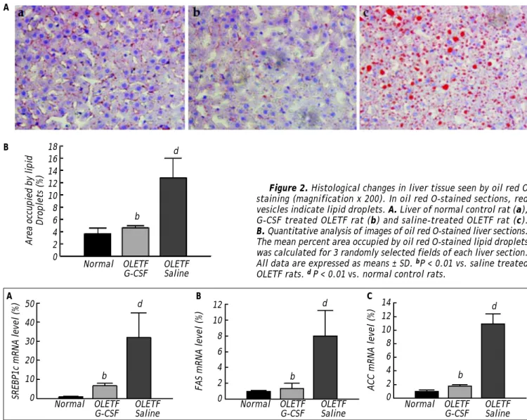
tigator. Fibrosis, collagen deposition and inflamma-tory cell infiltration did not evident, whereas lipid va-cuoles and mild ballooning in the hepatocytes were observed (data not shown). Four weeks after treat-ment, numerous cytoplasmic lipid vacuoles and mild ballooning were evident in the hepatocytes of the sali-ne-treated group. In contrast, no fat accumulation could be seen in the livers of the G-CSF-treated group, which were no different from those of the nor-mal control group (Figure 1). The area of lipid dro-plets as indicated by oil red O staining was much lower in the G-CSF-treated group than in the sali-ne-treated group (4.62 ± 0.38% vs. 12.80 ± 3.23%,
P < 0.01) and not significantly different from that in the normal control group (3.65 ± 0.94%) (Figure 2).
Expression of hepatic lipogenic genes
Levels of SREBP-1c, FAS, and ACC mRNA in the liver were measured by real-time PCR. The level of SREBP-1c mRNA was significantly lower in the G-CSF-treated group than in the saline-treated group (6.69 ± 1.28 vs. 32.06 ± 12.77, P < 0.01), and was equal to that in the normal control group (1.00 ± 0.20) (Figure 3A). The level of FAS mRNA in the G-CSF-treated group was also significantly lower
Figure 2. Histological changes in liver tissue seen by oil red O staining (magnification x 200). In oil red O-stained sections, red vesicles indicate lipid droplets. A. Liver of normal control rat (a), G-CSF treated OLETF rat (b) and saline-treated OLETF rat (c). B. Quantitative analysis of images of oil red O-stained liver sections. The mean percent area occupied by oil red O-stained lipid droplets was calculated for 3 randomly selected fields of each liver section. All data are expressed as means ± SD. bP < 0.01 vs. saline treated OLETF rats. d P < 0.01 vs. normal control rats.
Figure 3. Levels of mRNA in liver tissue after treatment as determined by real-time polymerase chain reaction (PCR) analysis. A-C. Sterol regulatory element-binding protein (SREBP)-1c, fatty acid synthase (FAS), and acetyl-CoA carboxylase mRNA levels, respectively. Mean values were obtained from the livers of four separate animals. PCR reactions were performed in duplicate. The transcript levels were normalized by comparison with GAPDH expression. All data are expressed as means ± SD. bP < 0.01
vs. saline treated OLETF rats. dP < 0.01 vs. normal control rats.
A
B 18
16 14 12 10 8 6 4 2 0
Area occupied by lipid
Droplets (%)
Normal OLETF OLETF G-CSF Saline
d
b
d d
b b
d
b
Normal OLETF OLETF
G-CSF Saline Normal OLETFG-CSF OLETFSaline Normal OLETFG-CSF OLETFSaline
SREBP1c mRNA level (%)
50
40
30
20
10
0
A B C
FAS mRNA level (%) ACC mRNA level (%)
12 10 8 6 4 2 0
than in the saline-treated group (1.33 ± 0.66 vs. 8.02 ± 3.20, P < 0.01), and was equal to that in the normal control group (1.00 ± 0.09) (Figure 3B). Furthermore, the level of ACC mRNA was signifi-cantly lower in the G-CSF-treated group than in the saline-treated group (1.77 ± 0.18 vs. 10.93 ± 1.46, P < 0.01), and was equal to that in the normal con-trol group (1.00 ± 0.23) (Figure 3C).
Metabolic parameters
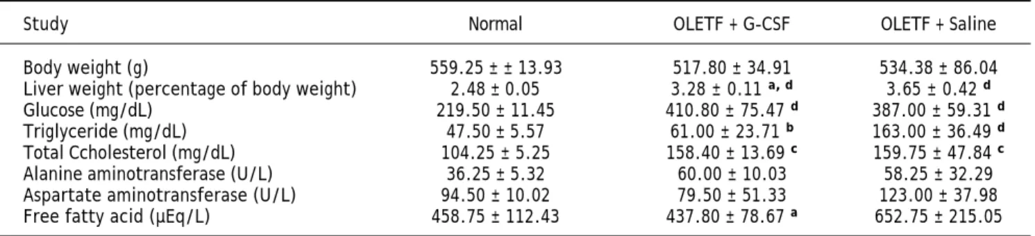
At the end of the experiment, liver weight relative to body weight was lower in the G-CSF-treated group than in the saline-treated group (3.28 ± 0.11% vs. 3.65 ± 0.42%, P < 0.05) but still higher than in the normal control group (2.48 ± 0.05%, P < 0.01). Levels of circulating TG and FFA were lower in the G-CSF-treated group than in the sali-ne-treated group (61.00 ± 23.71 mg/dL vs. 163.00 ± 36.49 mg/dL, P < 0.01 and 437.80 ± 78.67 µEq/L vs. 652.75 ± 215.05 µEq/L, P < 0.05, respectively). Circulating glucose and TC were similar in the G-CSF-treated and saline-treated groups, but appeared higher than in the normal control group. Body weight and circulating ALT and AST were similar in the three groups (Table 1).
DISCUSSION
In this study we showed that G-CSF ameliorates non-alcoholic hepatic steatosis in the OLETF rat, and reduces expression of the hepatic lipogenic ge-nes SREBP-1c, FAS, and ACC in the liver.
Histologically, we observed that G-CSF clearly de-creased lipid droplets and ballooning in hepatocytes (Figure 1); the administration of G-CSF decreased by about three fold the area of lipid droplets in the liver sections stained with oil red O (Figure 2), which is a well-established method of assessing the extent
of steatosis.23,27 Also, liver weight relative to body
weight was reduced by G-CSF without any change in body weight (Table 1).
Recently, several possible mechanisms of the ge-neral effect of G-CSF on various liver diseases have been suggested. Enhanced bone marrow cell homing towards damaged liver cells may induce trans-diffe-rentiation or a paracrine effect, contributing to the
regeneration of the damaged liver.15 In addition,
G-CSF may act directly on the liver cells
through G-CSF receptors.28 In this study, we only
determined the effect of G-CSF on non-alcoholic hepatic steatosis, which was found to be associated with down-regulation of SREBP-1c, FAS, and ACC. Further study is required to confirm the mechanis-ms underlying the effect of G-CSF on the liver.
Zhiyong Guo, et al. demonstrated that adminis-tration of diazoxide for 22 weeks reduced fat
depo-sition in the liver,29 and several groups have shown
that long-term exercise training has a beneficial
effect on non-alcoholic hepatic steatosis.30,31 Our
fin-dings suggest that G-CSF is effective with only a short-term treatment. In previous studies, the bene-ficial effects of treatment for non-alcoholic hepatic
steatosis were due to weight reduction,21,29 whereas
in our hands G-CSF ameliorated steatosis without weight reduction. Clinical studies of G-CSF have shown that it does not have any severe side effects,
though it may cause transient bone pain.32
According to previous studies, hepatic lipogenesis is responsible for the development of hepatic steato-sis. Hepatic lipogenesis is caused by an imbalance between input (uptake and synthesis) and output
(oxidation and degradation) of FFA.12,33 In our
stu-dy, G-CSF reduced circulating FFA down to the le-vel in the normal control group (Table 1).
SREBP-1c plays an important role in hepatic
lipo-genesis,33,34 and increased expression of SREBP-1c
accelerates hepatic lipogenesis by activating lipogenic
Table 1. Levels of metabolic parameters in the three groups.
Study Normal OLETF + G-CSF OLETF + Saline Body weight (g) 559.25 ± ± 13.93 517.80 ± 34.91 534.38 ± 86.04 Liver weight (percentage of body weight) 2.48 ± 0.05 3.28 ± 0.11 a, d 3.65 ± 0.42 d
Glucose (mg/dL) 219.50 ± 11.45 410.80 ± 75.47 d 387.00 ± 59.31 d
Triglyceride (mg/dL) 47.50 ± 5.57 61.00 ± 23.71 b 163.00 ± 36.49 d
Total Ccholesterol (mg/dL) 104.25 ± 5.25 158.40 ± 13.69 c 159.75 ± 47.84 c
Alanine aminotransferase (U/L) 36.25 ± 5.32 60.00 ± 10.03 58.25 ± 32.29 Aspartate aminotransferase (U/L) 94.50 ± 10.02 79.50 ± 51.33 123.00 ± 37.98 Free fatty acid (µEq/L) 458.75 ± 112.43 437.80 ± 78.67 a 652.75 ± 215.05
enzymes such as FAS, acetyl-CoA carboxylase (ACC), and stearoyl-CoA desaturase (SCD) (Figure 3).6,34,35 Also over-expression of SREBP-1c leads to
the development of hepatic steatosis in mice,36 and
SREBP-1c synergistically regulates expression of
li-pogenic genes such as FAS, ACC and SCD.35,37 Many
genes are associated with hepatic steatosis, and SREBP-1c, FAS, and ACC are comparatively known
as a key regulator of hepatic steatosis.35,38 In the
present study, we showed that G-CSF decreased the mRNA levels of SREBP-1c, FAS, and ACC mRNAs (Figures 3).
In other studies, hepatic fat accumulation closely
correlated with the oxidative stress.39,40 Oxidative
stress results from an imbalance between oxidant and antioxidant that leads to oxidative damage
in the liver.41 Thus, this study had limitations in
that the fatty acid oxidation in the liver and the he-patic mitochondrial enzymes were not analyzed. It is necessary to evaluate the changes in oxidative stress in future studies to clarify the mechanism by which G-CSF ameliorates hepatic steatosis.
Taken together our findings suggest that G-CSF reduces hepatic lipogenesis by inhibiting SREBP-1c production, which in turn reduces the accumula-tion of lipogenic enzymes such as FAS and ACC (Figure 4).
A reduction in circulating TG and FFA reduces the input of fatty acid to hepatocytes. SREBPs are well known as enhancers of FFA biosynthesis and
FFA uptake.42 We showed that administration of
G-CSF reduced the expression of SREBP-1c and circulating TG and FFA (Figure 3A and Table 1). Our analysis indicates that G-CSF reduces expression of SREBP-1c, resulting in less circulating TG and FFA. Further study is required to establish whether SREBP-1c is the main target of G-CSF, and to examine the expression of lipogenic enzymes other than FAS and ACC.
In summary, we have demonstrated that G-CSF ameliorates non-alcoholic hepatic steatosis in the OLETF rat model, and reduces the expression of SREBP-1c, which plays a key role in the development
of hepatic steatosis.33 We speculate that the reduction
of SREBP-1c expression by G-CSF is related to the improvement of the non-alcoholic hepatic steatosis. Despite the fact that the underlying mechanism is not known, our findings indicate that administration of G-CSF dramatically ameliorates non-alcoholic hepa-tic steatosis. To our knowledge, this is first report of the effect of G-CSF on non-alcoholic hepatic steatosis. In addition, we present evidence that the beneficial effect of G-CSF is associated with down-regulation of SREBP-1c. Therefore, our findings suggest that G-CSF is a novel approach to the treatment of non-alco-holic hepatic steatosis.
ABBREVIATIONS
• NAFLD: non-alcoholic fatty liver disease. • G-CSF: granulocyte colony-stimulating factor. • SREBP: sterol regulatory element-binding
protein.
• FAS: fatty acid synthase. • ACC: acetyl-CoA carboxylase.
• OLETF: Otsuka Long Evans Tokushima Fatty. • LETO: Long-Evans Tokushima Otsuka.
• H-E: hematoxylin and eosin. • PAS: periodic acid Schiff. • TC: total cholesterol. • TG: triglyceride.
• ALT: alanine aminotransferase. • AST: aspartate aminotrans. • FFA: free fatty acids.
• PCR: polymerase chain reaction. • SCD: stearoyl-CoA desaturase.
FINANCIAL SUPPORT
This work was supported by the grant for the Me-dical Research Center (2011-0028261) funded by the National Research Foundation of Korea (NRF) of the Ministry of Education, Science and Technology (MEST), Republic of Korea.
REFERENCES
1. Day CP, James OF. Hepatic steatosis: innocent bystander or guilty party? Hepatology 1998; 27: 1463-6.
2. Yamaguchi K, Yang L, McCall S, Huang J, Yu XX, Pandey SK, Bhanot S, et al. Inhibiting triglyceride synthesis improves hepatic steatosis but exacerbates liver damage and fibro-sis in obese mice with nonalcoholic steatohepatitis. Hepa-tology 2007; 45: 1366-74.
Figure 4. Schematic representation of the effects of G-CSF on hepatic lipogenesis.
Obesity
G-CSF SREBP-1c
FAS, ACC
Hepatic lipogenesis
3. Alba LM, Lindor K. Review article: Non-alcoholic fatty liver disease. Aliment Pharmacol Ther 2003; 17: 977-86. 4. Yoon KH, Lee JH, Kim JW, Cho JH, Choi YH, Ko SH, Zimmet
P, et al. Epidemic obesity and type 2 diabetes in Asia. Lan-cet 2006; 368: 1681-8.
5. Oliveira CP, Faintuch J, Rascovski A, Furuya CK Jr., Bastos Mdo S, Matsuda M, Della Nina BI, et al. Lipid peroxidation in bariatric candidates with nonalcoholic fatty liver disease (NAFLD)-preliminary findings. Obes Surg 2005; 15: 502-5. 6. Browning JD, Horton JD. Molecular mediators of hepatic
steatosis and liver injury. J Clin Invest 2004; 114: 147-52. 7. Vizzutti F, Arena U, Nobili V, Tarquini R, Trappoliere M,
Laffi G, Marra F, et al. Non-invasive assessment of fibrosis in non-alcoholic fatty liver disease. Ann Hepatol 2009; 8: 89-94.
8. Rector RS, Thyfault JP, Wei Y, Ibdah JA. Non-alcoholic fatty liver disease and the metabolic syndrome: an upda-te. World J Gastroenterol 2008; 14: 185-92.
9. Marceau P, Biron S, Hould FS, Marceau S, Simard S, Thung SN, Kral JG. Liver pathology and the metabolic syndrome X in severe obesity. J Clin Endocrinol Metab 1999; 84: 1513-7.
10. Powell EE, Cooksley WG, Hanson R, Searle J, Halliday JW, Powell LW. The natural history of nonalcoholic steatohe-patitis: a follow-up study of forty-two patients for up to 21 years. Hepatology 1990; 11: 74-80.
11. Day CP, James OF. Steatohepatitis: a tale of two “hits”?
Gastroenterology 1998; 114: 842-5.
12. Postic C, Girard J. The role of the lipogenic pathway in the development of hepatic steatosis. Diabetes Metab
2008; 34: 643-8.
13. Zohlnhofer D, Ott I, Mehilli J, Schomig K, Michalk F, Ibrahim T, Meisetschlager G, et al. Stem cell mobilization by granu-locyte colony-stimulating factor in patients with acute myocardial infarction: a randomized controlled trial.
JAMA 2006; 295: 1003-10.
14. Sato T, Suzuki H, Kusuyama T, Omori Y, Soda T, Tsunoda F, Shoji M, et al. G-CSF after myocardial infarction accelera-tes angiogenesis and reduces fibrosis in swine. Int J Car-diol 2008; 127: 166-73.
15. Jin SZ, Meng XW, Sun X, Han MZ, Liu BR, Wang XH, Sun LY, et al. Granulocyte colony-stimulating factor enhances bone marrow mononuclear cell homing to the liver in a mouse model of acute hepatic injury. Dig Dis Sci 2010; 55: 2805-13.
16. Fang B, Luo S, Song Y, Li N, Li H, Zhao RC. Intermittent dosing of G-CSF to ameliorate carbon tetrachloride-indu-ced liver fibrosis in mice. Toxicology 2010; 270: 43-8. 17. Lim YH, Joe JH, Jang KS, Song YS, So BI, Fang CH, Shin J,
et al. Effects of granulocyte-colony stimulating factor (G-CSF) on diabetic cardiomyopathy in Otsuka Long-Evans Tokushima Fatty rats. Cardiovasc Diabetol 2011; 10: 92. 18. Kilkenny C, Browne WJ, Cuthill IC, Emerson M, Altman DG.
Improving bioscience research reporting: the ARRIVE gui-delines for reporting animal research. PLoS Biol 2010; 8: e1000412.
19. Schroeder M, Zagoory-Sharon O, Shbiro L, Marco A, Hyun J, Moran TH, Bi S, et al. Development of obesity in the Ot-suka Long-Evans Tokushima Fatty rat. Am J Physiol Regul Integr Comp Physiol 2009; 297: R1749-R1760.
20. Kawano K, Hirashima T, Mori S, Saitoh Y, Kurosumi M, Na-tori T. Spontaneous long-term hyperglycemic rat with dia-betic complications. Otsuka Long-Evans Tokushima Fatty (OLETF) strain. Diabetes 1992; 41: 1422-8.
21. Rector RS, Uptergrove GM, Morris EM, Borengasser SJ, Laughlin MH, Booth FW, Thyfault JP, et al. Daily exercise
vs. caloric restriction for prevention of nonalcoholic fatty liver disease in the OLETF rat model. Am J Physiol Gastro-intest Liver Physiol 2011; 300: G874-G883.
22. Yeon JE, Choi KM, Baik SH, Kim KO, Lim HJ, Park KH, Kim JY, et al. Reduced expression of peroxisome proliferator-activated receptor-alpha may have an important role in the development of non-alcoholic fatty liver disease. J Gastroenterol Hepatol 2004; 19: 799-804.
23. Rector RS, Thyfault JP, Morris RT, Laye MJ, Borengasser SJ, Booth FW, Ibdah JA. Daily exercise increases hepatic fatty acid oxidation and prevents steatosis in Otsuka Long-Evans Tokushima Fatty rats. Am J Physiol Gastroin-test Liver Physiol 2008; 294: G619-G626.
24. Rector RS, Thyfault JP, Laye MJ, Morris RT, Borengasser SJ, Uptergrove GM, Chakravarthy MV, et al. Cessation of daily exercise dramatically alters precursors of hepatic steatosis in Otsuka Long-Evans Tokushima Fatty (OLETF) rats. J Physiol 2008; 586: 4241-9.
25. Chiu CC, Chen KP, Liu HC, Lu ML. The early effect of olan-zapine and risperidone on insulin secretion in atypical-nai-ve schizophrenic patients. J Clin Psychopharmacol 2006; 26: 504-7.
26. Ferno J, Vik-Mo AO, Jassim G, Havik B, Berge K, Skrede S, Gudbrandsen OA, et al. Acute clozapine exposure in vivo induces lipid accumulation and marked sequential changes in the expression of SREBP, PPAR, and LXR target genes in rat liver. Psychopharmacology (Berl) 2009; 203: 73-84. 27. Lerat H, Honda M, Beard MR, Loesch K, Sun J, Yang Y,
Okuda M, et al. Steatosis and liver cancer in transgenic mice expressing the structural and nonstructural proteins of hepatitis C virus. Gastroenterology 2002; 122: 352-65. 28. Ji Y, Dahmen U, Madrahimov N, Madrahimova F, Xing W,
Dirsch O. G-CSF administration in a small-for-size liver mo-del. J Invest Surg 2009; 22: 167-77.
29. Guo Z, Bu S, Yu Y, Ghatnekar G, Wang M, Chen L, Bu M, et al. Diazoxide prevents abdominal adiposity and fatty liver in obese OLETF rats at prediabetic stage. J Diabetes Complications 2008; 22: 46-55.
30. Narayan KA, McMullen JJ, Butler DP, Wakefield T, Calhoun WK. Effect of exercise on tissue lipids and serum lipopro-teins of rats fed two levels of fat. J Nutr 1975; 105: 581-7. 31. Gauthier MS, Couturier K, Latour JG, Lavoie JM. Concu-rrent exercise prevents high-fat-diet-induced macrovesicu-lar hepatic steatosis. J Appl Physiol 2003; 94: 2127-34. 32. Gabrilove JL, Jakubowski A, Scher H, Sternberg C, Wong
G, Grous J, Yagoda A, et al. Effect of granulocyte colony-stimulating factor on neutropenia and associated morbidi-ty due to chemotherapy for transitional-cell carcinoma of the urothelium. N Engl J Med 1988; 318: 1414-22.
33. Ferre P, Foufelle F. Hepatic steatosis: a role for de novo li-pogenesis and the transcription factor SREBP-1c. Diabe-tes Obes Metab 2010; 12(Suppl. 2): 83-92.
34. Kim YW, Kim YM, Yang YM, Kim TH, Hwang SJ, Lee JR, Kim SC, et al. Inhibition of SREBP-1c-mediated hepatic steato-sis and oxidative stress by sauchinone, an AMPK-activa-ting lignan in Saururus chinensis. Free Radic Biol Med
2010; 48: 567-78.
35. Sekiya M, Yahagi N, Matsuzaka T, Najima Y, Nakakuki M, Nagai R, Ishibashi S, et al. Polyunsaturated fatty acids ameliorate hepatic steatosis in obese mice by SREBP-1 su-ppression. Hepatology 2003; 38: 1529-39.
36. Sozio MS, Liangpunsakul S, Crabb D. The role of lipid meta-bolism in the pathogenesis of alcoholic and nonalcoholic hepatic steatosis. Semin Liver Dis 2010; 30: 378-90. 37. Dentin R, Pegorier JP, Benhamed F, Foufelle F, Ferre P,
re-quired for the synergistic action of ChREBP and SREBP-1c on glycolytic and lipogenic gene expression. J Biol Chem
2004; 279: 20314-26.
38. Zhang YL, Hernandez-Ono A, Siri P, Weisberg S, Conlon D, Graham MJ, Crooke RM, et al. Aberrant hepatic expression of PPARgamma2 stimulates hepatic lipogenesis in a mouse model of obesity, insulin resistance, dyslipidemia, and he-patic steatosis. J Biol Chem 2006; 281: 37603-15. 39. Oliveira CP, Gayotto LC, Tatai C, Della Nina BI, Lima ES,
Abdalla DS, Lopasso FP, et al. Vitamin C and vitamin E in prevention of Nonalcoholic Fatty Liver Disease (NAFLD) in choline deficient diet fed rats. Nutr J 2003; 2: 9.
40. idela LA, Rodrigo R, Araya J, Poniachik J. Insulin resis-tance and oxidative stress interdependency in non-al-coholic fatty liver disease. Trends Mol Med 2006; 12: 555-8.
41. Robertson G, Leclercq I, Farrell GC. Nonalcoholic steatosis and steatohepatitis. II. Cytochrome P-450 enzymes and oxidative stress. Am J Physiol Gastrointest Liver Physiol
2001; 281: G1135-G1139.