Evaluation the value of markers for
prediction of portal vein thrombosis after devascularization
Yang Fei,* Guang-quan Zong,* Jian Chen, Ren-min LiuDepartment of General Surgery, the 81st Hospital of P.L.A. Nanjing, JiangSu, China.
ABSTRACT
Aim. To evaluate the value of D-dimer and P-selectin in cirrhotic portal hypertension (PHT) patients for prediction of portal vein thrombosis (PVT) after devascularization. Material and methods. 137 patients with cirrhotic PHT who undergone devascularization from January 2012 to April 2014 were retrospectively re-viewed, all of them were divided into two groups (PVT group and non-PVT group) by Doppler ultrasonogra-phy (DU) examination. The level of D-dimer and P-selectin was tested during the peri-operative period. Results. 38 patients (27.7%) were found PVT by DU examination post-operatively. In contrast to the non-PVT group, the level of D-dimer and P-selectin in the non-PVT group was much higher significantly at 1, 3 and 7 days after devascularization (P < 0.05). However, in the 15 days after surgery, the difference of P-selectin between the two groups was not significant (P = 0.260). It was shown that the higher sensitivity of the two markers for PVT was D-dimer, the higher specificity belonged to P-selectin. The area under receiver ope-rating characteristic (ROC) curve of P-selectin was the bigger of the two markers. When the two markers were combined to be used to diagnose PVT, the sensitivity was increased to 0.911, with a slight drop of specificity to 0.715, the area under ROC curve was 0.919. Conclusion. The level of D-dimer and P-selectin might be good candidate predictive markers for PVT in patients with cirrhotic PHT after devascularization. The combined test of the two markers can increase the value of prediction.
Key words. D-dimer. P-selectin. Combination.
* First co-authors.
Correspondence and reprint request: Yang Fei, M.D.
Department of General Surgery, the 81st Hospital of PLA. Nanjing, JiangSu, China.
E-mail: [email protected]
Manuscript accepted: February 24, 2015. Manuscript accepted: March 08, 2015. INTRODUCTION
Portal vein thrombosis (PVT) includes thrombo-sis in the portal vein, splenic vein and superior me-senteric vein or intrahepatic portal vein branches as they form an interactive vascular system without valves. PVT following devascularization occurs in 6.3 to 39.0% of patients and is mostly an unpredicta-ble event.1,2 Complete or partial occlusion of the
portal venous system in cirrhotic portal hyperten-sion (PHT) patients may determine liver ischemia, with consequent deterioration of liver function, and a sudden increase in portal pressure. Some compli-cations of liver cirrhosis such as variceal hemor-rhage may develop and death eventually may
occur.3-5 It is therefore important to identify
mark-ers that can predict and prevent the occurrence of PVT.
D-dimer is a kind of fibrin degradation fragment, and is the molecular marker in the fibrinolysis cy-cle.7 P-selectin is a cell adhesion molecule that is
mainly produced by platelets and endothelial cells; it increases the activation of platelets and the adhe-sion of platelets to the endothelium.8 Recently,
D-dimer and P-selectin have been shown to play an important role in diagnostic indicator for thrombot-ic diseases.9,10 In this study, we investigated
wheth-er D-dimwheth-er and P-selectin had a bettwheth-er predictive value to PVT after devascularization in patients with cirrhotic PHT.
MATERIAL AND METHODS
Patients and study design
Two hundred sixty-seven consecutive patients who were diagnosed with hepatitis virus-related cir-rhosis PHT between January 2012 to April 2014 in
the 81st Hospital of PLA were included. The diag-noses of these patients were all confirmed by endos-copy and Doppler ultrasonography (DU). All of them underwent the surgery procedure. They accompa-nied with no serious cardiopulmonary diseases and concomitant chronic duodenal ulcers. Patients whose serum levels of D-dimer or P-selectin during the time of diagnosis and treatment were not availa-ble were excluded. Any patients who had been tak-ing warfarin or Aspirin would have also been excluded. Accordingly, one hundred and thirty-seven patients were enrolled into this study, which was approved by our hospital. The characteristics of the patients including age, gender, Child-Pugh’s score, preoperative biochemical tests were summarized in table 1. Biochemical tests were examined within 1-3 days preoperatively.
The study protocol conformed to the guidelines by the institutional Ethical Committee. All patients gave their informed consent to be included in the study. The research was carried out according to the principles of The Declaration of Helsinki.
PVT diagnosis
DU (ACUSON SequoiaTM 512, Color Doppler
equipment) examination of the portal vein was per-formed pre-/post-operationally,11 and was performed
in all cases by the same examiner to avoid interob-server variables. Spectral waveforms were obtained at measured angles of insonation of < 60°. The pre-operational examination was performed at three days before the surgery. The post-operational
exami-nation was performed at two, five, seven, fifteen, twenty-two and thirty days following the surgery. PVT was defined as the obstruction of more than 50% of the vascular lumen by a thrombus in the main portal vein, its large branches, or in the splenic vein. No patients had a diagnosed PVT prior to the operation. Then the samples were divided into two groups, including PVT group and non-PVT group.
D-dimer and P-selectin determinations
Peripheral venous blood sample (15 mL) was col-lected from each patient who had fasted for at least 12 h and mixed with 3.8% sodium citrate (ratio 9:1). Sample was centrifuged for 10 min at 2,000 g, and the supernatant was stored at -80°C until use. D-dimer and P-selectin were measured by automated latex enhanced immunoassay in citrated plasma on instrumentation Laboratory (Coagulation Systems ACL 9000) and quantified using enzyme linked im-munosorbent assay kits (ZhongShan Biotech, Bei-jing, China). Both of the two biomarkers were tested one day before surgery, the second test was per-formed in one day postoperationally. Thereafter, in three days, seven days and fifteen days after sur-gery, the test was performed again.
Surgical technique
The surgical technique was applied as described by Zong, et al.12 The same operative setup and
standardized technique were applied for all patients.
Table 1. Preoperative clinical characteristics of cirrhotic patients.
PVT (n = 38) Non-PVT (n = 99) t/χ2-value P-value
Age, mean ± SD (years) 48.1 ± 7.1 45.2 ± 8.4 0.622 0.231
Gender
Male 21 59 0.485 0.316
Female 17 40
Child-Pugh’s score 6.8 ± 1.3 7.0 ± 1.5 0.197 0.628
PT (sec) 14.7 ± 2.9 14.1 ± 2.3 0.206 0.595
TB (μmol/L) 26.3 ± 4.0 26.8± 4.7 0.146 0.742
ALT (IU/L) 29.1 ± 7.8 27.6 ± 6.3 0.323 0.382
AST (IU/L) 27.7 ± 7.1 28.5 ± 6.4 0.272 0.503
Operative time (min) 248.1 ± 71.2 231.0 ± 63.6 0.375 0.377
Intraoperative blood loss (mL) 634.6 ± 68.5 592.7 ± 89.3 0.571 0.259
FPP (cmH2O) 39.8 ± 7.6 36.3 ± 8.9 0.363 0.498
Procedures were performed by the same team of spe-cialists in gastrointestinal and hepatobiliary surgery.
Statistical analysis
All statistical analyses were performed using SPSS15.0 software (SPSS, Chicago, IL). Continuous data were expressed as mean values ± standard de-viation (SD). Significant differences between groups were determined by chi-squared analysis and unpaired Student’s t test. Dichotomous variables were created out of continuous variables by using clinically important cutoff points. Receiver operat-ing characteristic (ROC) curves, sensitivity (Sen), specificity (Spe), and positive and negative predic-tive values (PPV, NPV) were determined to assess the diagnostic value of D-dimer and P-selectin. P-values < 0.05 were considered statistically significant.
RESULTS
Baseline characteristics
DU examination showed thirty-eight patients developed PVT post-operatively (38/137, 27.7%), the occurrence of PVT was in 5.72 ± 1.73 days after devascularization, moreover, 86.8% PVT (33/ 38) occurred between day three and seven post-operationally.
There were no significant difference between the PVT group and the non-PVT group of the preopera-tive database including age, gender, Child-Pugh’s score, blood routine biochemical tests, mean operative time, intra-operative blood loss and intra-operative freedom portal vein pressure. The database also including the level of D-dimer and P-selectin (P > 0.05) (Table 2). The two groups were well balanced in the distribution of pre- and intra-operational characteristics.
The level of D-dimer and P-selectin
As shown in table 2, in contrast to the non-PVT group, the level of D-dimer in the PVT group was much higher significantly in one day, three days, one week and fifteen days after devascularization (P < 0.05). The similar situation could be found in the level of P-selectin. The significantly more al-tered was in the PVT group than in the non-PVT group postoperationally (P < 0.05). However, in the fifteen days after surgery, the difference of P-selectin between the two groups was not significant
(P = 0.260). Table 2.
The level of D-dimer and P-selectin pre/post-operation.
Pre-1
Post-1
Post-
3
Post-7
Post-15
PV
T
Non-PVT
PV
T
Non-PVT
P
VT
Non-PVT
PV
T
Non-PVT
PV
T
Non-PVT
D-dimer
230.6 ± 58.4
218.2 ± 68.3
793.5 ± 65.8
362.1 ± 52.9
941.0
±
67.2
391.8 ± 83.0
892.5 ± 82.5
398.5 ± 72.4
737.3 ± 59.5
351.5 ± 63.9
(ng/mL) P-selectin
29.1 ± 7.2
30.8 ± 6.1
77.2 ± 6.8
50.8 ± 5.3
106.7 ± 8.3
59.2 ± 6.7
97.3 ± 7.1
58.3 ± 7.0
47.9 ± 5.2
44.1 ± 6.2
(ng/mL) P-value
0.206
a
0.383
b
0.041
a
0.048
b
0.018
a
0.022
b
0.039
a
0.036
b
0.048
a
0.260
b
PVT: portal venous thrombosis. Pre: preoperation. Post: postoperation.
a Compare PVT with non-PVT group of D-dimer level. b Compare PVT with non-PVT group
of
P
-selectin
Value of D-dimer and P-selectin on PVT
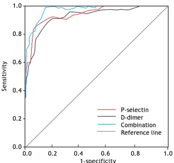
By using the change in D-dimer level and P-selec-tin level as the diagnostic standard for PVT in cir-rhosis patients after devascularization, we achieved the Sen, Spe, PPV and NPV in predicting incidence of PVT (Table 3). It showed that the higher Sen of the two markers for PVT was D-dimer, the higher Spe belonged to P-selectin. Based on the different changes in D-dimer, P-selectin levels and the false positive rates and Sen rates in different D-dimer, P-selectin levels, a ROC curve was plotted. Using the ROC analysis, the area under the ROC curve were 0.826 and 0.869, respectively. When the two mark-ers were combined to diagnose PVT, the Sen was in-creased to 0.911, with a slight drop of Spe to 0.715, the area under the ROC curve was 0.919 (Figure 1).
DISCUSSION
The prevalence of PVT in cirrhotic PHT had been reported as values ranging from 0.6 to 2.1%.13
How-ever, PVT has been recognized as a potential compli-cation following devascularization for PHT, some literatures showed a frequency from 6.3 to 39.0%.1,2
In our study, the incidence of PVT after devasculari-zation was 8.6%. PVT is often an occasional diagno-sis in asymptomatic patients, but it may rapidly progress leading to massive thrombosis of intestinal veins, even fatal consequences.14 Therefore, early
di-agnosis of PVT may be useful to prevent uneventful outcome. The early predictive indicator of postoper-ative PVT may be the most focus.
At present, some methods were performed to pre-vent the occurrence of PVT.
• Systemic or topical injection heparin for prophy-laxis intraoperatively or immediate postoperative-ly.15 But not all patients may occur PVT or
accept the treatment of injection heparin.
• Anticoagulant therapy base on the testing plate-let count, since this method reflects only the number of platelets, ignores the function of
plate-let count. Some reports showed that the level of platelet count was not more than the level of 500 x 109/L until portal vein thrombosis formation.16
• Imaging examination especially DU can provide information on the thromboses via echo analysis, detect the presence of post-stenotic dilatation, as well as flow defects and turbulence.
Although PVT can be accurately diagnosed by DU, the formation of thrombosis was a dynamic process, only when the appearance of PVT, the ul-trasound can play a valuable role, so it can’t predict thrombosis.
D-dimer is a small peptide fragments of early thrombosis in the role of fibrinogen degradation of plasmin formation, the presence of secondary fiber
in vivo dissolved and specific marker of thrombosis.17
Risch thought that D-dimer could be used as a diag-nostic specificity of acute deep vein thrombosis indica-tors.18 Palareti found the change of D-dimer level after
anticoagulation therapy for patients may predict the risk of thrombosis recurrence.19 In recent years,
some studies have shown that D-dimer level as sensi-tive indicator for coagulation system and fibrinolytic system could predict the formation of PVT.20 Our
study showed that the specificity of D-dimer was 77.3% in predicting PVT incidence, the sensitivity was 84.6%, and it may be regarded as a stable and good indicator. Increased D-dimer after operation sug-gested that activation of coagulation in vivo and hepatic clearance of tissue plasminogen activator were related to the reduction of fibrinolysis inhibitor protein synthesis.
The study on deep venous thrombosis showed that P-selectin involved in platelet/endothelial cell adhesion and inflammation in vascular wall, which may lead to a new pathogenesis of venous thrombo-sis.21 Blenn, et al. found that soluble P-selectin level
in patients with deep vein thrombosis was signifi-cantly higher, which may be related to excessive platelet activation.22 Myers’s experiment showed
that P-selectin could enlarge the effect of vein wall inflammation caused by thrombotic stimuli and promoting venous thrombosis. The higher level
Table 3. Evaluation the value for PVT of D-dimer and P-selectin.
Cut-off value Sen Spe PPV NPV AUC
D-dimer 500 ng/mL 0.838 0.760 0.813 0.807 0.826
P-selectin 80 ng/mL 0.793 0.906 0.919 0.763 0.869
Combination — 0.911 0.715 0.747 0.886 0.919
P-selectin, the more inflammatory cells in vein wall, the bigger intravenous clot generated simultaneously in model mice.23 Therefore, as an indicator of the
degree or function of platelet activation, P-selectin can be regarded as a marker to evaluate the pro-thrombotic state, coagulation tendency and forma-tion of thrombosis in vivo. Our study showed that compared with non-PVT group, the level of P-selec-tin was higher in PVT group post-operationally, es-pecially in three days after surgery. When 80 ng/mL was set as a cut-off point, P-selectin had an 87% sensitivity and specificity for PVT.
As a dynamic process, the formation of thrombo-sis was monitored by our DU continuously. And the two indicators including D-dimer and P-selectin were tested continuously for assessing the predictive ability. DU examination showed the occurrence of PVT was 5.72 ± 1.73 days (2-7 days) after devascu-larization. The two indicators showed significant difference between PVT group and non-PVT group in one, three and seven days after operation (P < 0.05), however the differences could not be found in the fifteen days post-operatively. By this token, the best prediction PVT critical point should be set as early as possible, and should be lasted three days at least. From the ROC curve figure, the area under curve of P-selectin was the bigger. When the two indicators were combined together, the area may be up to 0.919, the value of early prediction can be improved. Then clinical interventions can be performed in time and safely.
CONCLUSION
The level of D-dimer and P-selectin might be good candidate predictive markers for PVT in patients with cirrhotic PHT after devascularization. The combined test of the two markers can increase the value of prediction.
ABBREVIATIONS
• DU: Doppler ultrasonography. • NPV: negative predictive values. • PHT: portal hypertension. • PPV: positive predictive values. • PVT: portal vein thrombosis.
• ROC: receiver operating characteristic.
CONFLICT OF INTEREST
The authors declare no conflict of interest.
REFERENCES
1. Winslow ER, Brunt LM, Drebin JA. Portal vein thrombosis after splenectomy. Am J Surg 2002; 184: 631-5.
2. Webster GJ, Burroughs AK, Riordan SM. Review article: portal vein thrombosis-new insights into aetiology and management. Aliment Pharmacol Ther 2005; 21: 1-9. 3. de Franchis R. Evolving consensus in portal hypertension.
J Hepatol 2005; 43: 167-76.
4. Grace ND, Groszmann RJ, Garcia-Tsao G, Burroughs AK, Pagliaro L, Makuch RW. Portal hypertension and variceal bleeding: an AASLD single topic symposium. Hepatology 1998; 28: 868-80.
5. Garcia-Tsao G, Bosch J, Groszmann R. Portal hypertension and variceal bleeding, unresolved issues. Summary of an American Association for the study of liver disease and Eu-ropean Association for the study of the liver single-topic conference. Hepatology 2008; 47: 1764-72.
6. van der Bom JG, Heckbert SR, Lumley T, Holmes CE, Cush-man M, Folsom AR, Rosendaal FR, et al. Platelet count and the risk for thrombosis and death in the elderly. J Thromb Haemost 2009; 7: 399-405.
7. Gosselin RC, Wu JR, Kottke-Marchant K, Peetz D, Christie DJ, Muth H, Panacek E. Evaluation of the Stratus CS Acute Care D-dimer assay (DDMR) using the Stratus CS STAT Fluorometric Analyzer: a prospective multisite study for exclusion of pulmonary embolism and deep vein thrombo-sis. Thromb Res 2012; 130: e274-e278.
8. Patel KN, Soubra SH, Bellera RV, Dong JF, McMullen CA, Burns AR, Rumbaut RE. Differential role of von Willebrand factor and P-selectin on microvascular thrombosis in en-dotoxemia. Arterioscler Thromb Vasc Biol 2008; 28: 2225-30.
9. Smith K. Endoscopy: ESD is associated with a moderate risk of deep vein thrombosis that may be determined by D-dimer levels. Nat Rev Gastroenterol Hepatol 2011; 8: 538. 10. Gremmel T, Ay C, Seidinger D, Pabinger I, Panzer S,
Kop-pensteiner R. Soluble p-selectin, D-dimer, and high-sensi-tivity C-reactive protein after acute deep vein
Figure 1. The receiver operating characteristic (ROC) curve
for diagnosis PVT by D-dimer and P-selectin. 1.0
0.8
0.6
0.4
0.2
0.0
0.0 0.2 0.4 0.6 0.8 1.0
1-specificity
P-selectin D-dimer Combination Reference line
thrombosis of the lower limb. J Vasc Surg 2011; 54(6 Sup-pl.): 48S-55S.
11. Zong GQ, Fei Y, Chen J, Liu RM, Xu YF. Effects of selec-tive double portazygous disconnection and devasculariza-tion on hemodynamics of the portal venous system. Med Ultrason 2014; 16: 291-7.
12. Zong GQ, Fei Y, Chen J, Liu RM. Selective double discon-nection for cirrhotic portal hypertension. J Surg Res 2014; 192: 383-9.
13. Sobhonslidsuk A, Reddy KR. Portal vein thrombosis: a con-cise review. Am J Gastroenterol 2002; 97: 535-41. 14. Reit MV, Burger JW, van Muiswinkel JM. Diagnosis and
treatment of portal vein thrombosis following splenecto-my. Br J Surg 2000; 87: 1229-33.
15. Lisman T. Low molecular weight heparin in management and prevention of portal vein thrombosis. Thromb Res 2014; 134: 761-2.
16. Jensen MK, de Nully Brown P, Lund BV, Nielsen OJ, Hassel-balch HC. Increased circulating platelet-leukocyte aggre-gates in myeloproliferative disorders is correlated to previous thrombosis, platelet activation and platelet count. Eur J Haematol 2001; 66: 143-51.
17. Martin K, Kia L, Parikh-Neehar D, Kulik L, McMahon B. Heparin-induced thrombocytopenia testing is
over-utilized in cirrhosis & correlates with poor clinical out-comes. Ann Hepatol 2014; 13: 548-54.
18. Risch L, Monn A, Lüthy R, Honegger H, Huber AR. The pre-dictive characteristics of D-dimer testing in outpatients with suspected venous thromboembolism: a Bayesian ap-proach. Clin Chim Acta 2004; 345: 79-87.
19. Palareti G, Cosmi B, Legnani C, Tosetto A, Brusi C, Iorio A, Pengo V, et al. D-dimer testing to determine the duration of anticoagulation therapy. N Engl J Med 2006; 355: 1780-9. 20. Cosmi B, Legnani C, Tosetto A, Pengo V, Ghirarduzzi A,
Testa S, Prisco D, et al. Usefulness of repeated D-dimer testing after stopping anticoagulation for a first episode of unprovoked venous thromboembolism: the PROLONG II prospective study. Blood 2010; 115: 481-8.
21. Ramacciotti E, Blackburn S, Hawley AE, Vandy F, Ballard-Lipka N, Stabler C, Baker N, et al. Evaluation of soluble P-selectin as a marker for the diagnosis of deep venous thrombosis. Clin Appl Thromb Hemost 2011; 17: 425-31. 22. Blann AD, Noteboom WM, Rosendaal FR. Increased soluble
P-selectin levels following deep venous thrombosis: cause or effect. Br J Haematol 2000; 108: 191-3.