Full Terms & Conditions of access and use can be found at
http://www.tandfonline.com/action/journalInformation?journalCode=ljlc20
Download by: [Uni Autonoma del Estado de Hidalgo] Date: 21 October 2016, At: 10:58
Journal of Liquid Chromatography & Related
Technologies
ISSN: 1082-6076 (Print) 1520-572X (Online) Journal homepage: http://www.tandfonline.com/loi/ljlc20
Dispersive solid-phase extraction based on
butylamide silica for the determination of
sulfamethoxazole in milk samples by capillary
electrophoresis
Gabriela Islas, José A. Rodríguez, M. Elena Páez-Hernández, Silvia
Corona-Avendaño, Alberto Rojas-Hernández & Enrique Barrado
To cite this article: Gabriela Islas, José A. Rodríguez, M. Elena Páez-Hernández, Silvia Corona-Avendaño, Alberto Rojas-Hernández & Enrique Barrado (2016) Dispersive solid-phase extraction based on butylamide silica for the determination of sulfamethoxazole in milk samples by capillary electrophoresis, Journal of Liquid Chromatography & Related Technologies, 39:14, 658-665, DOI: 10.1080/10826076.2016.1230551
To link to this article: http://dx.doi.org/10.1080/10826076.2016.1230551
Accepted author version posted online: 13 Sep 2016.
Published online: 13 Sep 2016.
Submit your article to this journal
Article views: 18
View related articles
2016, VOL. 39, NO. 14, 658–665
http://dx.doi.org/10.1080/10826076.2016.1230551
Dispersive solid-phase extraction based on butylamide silica for the determination
of sulfamethoxazole in milk samples by capillary electrophoresis
Gabriela Islasa, José A. Rodrígueza, M. Elena Páez-Hernándeza, Silvia Corona-Avendañob,
Alberto Rojas-Hernándezc, and Enrique Barradod
aÁrea Académica de Química, Universidad Autónoma del Estado de Hidalgo, Pachuca, Hidalgo, Mexico; bDepartamento de Materiales, Área Ingeniería
de Materiales, Universidad Autónoma Metropolitana-Azcapotzalco, Ciudad de Mexico, Mexico; cDepartamento de Química, Área de Química
Analítica, Universidad Autónoma Metropolitana-Iztapalapa, Ciudad de Mexico, Mexico; dDepartamento de Química Analítica, Facultad de Ciencias,
Universidad de Valladolid, Valladolid, Spain
ABSTRACT
A new method based on the combination of dispersive solid-phase extraction and capillary electrophoresis is proposed for the determination of sulfamethoxazole in milk samples. Butylamide silica was synthesized and used as extractant. Factors involved in sample treatment method such as: butylamide silica amount, NaOH concentration in methanol, sample volume, and dispersion time were evaluated using a Taguchi parameter design. Under optimal conditions, average recoveries ranged from 73 to 85% with a limit of detection of 0.05 mg L 1 were achieved. The proposed method is a useful technique for cleanup milk samples.
GRAPHICAL ABSTRACT
KEYWORDS
Butylamide silica; capillary electrophoresis; dispersive solid-phase extraction; milk; sulfamethoxazole
Introduction
The use of subtherapeutic doses of antibiotics in animals intended for human consumption is a common practice to prevent diseases and promote weight gain.[1] One group of synthetic antibiotics most frequently used for these purposes are sulfonamides.[2] In consequence, residues of sulfonamides can be found in animal tissues, milk, and also in environ-mental samples such as surface waters causing adverse effects in humans such as allergic reactions in hypersensitive people besides to induce high levels of bacterial resistance.[3–6] The sulfamethoxazole (SMX) is a sulfonamide commonly adminis-tered in animals and humans. As a result, several methodolo-gies for the analysis of SMX residues have been developed, including spectroscopic,[7] electrochemical,[8] or separation techniques based on capillary electrophoresis[9] and high- performance liquid chromatography.[10]
On the other hand, pretreatment of milk samples represents a challenge during the development of an analytical method-ology for the determination of antibiotic residues. The milk matrix is complex and it requires different steps during the
treatment of the sample, some of which are precipitation of proteins by decreasing the pH value or adding organic solvents (methanol or acetonitrile), filtration, and centrifugation. Additionally, preconcentration of analytes may be necessary, a liquid–liquid extraction[11,12] or solid-phase extraction (SPE)[13,14] is required. SPE is the sample preparation tech-nique most usually used for the analysis of antibiotic residues in foods; it is based on the passage of the sample through a solid extractant contained in a cartridge where the analytes are retained and subsequently eluted with a suitable solvent.[15,16]
Dispersive solid-phase extraction (DSPE)[17,18] was described as an alternative to SPE. To improve retention of the analytes, the solid phase is added to the liquid sample to form dispersion. The contact between the analyte and the solid extractant is higher than in classic SPE, providing a better interaction during analyte extraction, and in consequence, a time reduction of sample treatment. Additionally, DSPE has the advantage of being a microscale extraction method, which makes it an attractive alternative to preconcentrate different analytes in complex matrices.[17–19]
CONTACT José A. Rodríguez [email protected] Área Académica de Química, Universidad Autónoma del Estado de Hidalgo, Carr. Pachuca Tulancingo km 4.5, Pachuca, Hidalgo 42076, Mexico.
Some applications of DSPE for the determination of sulfo-namides have been developed. It has been described by its application during the analysis of mineral water and pork tissue samples where the extractant phase was composed of carbon nanotubes which contributes to adsorption of the anti-biotic through π–π interactions.[17,18] Recently, to enhance the use of solid phases, it has been proposed separation methodol-ogies using solid phases that contain functional groups which are different from those conventionally used. As a result, the analytes can be retained by mixed mode interactions as hydro-gen bonding and dipole–dipole interactions.[20–22] These solid phases have been applied for the analysis of some drugs[10,23,24] as well as pesticides.[19] Milk samples have been analyzed through DSPE–RP–HPLC using silica modified with
N-propyl-ethylenediamine as extracting phase for the multire-sidue determination of pesticides. This work presents the synthesis and evaluation of the butylamide silica in SMX determination in milk samples by DSPE combined with capillary electrophoresis.
Experimental
Reagents and chemicals
All solutions were prepared by dissolving the respective ana-lytical grade reagent in deionized water with a resistivity not less than 18.0 MΩ cm, Milli-Q system (Millipore, Bedford, MA, USA). Sodium hydroxide, sodium phosphate monobasic, and acetic acid were obtained from Sigma-Aldrich (Steinheim, Germany). Methanol, acetonitrile, anhydrous toluene, and ethanol were from J.T. Baker (Phillipsburg, NJ, USA). 3-(Aminopropyl) trimethoxysilane (99%, APTMS), tetra-methylorthosilicate (98%, TMOS), triethylamine (99%), methyl acrylate (99%), and butylamine (99.5%), triton X-100, and cetyltrimethyl ammonium bromide (CTAB) were obtained from Sigma-Aldrich (St. Louis, MO, USA).
Sulfamethoxazole (99.9%) was obtained from Sigma- Aldrich (Steinheim, Germany). The standard solution was pre-pared daily by the dilution of a stock solution (100 mg L 1) by dissolving the pure substance in methanol. The solution was stored in the dark and refrigerated at 4°C and renewed weekly. Doxycycline hyclate (98%, from Sigma-Aldrich) was used as the internal standard (IS).
Synthesis of the butylamide silica adsorbent
Synthesis of butylamide silica was performed on the basis of sol–gel process. TMOS, 0.02 moles (5.17 mL), was previously solubilized in 25 mL of a solution containing 2.0% (w/v) Tri-ton X-100, 0.02% (w/v) CTAB, 12.5% (v/v) methanol, and 1.0 mL of NH3 28% (w/w) as a catalyst. The mixture was
refluxed for 16 h with stirring.[25] Silica phase was then washed with distilled water, followed by ethanol (3 �10 mL each one), and dried at 100°C for 24 h. In order to activate it, the silica (3.0 g) was immersed in 40 mL of HCl 3 M and refluxed for 8 h; later it was filtered, washed with distilled water, and dried at 120°C all night. Activated silica was mixed with 30 mL of anhydrous toluene, 0.5 mL of triethylamine, and 3 mL of APTMS; the mixture was refluxed for 24 h. Propylamine
functionalized silica was filtered, washed with toluene followed by ethanol (3 �10 mL each one), and dried at 60°C for 24 h. Concluded the reaction time, propylamine functionalized silica was mixed with 60 mL of methyl acrylate/methanol solution (1:1, v/v) and stirred under a nitrogen atmosphere at 50°C for 2 h. The product was filtered and washed with methanol to obtain methylester functionalized silica. Secondary amide was obtained by the reaction of methylester functionalized solid with 15 mL of butylamine in 15 mL of methanol; the mixture was stirred at 50°C for 8 h. The butyla-mide silica was filtered, washed with ethanol (3 �10 mL), and dried at 60°C for 24 h.[26]
Apparatus
Infrared characterization of the butylamide silica synthesized was performed in a Perkin-Elmer Fourier transform infrared (FTIR) spectrophotometer model IRDM. The samples were analyzed in KBr (1%) sample pellets. The morphological analysis of the solid phase was performed using a scanning electron microscopy (SEM, FEI Model Quanta 200 F, Holland).
Electrophoresis was performed using a Beckman Coulter PA 800 plus (Fullerton, CA, USA) with a photo diode array detector. Data were collected and analyzed with Beckman PA system 10.1 version 32 Karat software. Separation of the SMX was performed in a fused silica capillary (21.5 cm � 75 µm I.D.) using an electrolyte solution consisted of a phos-phate buffer solution (30 mM, pH 7.0). At the beginning of each working day, the capillary was activated with NaOH 1.0 M at 25°C for 20 min, followed by NaOH 0.1 M for 10 min and deionized water at 25°C for 10 min and then elec-trolyte solution at 25°C for 10 min. The capillary was washed out between sample analysis using: NaOH 1.0 M for 3 min, NaOH 0.1 M for 1 min, deionized water for 2 min, and electrolyte solution for 2 min. All flushing procedures were performed at a pressure of 20 psi.
The wavelength detector (λ) was set at 214 nm to monitor SMX separation. Samples were injected in hydrodynamic mode under a pressure of 0.5 psi for 5 s. The capillary was kept at 25°C, and a voltage of 15 kV was applied to separate the analytes. The peak area ratio of analyte/internal standard was used in the quantification. The different peaks were ident-ified by migration times, coinjection of standard solutions, and UV spectrum. A pH/ion analyzer model 450 from Corning (Corning Science Products, NY, USA) was used to accurately adjust the pH of the electrolyte solution to 0.01 pH unit. A Cole-Parmer Ultrasonic system (Vernon Hills, Illinois, USA) model 8891 was used in the dispersion of samples. Finally, a Maxi-Mix I (Barnstead/Thermolyne, IA, USA) model M16715 was used as a vortex mixer in the dispersion of butyl silica.
Sample analysis
Milk samples (10 mL) were fortified with internal standard (doxycycline, 15 mg L 1) in polypropylene tubes. Proteins were precipitated by adding 2.0 mL of acetic acid 2% (v/v), fol-lowed by heating for 5 min (65°C) and centrifuging at
3200 rpm for 15 min. Deproteinization helps to prevent the emulsion formation during sample treatment; additionally, the obtained aqueous matrix (acidic pH value) is suitable for extraction of the SMX by DSPE.[27,28] Subsequently, 1.0 mL of the liquid phase was taken, adjusted the pH value at 2.0 (adding H3PO4 1.0 M), and diluted to 10 mL with deionized
water in a calibration flask (solution A).
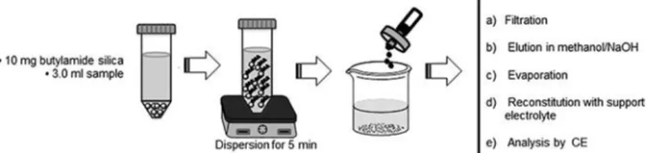
10 mg of butylamide silica was placed in polypropylene centrifuge tubes (15 mL) together with 3 mL of solution pre-pared earlier (solution A). The mixture was dispersed for 5 min in a vortex and filtered through a 0.45 µm nylon mem-brane, the solid phase (containing the analyte and the IS) was isolated and SMX and IS were eluted from the butylamide silica by passing 3.0 mL of NaOH 0.1 M in methanol through the membrane. The resulting solution was evaporated to dryness with moderate heating; subsequently the residue was reconstituted in 1.0 mL of support electrolyte (phosphate buffer, 30 mM, pH 7.0) and filtered through a nylon membrane, and finally analyzed by CE (Figure 1).
Results and discussion
Characterization of the solid adsorbent
The butylamide silica synthesized was characterized by FTIR spectroscopy (Figure 2). Infrared spectroscopy is a useful tool to identify the functional groups in silica phases. Figure 2a
shows the spectrum of unmodified silica, a stretching band at 3600–3500 cm 1 was attributed to the vibration of the sila-nol group (Si–OH), a bending band at 1645 cm 1 was attrib-uted to the water contained in the silica gel. In addition, a stretch band at 1300–1000 cm 1 belonging to the vibration of the siloxane group (Si–O–Si) as well as a band in 800 cm 1
for the siloxide group (Si–O). On the other hand, spectrum of
Figure 2b for butylamide silica solid shows a band at 1650– 1515 cm 1 attributed to the flexion of N–H and another characteristic band of the amide group (C=O) corresponding to the vibration at 1710 cm 1.[29]
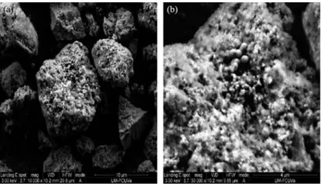
The morphology of the synthesized butylamide silica was analyzed by SEM. Figure 3 shows that solid has a granular morphology with spherical agglomerates. Particle sizes are irregular between 4 and 10 µm (Figure 3a), while the solid sur-face has granules of about 0.25 µm (Figure 3b). This botryoidal phase contributes to form porous and in consequence the solid possesses high surface area.[30]
Optimization of the DSPE method
The DSPE involves diverse control variables which can affect the extraction and elution processes such as: composition and volume of sample and eluent, amount and type of solid phase and contact time during extraction and elution. To evaluate the effect of some control parameters in the extraction of SMX by DSPE, the effect of pH value and sample volume was studied (Figure 4). The pH value plays an important role in the extraction and isolation of the SMX[17,18] because it has two ionizable groups (pKa values, 1.49, 5.48).[31] Extraction experiments were performed in a pH range from 1.0 to 10.0.
Figure 4 shows that at pH 2.0, the neutral form of SMX has higher affinity, indicating that charged SMX species (cationic at pH values below 1.49, and anionic above 5.48) causes lower extractions.
The influence of sample volume on the extraction of SMX (Figure 4) was studied in the range of 3 at 25 mL using 30 mg of butylamide silica. The amount of SMX extracted were stable in all range evaluated, resulting in an extraction capacity of 0.56 �0.02 µgSMXmg 1solid.[32] A sample volume of 3 mL was
chosen for further analysis.
Once the appropriate extraction conditions, it was evalu-ated the following solvents in the elution of SMX from the solid: methanol, acetic acid (0.01 M in methanol), NaOH (0.01 M in methanol), acetonitrile, acetic acid (0.01 M in acet-onitrile), and NaOH (0.01 M in acetonitrile). Figure 5 shows the recovery of SMX using each of the elution solutions; better recoveries are obtained when basified solutions are used (73– 85% with NaOH in acetonitrile and methanol, respectively). This behavior is associated with electrostatic repulsion at pH values above 5.48. Remain siloxide group acquires a negative charge, promoting a repulsion of SMX in its anionic form.[31] According to the results, the following conditions were selec-ted to perform the extraction-elution of SMX in milk samples: pH value, 2.0; sample volume, 3 mL; dispersion time, 3 min;
Figure 1. Schematic procedure for the isolation of SMX from milk samples using DSPE.
amount of solid phase, 30 mg; and eluent composition, meth-anol-basified. These parameters were fixed to continue the optimization of SMX elution.
Taking into account the possible interaction between con-trol factors, DSPE methodology was optimized using a frac-tional factorial design (Taguchi parameter design) because it can discriminate the effects of control factors, with a mini-mum of experiences. Taguchi parameter design uses matrices (orthogonal arrays) in which the columns (factors and their interactions) and the rows (experience) are placed in the properly manner, indicating the combination of factors and levels of each experiment. In this work, the orthogonal array L9(34) was used to evaluate four control factors at three levels
each.[33] Control variables were selected according to the previous results: amount of solid phase, dispersion time, eluent composition, and eluent volume. The amount of butylamide silica (from 10 to 50 mg) was chosen because of it affects the efficiency in the extraction; the concentration of NaOH in methanol (0.001–0.1 M) was considered because it is associa-ted with the electrostatic repulsion between the solid phase and the SMX; elution volume (1–3 mL) and dispersion time
(1–5 min) were selected to ensure the maximum retention of SMX in the butylamide silica.[34]
All experiments were performed analyzing 10 mL of a milk sample doped with SMX at 7 mg L 1. Once the extraction is complete, the retained SMX was eluted under conditions previously described in the experimental section and analyzed by CE. Output variable was the relationship of signal areas (SMX/IS) which is desired to be maximum.
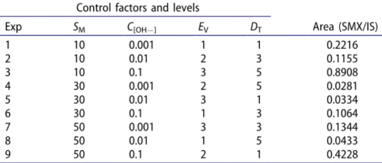
Table 1 shows the design matrix and the area ratio obtained for each trial. All experiments were performed in duplicate and the total number of experiments was 18 (9 experiments �2 replicates).
Table 2 shows that values of the variance ratio (Fcalculated)
are higher than critical variance (Fcritical ¼4.26 for a 95%
con-fidence level), which denote that all the control factors evalu-ated are critical. The factors of higher influence on the response are NaOH concentration in methanol and amount of butylamide silica, which accounted 46.75 and 30.63%, respectively. The contribution of residual error was 0.05%; this value is associated with the proper selection of control factors.
Figure 6 shows the analysis of means obtained for DSPE methodology. Optimal levels selected were butylamide silica amount of 10 mg, NaOH concentration of 0.1 M in methanol, 3 mL of elution volume, and 5 min of dispersion time. An increase in the butylamide silica amount would result in a
Figure 3. Micrographs for the butylamide silica.
Figure 4. Effect of pH and volume on the extraction of SMX. Conditions:
butylamide silica 30 mg; SMX 7 mg L 1. Figure 5. Elution solvent evaluated for DSPE experiments.
lower dispersion between solid and phases and also higher desorption solution volumes would be needed.[32,35] In the case of volume and dispersion time variables, highest values are needed to promote the contact between the analyte and the solid. Additionally, the level selected of NaOH concen-tration in methanol must be highest to form negative charges which promote repulsion between the solid and SMX. The results demonstrate the existence of possible hydrogen bond interactions between the solid and SMX.
Validation and application of method
Under the optimal conditions, analytical parameters of the DSPE–CE method were evaluated using a sample volume of 10.0 mL of spiked milk with SMX in the range of concen-tration of 0.10–34.0 mg L 1. Each standard was prepared and analyzed in triplicate. The regression parameters of the cali-bration lines are shown in Table 3. A linear dependence of the peak area ratio and the concentration of SMX, in the doped milk sample, were obtained. The limit of detection was calculated from the relation Se/b1 (3.29), where Se is the
square root of the residual variance of the standard curve, and b1 is the slope, in accordance with the recommendations
of IUPAC.[36]
The accuracy of the method was determined by the absolute recovery of SMX added to a sample of blank milk (previously analyzed by the proposed method) at two concentration levels with three replicates for each level (7 and 23 mg L 1). The average recovery obtained for the SMX in doped milk samples was found in a range of 70% at 85% with relative standard deviations (RSD) of 1.85 and 2.3%, respectively. Precision is adequate for the analysis of complex samples (RSD <5%) in all cases using the proposed DSPE–CE methodology.
Electropherograms obtained in the determination of SMX in doped milk samples under optimal conditions are shown in Figure 7. Figure 7a shows interferences presented by the components of a real milk sample during SMX analysis with-out DSPE treatment. Figure 7b shows a blank milk sample treated by a proposed DSPE–CE methodology (without IS).
Figure 7c displayed the electropherogram of the analysis of a spiked milk sample. An effective cleanup and separation of
Figure 6. Effects of control factors on the output variable during DSPE–CE. SM,
solid mass; C[OH ], NaOH concentration in methanol; EV, elution volume; DT,
dispersion time.
Table 3. Regression parameters of the calibration lines obtained in doped milk sample.
Regression parameter Value
Correlation coefficient, r2 0.998
Square root of residual variance, Se 0.034
Intercept, b0 ( mg L 1) 0.105 �0.001
Slope, b1 (Abs L mg 1) 1.882 �0.051
Repeatability intraday (RSD (%) n ¼3, 15 mg L 1) 1.17
Repeatability interday (RSD (%) n ¼3, 15 mg L 1) 0.95 Linearityrange ( mg L 1) 0.10–34.0
Limit of detection ( mg L 1) 0.05 Limit of quantification ( mg L 1) 0.10
Area SMX: Area IS vs [SMX]: [IS] in concentration ( mg L 1).
Figure 7. Electropherograms obtained from the analysis of SMX by DSPE–CE: (a) milk sample, (b) blank milk sample, and (c) spiked milk sample (7 mg L 1);
1 doxycycline (IS), 2 sulfamethoxazole. Table 1. Orthogonal design matrix L9(34) used for the optimization of the DSPE
methodology proposed (n ¼2). Control factors and levels
Area (SMX/IS)
Exp SM C[OH ] EV DT
1 10 0.001 1 1 0.2216
2 10 0.01 2 3 0.1155
3 10 0.1 3 5 0.8908
4 30 0.001 2 5 0.0281
5 30 0.01 3 1 0.0334
6 30 0.1 1 3 0.1064
7 50 0.001 3 3 0.1344
8 50 0.01 1 5 0.0433
9 50 0.1 2 1 0.4228
SM, butyl silica amount (mg); C[OH ], NaOH concentration in methanol ( mol L 1); EV, elution volume (mL); DT, dispersion time (min).
Table 2. Regular variance analysis (pooled ANOVA). Variance
source
Degree of freedom
Sum of
squares Variance
Variance ratio (F)a
Influence (%)b
SM 2 0.38 0.19 2671.88 30.63
C[OH ] 2 0.58 0.29 4078.13 46.75
EV 2 0.16 0.08 1125.00 12.90
DT 2 0.12 0.06 843.75 9.67
Residual 9 6.4 �10 4 7.11 �10 5 0.05
Total 17 1.24 0.07 100.00
aThe critical variance ratio for a 95% confidence level is 4.26 (2,9 d.f.). b
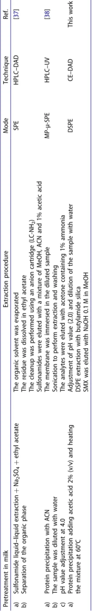
SMX from milk samples is verified, demonstrating the selec-tivity of the butylamide silica during DSPE–CE analysis. Milk cleanup using the butylamide silica is similar than other SPE techniques based on anion-exchange (–NH2) mechanism and
polymeric membrane in combination with HPLC (Table 4). The proposed DSPE–CE methodology has the advantage of low consumption of organic solvents.
The proposed methodology was applied to the determi-nation of SMX in 12 commercial milk samples from different brands. Three replicate determinations were performed on each sample. According to the results obtained by analysis of SMX in milk, 1 of the 12 samples tested was positive with con-centration of 145.3 �0.04 µg kg 1.
Conclusions
The proposed methodology DSPE–CE, based on butylamide silica, was applied successfully in the cleanup/extraction and analysis of SMX in milk samples. Electrostatic interactions are critical in the methodology proposed. Butylamide silica retains SMX through hydrogen bond interactions. The DSPE–CE technique has advantages such as similar cleanup capacity, lower reagent consumption, and analysis time respect to HPLC methods.
Funding
The authors wish to thank PRODEP (Project RedNIQAE-2015) and Junta de Castilla y Leon, (project VA171U14) for the financial support.
References
[1] García-Galán, M. J.; Díaz-Cruz, M. S. Identification and Determi-nation of Metabolites and Degradation Products of Sulfonamides Antibiotics. TrACTrends Anal. Chem. 2008, 27, 1008–1022. [2] Kaneene, J. B., Miller, R. Description and Evaluation of the
Influ-ence of Veterinary PresInflu-ence on the Use of Antibiotics and Sulfona-mides in Dairy Herds. J. Am. Vet. Med. Assoc. 1992, 201, 68–71. [3] Shahani, K. M., Whalen, P. J. Significance of Antibiotics in Foods
and Feeds. In: Agricultural Uses of Antibiotics. ACS Symposium Ser-ies; Moats, W. A. Ed.; American Chemical Society: Washington, 1986, pp. 88–99.
[4] Pereira, A. V., Cass, Q. B. High-Performance Liquid Chromato-graphy Method for the Simultaneous Determination of Sulfa-methoxazole and Trimethoprim in Bovine Milk Using an On-Line Column. J. Chromatogr. B 2005, 826, 139–146.
[5] Cai, M., Zhu, L., Ding, Y., Wang, J., Li, J., Du, X. Determination of Sulfamethoxazole in Foods Based on CeO2/Chitosan Nanocompo-site-Modified Electrodes. Mater. Sci. Eng. C 2012, 32, 2632–2627. [6] Iglesias, A., Nebot, C., Miranda, J. M., Vázquez, B. I., Cepeda, A.
Detection and Quantitative Analysis of 21 Veterinary Drugs in River Water Using High-Pressure Liquid Chromatography Coupled to Tandem Mass Spectrometry. Environ. Sci. Pollut. Res. 2012, 19, 3235–3249.
[7] Upadhyay, K., Asthana, A., Tiwari, N. Solid Phase Extractive Spec-trophotometric Determination of Some Sulfa Drugs. Asian J. Pharm. Clin. Res. 2012, 5, 222–226.
[8] Conzuelo, F., Gamella, M., Campuzano, S., Pinacho, D. J., Reviejo, A. J., Marco, M. P., Pingarron, J. M. Disposable and Integrated Amperometric Immunosensor for Direct Determination of Sulfona-mide Antibiotics in Milk. Biosens. Bioelectron. 2012, 36, 81–88. [9] Wang, L., Wu, J., Wang, Q., He, C., Zhou, L., Wang, J., Pu, Q. Rapid
and Sensitive Determination of Sulfonamides Residues in Milk and
Chicken Muscle by Microfluidic Chip Electrophoresis. J. Agric. Food Chem. 2012, 60, 1613–1618.
[10] Ibarra, I. S., Miranda, J. M., Rodríguez, J. A., Nebot, C., Cepeda, A. Magnetic Solid phase Extraction Followed by High-Performance Liquid Chromatography for the Determination of Sulfonamides in Milk. Food Chem. 2014, 157, 511–517.
[11] Liu, J., Jiang, M., Li, G., Xu, L., Xie, M. Miniaturized Salting-Out- Liquid-Liquid Extraction of Sulfonamides from Different Matrices. Anal. Chim. Acta 2010, 679, 74–80.
[12] Haller, M. Y., Muller, S. R., McArderll, C. S., Alder, A. C., Suter, M. J. F. Quantification of Veterinary Antibiotics (Sulfonamides and Tri-methoprim) in Animal Manure by Liquid Chromatography Mass Spectrometry. J. Chromatogr. A 2002, 952, 111–120.
[13] Zayas-Blanco, F., García-Falcón, M. S., Simal-Gándara, J. Determi-nation of Sulfamethazine in Milk by Solid Phase Extraction and Liquid Chromatographic Separation with Ultraviolet Detection. Food Control 2004, 15, 375–378.
[14] Zhang, Y., Liu, H., Zhang, X., Lei, H., Bai, L., Yang, G. On-Line Solid Phase Extraction Using Organic-Inorganic Hybrid Monolithic Columns for the Determination of Trace β-Lactam Antibiotics in Milk and Water Samples. Talanta 2013, 104, 17–21.
[15] Vidal, L., Riekkola, M. L., Canals, A. Ionic Liquid-Modified Materi-als for Solid-Phase Extraction and Separation: A Review. Anal. Chim. Acta 2012, 71, 19–41.
[16] Plotka-Wasylka, J., Szczepanska, N., De la Guardia, M., Namiesnik, J. Miniaturized Solid-Phase Extraction Techniques. Trends Analyt. Chem. 2015, 73, 19–38.
[17] Herrera-Herrera, A. V., Hernández-Borges, J., Afonso, M. M., Palenzuela, J. A., Rodriguez-Delgado, M. A. Comparison between Magnetic and Nonmagnetic Multi-Walled Carbon Nanotubes-Dis-persive Solid-Phase Extraction Combined with Ultra-High of Sulfo-namide Antibiotics in Water Samples. Talanta 2013, 116, 695–703. [18] Xiao-Lin, H., Yin-Liang, W., Ting, Y., Xiang-Dang, D. Multi-Walled Carbon Nanotubes-Dispersive Solid-Phase Extraction Combined with Liquid Chromatography-Tandem Mass Spectrometry for the Analysis of 18 Sulfonamides in Pork. J. Chromatogr. B 2013, 929, 107–115. [19] Dagnac, T., Garcia-Chao, M., Pulleiro, P., Garcia-Jares, C.,
Llompart, M. Dispersive Solid-Phase Extraction Followed by Liquid Chromatography-Tandem Mass Spectrometry for the Multi-Residue Analysis of Pesticides in Raw Bovine Milk. J. Chromatogr. A 2009, 1216, 3702–3709.
[20] Snyder, L. R., Dolan, J. W., Carr, P. W. The Hydrophobic-Subtrac-tion Model of Reversed-Phase Column Selectivity. J. Chromatogr. A
2004, 1060, 77–116.
[21] Rimmer, C. A., Sander, L. C. Shape Selectivity in Embedded Polar Group Stationary Phases for Liquid Chromatography. Anal. Bioanal. Chem. 2009, 394, 285–291.
[22] Kirkland, J. J. Development of Some Stationary Phases for Reversed- Phase HPLC. J. Chromatogr. A 2004, 1060, 9–21.
[23] Aturki, Z., D’Orazio, G., Rocco, A., Si-Ahmed, K., Fanali, S. Investigation of Polar Stationary Phases for the Separation of Sympathomimetic Drugs with Nano-Liquid Chromatography in Hydrophilic Interaction Liquid Chromatography Mode. Anal. Chim. Acta 2011, 685, 103–110.
[24] Martínez-Villalba, A., Moyano, E., Galceran, M. T. Analysis of Amprolium by Hydrophilic Interaction Liquid Chromatography- Tandem Mass Spectrometry. J. Chromatogr. A 2010, 1217, 5802–5807.
[25] Ibarra, I. S., Rodriguez, J. A., Miranda, J. M., Vega, M., Barrado, E. Magnetic Solid Phase Extraction Base on Phenyl Silica Adsorbent for the Determination of Tetracyclines in Milk by Capillary Electro-phoresis. J. Chromatogr. A 2011, 1218, 2196–2202.
[26] Islas, G., Rodríguez, J. A., Cruz-Borbolla, J., Vásquez-Pérez, J. M., Barrado, E. Synthesis and Characterization of Amide Stationary Phases for the Determination of Sulfonamides by Sequential Injec-tion Chromatography. Anal. Lett. 2016, 49, 676–689.
with Diode-Array Detection. J. Chromatogr. A 2009, 1216, 2263–2269.
[28] Granja, R. H. M. M., de Lima, A. C., Salerno, A. G., Wanschel, C. B. A. Validation of a Liquid Chromatography with Ultraviolet Detection Methodology for the Determination of Sulfonamides in Bovine Milk According to 2002/657/EC. Food Control 2012, 28, 304–308.
[29] Xu, L., Peng, R., Guan, X., Tang, W., Liu, X., Zhang, H. Preparation, Characterization, and Application of a New Stationary Phase Containing Different Kinds of Amine Groups. Anal. Bioanal. Chem.
2013, 405, 8311–8318.
[30] Huang, Z., Zhou, H., Chen, Z., Zeng, F., Chen, L. Facile Synthesis of Porous Pt Botryoidal Nanowires and their Electrochemical Properties. Electrochim. Acta 2014, 147, 643–649.
[31] Sanli, S., Altun, Y., Sanli, N., Alsancak, G., Beltran, J. L. Solvent Effects on pKa Values of Some Substituted Sulfonamides in Acetonitrile-Water Binary Mixtures by UV-Spectroscopy Method. J. Chem. Eng. 2009, 54, 3014–3020.
[32] Liu, F. J., Liu, C. T., Li, W., Tang, A. N. Dispersive Solid-Phase Microextraction and Capillary Electrophoresis Separation of Food Colorants in Beverages Using Diamino Moiety Functionalized Silica Nanoparticles as Both Extractant and Pseudostationary Phase. Talanta 2015, 132, 366–372.
[33] Massart, D. L., Vandeginste, B. G. M., Buydens, L. M. C., De Jong, S., Lewi, P. J., Smeyers-Verbeke. Handbook of Chemometrics and Qualimetrics Part A. Editorial Elsevier: Amsterdam, 1997; Vol. 20A, 799–803 pp.
[34] Zhang, M., Chen, J., Mallik, A. K., Qiu, H., Jiang, S., Ihara, H. Preparation and Chromatographic Evaluation of New Branch-Type Diamide-Embedded Octadecyl Stationary Phases with Enhanced Shape Selectivity. Anal. Chim. Acta 2014, 833, 48–55.
[35] Pardasani, D., Kanaujia, P. K., Purohit, A. K., Shrivastava, A. R., Dubey, D. K. Magnetic Multi-Walled Carbon Nanotubes Assisted Dispersive Solid Phase Extraction of Nerves Agents and their Markers from Muddy Water. Talanta 2011, 86, 248–255.
[36] Currie, L. A. Nomenclature in Evaluation of Analytical Methods Including Detection and Quantification Capabilities (IUPAC Recommendation). Pure Appl. Chem. 1995, 67, 1699–1723. [37] Yin-Liang, W., Cun, L., Yong-Jun, L., Jian-Zhong, S. Validation
Method for the Determination of Sulfonamide Residues in Bovine Milk by HPLC. Chromatographia 2007, 66, 191.
[38] Jiangeng H., Juanjuan L., Cong Z., Jiaojiao W., Li M., Shan Y., Gao L., Li X. 2012. Determination of Sulfonamides in Food Samples by Membrane-Protected Micro-Solid Phase Extraction Coupled with High Performance Liquid Chromatography. J. Chromatogr. A 2012, 1219, 66–74.