pdf elaborado por medigraphic Rev Biomed 2005; 16:239-246.
Corresponding address: Elizabeth Abrahams-Sandí. Department of Parasitology, University of Costa Rica, San Pedro/Mts. Oca, Costa Rica. Fax: +506-225-2374 E-mail: eabraham@cariari.ucr.ac.cr eabraham20@yahoo.com
Received September 21, 2005; Accepted November 15, 2005.
Original Article
This paper is also available at http://www.uady.mx/sitios/biomedic/revbiomed/pdf/rb051643.pdf
Specific antibody production
against different life cycle
stages during an experimental
A. costaricensis
infection in
mice.
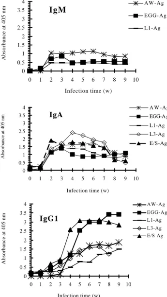
Results and Discussion.- The production of different antibody isotypes was directed against all the different antigen preparations, with a predominance of IgG1 antibodies and a significant increase of IgA antibody levels after maturation of worms and egg deposition. The highest parasite-specific IgA antibody levels were detected against E/S- and L3-antigen at 2 and 3 weeks p.i., respectively. On the other hand, the highest IgG1 antibody response was measured against E/S- and egg-antigen at 3 weeks p.i. and remained high until the end of the experiment. Values for parasite-specific IgG2a, determined by ELISA, reached only low levels against all antigen preparations. We propose further studies with human sera in order to investigate the usefulness of egg and E/S-antigen for the diagnosis of human abdominal angiostrongyliasis.
(Rev Biomed 2005; 16:239-246)
Elizabeth Abrahams-Sandí1-2, Stefan Michael Geiger2,3, Katherine Fernández-Quesada4, Hartwig
Schulz-Key3.
1Department of Parasitology, University of Costa Rica, San Pedro/Mts. Oca, Costa Rica.
2Institute of Tropical Medicine, University of Tübingen, Wilhelmstrasse 27, 72074 Tübingen, Germany
3Centro de Pesquisas René Rachou-Fiocruz Av. Augusto de Lima 1715 Barro Preto Belo
Horizonte-MG CEP: 30190-002
4Microbiology Department. Boston Scientific Costa Rica. 302 Global Parkway, Heredia, Costa Rica.
SUMMARY.
Objectives.- In the present study we aimed at investigating the humoral immune response against different Angiostrongylus costaricensis life-cycle stages- L1, L3, eggs and adult worms- during an experimental infection in C57BL/6 mice.
Materials and Methods.- C57BL/6 mice were experimentally infected with six third- stage larvae
(L3) Angiostrongylus costaricensis. Blood
pdf elaborado por medigraphic Keywords: Angiostrongylus costaricensis
antigen, antibody production, C57BL/6, ELISA.
RESUMEN.
Producción de anticuerpos específicos contra diferentes estadios del ciclo de vida de Angiostrongylus costaricensis durante una infección experimental.
Objetivos.- El propósito de este estudio fue analizar la respuesta inmune humoral contra diferentes estadios evolutivos de Angiostrongylus costaricensis durante una infección experimental en ratones de la cepa C57BL/6.
Materiales y Métodos.- Se infectaron ratones C57BL/6 s en el laboratorio con 6 larvas de tercer estadio (L3). Semanalmente se obtuvieron muestras de sangre de cada animal por punción del plexo venoso retroorbital, y se almacenó el suero a -20ºC. Se preparó antígeno soluble a partir de las formas adultas, sus productos de excreción–secreción (E/ S), larvas de primer y tercer estadio (L1, L3) y huevecillos. Mediante SDS-PAGE se procedió a revelar el perfil proteico de cada uno de ellos y se monitoreó la producción de anticuerpos en ratones infectados contra estos antígenos durante 9 semanas p.i. mediante un ELISA.
Resultados y Discusión.- La producción de los diferentes isotipos de anticuerpos estuvo dirigida contra todas las preparaciones de antígeno, con predominio de anticuerpos del tipo IgG1 y un significativo aumento de la IgA después de la maduración de las formas adultas y el inicio de la oviposición. Los niveles más altos de IgA parásito-específico se detectaron para los productos E/S y L3, entre la 2da. y 3era. semana p.i. Por otro lado, la respuesta más alta de anticuerpos tipo IgG1 se determinó a partir de la 3era. semana p.i. y al utilizar antígeno de E/S y de huevecillos, permaneciendo con niveles elevados hasta el final del experimento.
Los niveles de IgG2a determinados por ELISA
fueron muy bajos para cada uno de los antígenos. Nosotros proponemos estudios con sueros humanos para investigar la utilidad de los huevecillos y productos de E/S en el diagnóstico de la
angiostrongiliasis abdominal humana.
(Rev Biomed 2005; 16:239-246)
Palabras clave:Angiostrongylus costaricensis, antígeno, producción de anticuerpos, C57BL/6.
INTRODUCTION.
Angiostrongylus costaricensis is the etiological agent of human abdominal angiostrongyliais. The disease has been reported from the United States to Argentina with a widespread occurrence of the nematode throughout Central and South America, (1-3). Clinical cases are usually diagnosed postoperatively by anatomo-pathological examination of biopsies or surgical specimens, since no other diagnostic method is available (4). In these biopsies a severe inflammatory response to adult worms, eggs, and larvae has been observed, accompanied with massive eosinophilic infiltration of the intestinal wall and granulomatous reaction.
The natural life cycle of A. costaricensis
mainly involves the rodents Sigmodon hispidus and
Oryzomys spp. as the definitive vertebrate hosts, and veronicellid slugs as the main intermediate hosts.
In the course of Angiostrongylus costaricensis
infection, the parasitic nematode carries out a complex life cycle, involving the migration and development of different life-cycle stages through the definitive host. In some helminthiases each of these parasite stages has its own antigenic characteristics and is capable of eliciting its characteristic T-cell reponse (5, 6) or its humoral immune response that differs in terms of quantity of antibodies, isotype profile, and affinity of the produced antibodies (7). Experimental studies on
pdf elaborado por medigraphic
costaricensis infection in C57BL/6 mice.
MATERIAL AND METHODS.
Animals
C57BL/6 mice were kept under standard laboratory conditions, as described (9). At the time of infection the C57BL/6 mice were between 2 and 4 months old (n=11). All animal experiments were conducted in accordance with German law.
Parasites and infection
A. costaricensis infective third-stage larvae
(L3) were isolated from infected Biomphalaria
glabrata, as previously described (10). Each mouse was infected with six L3 via a stomach tube.
Parasitological examinations
Following the administration of the L3, the survival time of the infected animals was monitored daily. Beginning with 25 days post-infection (p.i.), the excretion of first stage larvae (L1) in feces was determined weekly until the end of patency
according to Geiger et al.(11). The recovery of
adult worms from the different organs was determined after host death, and in surviving animals at the end of the patency period. Heart, aorta dorsalis, liver, and mesenteric arteries were examined under a stereomicroscope, and parasites were extracted with a fine forceps. A. costaricensis
adult worms were washed with phosphate buffered saline (PBS) three times and stored at -20ºC until used for antigen preparation.
Antigen preparation
Adult somatic antigen (AW-Ag) was prepared
from fertile male and female A. costaricensis
worms as previously described (10).
Soluble egg antigen (EGG-Ag) was extracted
from homogenized eggs collected from in vitro
cultivated A. costaricensis fertile females. Briefly, in repeated cultures 20 fertile females were collected with blunt forceps from the mesenterial arteries of
infected cotton rats (Sigmodon hispidus), and
washed three times with sterile PBS. Subsequently,
female worms were incubated in Waymouth’s medium (GIBCO) supplemented with 100 U/ml penicillin, 100 µg/ml streptomycin, and 0.25 µg/ml
amphotericin B, at 37ºC, 5%CO2 for 96 hrs. The
culture medium was changed daily, pooled, and centrifuged for 10 min at 1000 g at 4ºC.The egg pellet was washed three times with PBS, then pooled, and the suspension was sonicated four times on ice for 3 min (30% cycle, model 250 Branson Ultrasonics, Danbury, Conn) and centrifuged for 20 min at 16,000 g and 4ºC. The protein concentration was determined by the Bradford assay (Bio Rad, Munich, Germany) using bovine serum albumin as standard.
Soluble first-stage larval antigen (L1-Ag): L1 were isolated with the Baermann technique from feces of infected cotton rats according to Geiger et al. (11). The L1 were washed three times in sterile PBS at 4ºC and disrupted by sonication as described above. The soluble antigen fraction was collected after centrifugation for 20 min at 16,000 g and 4ºC. The protein concentration was determined as described above.
Soluble third-stage larval antigen (L3-Ag): L3
were isolated from B. glabrata by enzymatic
digestionas previously described (10) and larval
soluble antigens were prepared as described for L1-Ag.
pdf elaborado por medigraphic
Bedford, USA). The E/S products were stored in aliquots at –70ºC until used.
Sodium dodecyl sulfate-polyacrylamide gel electrophoresis (SDS-PAGE).
Fractionation of all soluble antigen preparations by SDS-PAGE was performed as described by
Geiger et al. (10). The separated proteins were
visualized using a silver staining method as described by Heukeshoven and Dernick (12).
Determination of parasite-specific immunoglobulins by enzyme-linked immunosorbent assay (ELISA) in the sera of infected mice.
Blood samples from infected mice were obtained by puncture of the retro-orbital venous plexus under ether anesthesia. Individual serum samples were stored at –20ºC until further use. A. costaricensis-specific antibodies in a serum pool of infected mice were determined by indirect ELISA, as described previously (5). Microtitre plates (Costar, Cambridge USA) were coated and incubated overnight at 4ºC with 1 µg/ml of EGG-Ag, 5 µg/ml L1-EGG-Ag, 2 µg/ml L3-EGG-Ag, and 2 µg/ml E/ S-Ag, respectively. The class and subclass specific antibodies were used in the following dilutions: IgM, IgA 1:1000 (Sigma, Munich, Germany); IgG1 1:2000, and IgG2a 1:500 (Rockland, Gilbertsville, USA). Only serum samples from the animals that survived the experiment were used. The plates were read at 405 nm in a microplate autoreader (Biotek, Winooski, Belgium) and the results were expressed as mean optical densities for duplicate determinations from a pool of sera from infected animals.
RESULTS.
The mortality observed in the C57BL/6 mice throughout the 11 weeks of infections was 81.8%, with an increased mortality between 2 and 5 weeks p.i. In our study, in spite of the low dose of inoculated L3, adult worms were found in all animals. The mean recovery of adult worms in
infected animals was 48.5%, which is equivalent to nearly three adult worms per infected animal.
Fecal L1 excretion by infected mice showed a high variation among different animals (data not shown). L1 were detected from 25 days p.i. onwards and showed a maximum L1 excretion at 39 days p.i. The mean patency period in C57BL/6 mice was 5 weeks.
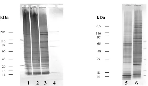
Sodium dodecyl sulfate-polyacrylamide gel electrophoresis (SDS-PAGE).
The protein profile of the L1, adult worms, and adult E/S products were analyzed and characterized by polyacrylamide gel electrophoresis in sodium dodecyl sulfate under reducing conditions (fig. 1). After SDS-PAGE and silver staining, the analysis of L1-Ag revealed several bands with molecular weights ranging from 66 kDa to 10 kDa. A band duplet by 62-57 kDa and several bands below 25 kDa were common in adult somatic antigen (fig. 1, lane 6). With E/S antigen, five distinct bands at 15, 83, 106, 195, and at 205 kDa were obtained (fig. 1, lane 4). All of those bands were also present in adult somatic antigen, mainly in female somatic extracts. Due to an insufficient amount of antigen, it was not possible to obtain a protein profile from extracts of eggs and L3.
Antibody responses in the course of A. costaricensis infection.
The production of specific antibodies against
A. costaricensis was monitored during 9 weeks post-infection. Results summarized in figure 1 indicate a weak signal for specific antibodies one week p.i. Because of antigen shortage, it was not possible to determine the production of parasite-specific IgM against L3- and E/S-Ag.
pdf elaborado por medigraphic
The specific antibodies against EGG-Ag and E/S-Ag detected from week 3 p.i. until the end of the experiment were predominantly IgG1 (fig. 2), whereas parasite-specific IgA was highly reactive to larval antigens. For parasite-specific IgG2a antibodies, only low optical densities were detected throughout the experiment (data not shown).
DISCUSSION.
The protein profile of somatic extracts from the different A. costaricensis developmental stages and excretory-secretory products of adult worms was characterized by the presence of stage-specific as well as shared proteins. The L1, male, and female somatic extracts were highly complex, each consisting of more than 20 different protein bands with molecular weights varying form 200 Da to14 kDa. The protein profiles of male and female adult worms were similar, with some differences, mainly in the range of 70 kDa. Additionally, the protein profile of adult somatic extracts showed a similar pattern when compared with the protein profile of L1 extracts, mainly below 60 kDa. In the case of the E/S products, allprotein bands revealed in the electrophoresis were also observed in adults, but at a higher concentration, especially in somatic
extracts of adult female worms. This may indicate a production and release of these products by the adult worms. Similar results have been described in previous studies with A. cantonensis (13-15). In these studies, the authors found common polypeptides in more than one developmental stage and demonstrated, by radioimmunoprecipitation, an antigenic cross reaction between them.
In order to study the humoral immune response to different A. costaricensis life-cycle stages, the production of parasite specific antibodies was monitored during 9 weeks p.i. in C57BL/6 infected mice. At one week post-infection a low level of antibodies against all antigens was observed. After week 2 p.i. a strong increase in the level of parasite-specific immunoglobulin against all developmental stages was measured, although at this point of the infection only adult worms and eggs were present in the host intestine. These results suggest that common components in larvae, especially L3, and adult somatic and E/S extracts are the immunodominant antigens recognized, especially by IgA, in infected mice. In mammals most of the IgA is found in mucosal secretions. A role of this immunoglobulin, mainly secreted IgA, in an eosinophil-mediated defense mechanism ocurring in 1 2 3 4
205
116
97
66
29 18 14 48
116 97
66
29
18 14 48
5 6
205
kDa kDa
Figura 1.- SDS-PAGE analysis of soluble proteins extracted from different life-cycle stages of A. costaricensis. Lane
1-2, adult worms, female and male respectively; Lane3,6,mix of female and male adult worms;Lane4,
pdf elaborado por medigraphic
parasite-specific IgA. This increase correlates with
a decrease of excreted larvae (21, 22). In a A.
cantonensis infection, an increase in the level of parasite-specific IgA and IgG1 in serum and cerebrospinal fluid has been correlated with staining adult worms in the host brain (23). At present, the role of parasite-specific IgA during the course of
A. costaricensis infection is not yet clear and further studies are necessary.
A strong increase in the level of parasite-specific IgG1 was determined at 3-4 week p.i. for antigen preparations of all life-cycle stages, but especially against EGG-Ag and E/S-Ag. The substantial increase in levels of IgG1 against E/S-Ag antigens and the higher antibody levels as compared to AW-Ag indicates that, either the metabolic products of the worms are continuously released, or are more antigenic than the adult somatic antigens. Nacapunchai et al. (24) reported similar results in an A. costaricensis infection in ddY mice. In the present work, it is of interest to point out that the increase in the antibody level coincides with the oviposition and hatching of L1 in the life cycle of the parasite. An intense production of IgG1 after egg deposition associated with a weak IgG2a
response has also been described during an S.
mansoni infection in mice (25, 26). For this parasitosis, it is known that the switch to a T helper 2 immune response is triggered by egg antigens (27).
Previous studies on an experimental A .
costaricensis infection indicate that the onset of egg laying plays an important role in the induction of the immune response against A. costaricensis and have been correlated with an increase in mortality during the acute phase of the infection (9, 10). Recently, Bender et al. (28) described a strong recognition to eggs and reproductive organs of female
A. costaricensis by human sera of patients with abdominal angiostrongyliasis, which could make them potential antigens for the immunodiagnosis of acute cases of the disease. Further studies on the contribution of eggs to the humoral and cellular
immune response in the course of an A .
costaricensis infection would provide valuable
Figure 2.- Parasite-specific antibodies to A. costarricensis
antigens in C57BL/6 mice infected with six third-stage larvae. In order to compare the humoral immune response to different life-cycle stages only serum samples from animals that survived throughout the experiment were used. Data correspond to the values recorded from a pool of sera from seven animals (means of duplicate analysis).
IgM
0 0.5 1 1.5 2 2.5 3 3.5 4
0 1 2 3 4 5 6 7 8 9 10
Infection time (w)
Absorbance at 405 nm
A W - A g
E G G - A g
L1-Ag
IgA
0 0.5 1 1.5 2 2.5 3 3.5 4
0 1 2 3 4 5 6 7 8 9 10
Infection time (w)
Absorbance at 405 nm
A W -Ag
EGG-A g
L1-Ag
L3-Ag
E/S-Ag
IgG1
0 0.5 1 1.5 2 2.5 3 3.5 4
0 1 2 3 4 5 6 7 8 9 10
Infection time (w)
Absorbance at 405 nm
AW-Ag EGG-Ag L1-Ag L3-Ag E/S-Ag
the intestinal tract, has been described in some human parasitic infections (16, 17). During the immune response against schistosomes, a participation of IgA in the maturation, fertility, and development of eggs has been described (18-20). Likewise, infection with Strongyloides stercoralis
pdf elaborado por medigraphic
information to elucidate factors of pathogenesis and immunoregulation.
Studies to demonstrate the usefulness of EGG-and E/S-antigen in the diagnosis of abdominal angiostrongyliasis are currently being performed in our laboratory.
ACKNOWLEDGEMENTS.
This study was supported by Förtune program of the University of Tübingen. Financial support for the exchange of scientists was received from German Academic Exchange Service (DAAD) and “Conselho para Apoio na Pesquisa” (CAPES) in Brazil.
REFERENCES.
1.- Morera P. Abdominal angiostrongyliasis: a problem of public health. Parasitol Today 1985; 1:173–5.
2.- World Health Organization. Prevention and control of intestinal parasitic infections. Tech Rep Ser 1987; 749:21– 8.
3.- Graeff-Teixeira C, Camillo-Coura L, Lenzi HL. Angiostrongíliase abdominal nova parasitose no sul do Brasil R. AMRIGS 1991; 35:91–8.
4.- Graeff-Teixeira, C., Agostini, A.A., Camillo-Coura, L., Ferreira-Da-Cruz, M.F. Seroepidemiology of abdominal a n g i o s t r o n g y l i a s i s : t h e s t a n d a r d i z a t i o n o f a n immunoenzymatic assay and prevalence of antibodies in two localities in southern Brazil. Trop Med Int Health 1997; 2: 254–60.
5.- Lawrence RA, Allen JE, Osborne J, Maizels. Adult and
microfilarial stages of the filarial parasite Brugia malayi
stimulate contrasting cytokine and Ig isotype responses in BALB/c mice. J Immunol 1996; 153: 1216-1224.
6.- Pearce EJ, Caspar P, Grzych JM, Lewis FA, Sher A. Down regulation of Th1 cytokine production accompanies induction of Th2 responses by a parasite helminth,
Schistosoma mansoni. J Exp Med 1991; 173: 159-66.
7.- Parkhouse RME, Harrison LJS. Antigens of parasitic helminths in diagnosis, protection and pathology. Parasitology 1989; 99: S5-S19.
8.- Abrahams-Sandí E, Hoffmann WH, Graeff-Teixeira C, Schulz-Key H, Geiger SM. Long-term observations on m o u s e s t r a i n s e x p e r i m e n t a l l y i n f e c t e d w i t h
Angiostrongylus costaricensis. Parasitol Res 2004; 93:
230-4.
9.- Geiger SM, Abrahams-Sandí E, Soboslay PT, Hoffmann WH, Pfaff AW, Graeff-Teixeira C, Schulz-Key H. Cellular immune responses and cytokine production in BALB/c a n d C 5 7 B L / 6 m i c e d u r i n g t h e a c u t e p h a s e o f
Angiostrongylus costaricensis. Acta Trop 2001; 80:
59-68.
10.- Geiger SM, Graeff-Teixeira C, Soboslay PT,
Schulz-Key H. Experimental Angiostrongylus costaricensis
infection in mice: immunoglobulin isotype responses and parasite-specific antigen recognition after primary low-dose infection. Parasitol Res 1999; 85: 200-5.
11.- Geiger SM, Hoffmann WH, Soboslay PT, Pfaff AW,
Graeff-Teixeira C, Schulz-Key H. Angiostrongylus
costaricensis infection in C57BL/6 mice: MHC-II
deficiency results in increased larval elimination but unaltered mortality. Parasitol Res 2003; 90:415–20.
12.- Heukeshoven J, Dernick R. Characterization of a solvent system for separation of water-insoluble poliovirus proteins by reversed-phase high-performance liquid chromatography. J Chromatogr 1985; 326:91-101.
13.- Fujii T. Angiostrongylus cantonensis:immunoblot
analysis of the antigens recognized by rats. Parasitol Res 1987; 73: 366-74.
14.- Fujii T. Immunoblot analysis of the circulating a n t i g e n s o c c u r r i n g i n s e r u m r a t s i n f e c t e d w i t h
Angiostrongylus cantonensis. Parasitol Res 1988; 74:
476-83.
15.- Dharmkrong-AT A, Sirisinha S. Analysis of antigens
from different developmental stages of A. cantonensis.
Southeast Asian J Trop Med Pub Hlth 1983; 14: 154-62.
16.- Fujisawa T, Abu-Ghazaleh R, Kita H, Sanderson CJ, Gleich GJ. Regulatory effect of cytokines on eosinophil degranulation. J Immunol 1990; 114: 642-6.
17.- Pritchard DI. Immunity to helminths: is too much IgE parasite-rather than host protective?. Parasite Immunol 1993; 15: 5-9.
pdf elaborado por medigraphic
19.- Dunne DW. The use of mouse/ human chimaeric antibodies to investigate the roles of different antibody
isotype, incluiding IgA2, in the killing of Schistosoma
mansoni by eosinophils. Parasite Immunol 1993; 15:
181-5.
20.- Grezel D, Capron M, Grzych JM. Protective immunity induced in rat schistosomiasis by a single dose of the Sm28GST recombinant antigen: effector mechanism involving IgE and IgA. Europ J Immunol 1993; 23:454-60.
21.- Atkins N S, Lindo JF, Lee MG, Conway DJ, Bailey JW, Robinson R, Bundy DAP. Humoral responses in human strongyloidiasis: correlations with infection chronicity. Trans Roy Soc Trop Med Hyg 1997; 91: 609-13.
22.- Atkins NS, Conway DJ, Lindo JF, Bailey JW, Bundy DAP. L3 antigen-specific antibody isotype responses in human strongyloidiasis: correlations with larval output. Parasite Immunol 1999; 21: 517-26.
23.- Sugaya H, Aoki M, Yoshida T, Takatsu K, Yoshimura K Eosinophilia and intracranial worm recovery in interleukin-5 transgenic and interleukin-5 receptor
chain-knockout mice infected with A n g i o s t r o n g y l u s
cantonensis. Parasitol Res 1997; 83: 583-90.
24.-Nacapunchai D, Ishii AI, Terada M, Kino H, Sano M. Humoral immune responses in mice infected with
Angiostrongylus costaricensis. Serodiagnosis and
Immunotherapy in Infectious Disease 1989; 3:51-6.
25.- Mountford AP, Fisher A, Wilson RA. The profile of IgG1 and IgG2a antibody responses in mice exposed to
Schistosoma mansoni. Parasite Immunol 1994; 16: 521-7.
26.- Poulain-Godefroy O, Gaubert S, Lafitte S. Immunoglobulin A response in murine schistosomiasis: stimulatory role of egg antigens. Infect Immun 1996; 64: 763-8.
27.- Taverne J, Bradley JE. Immunity to protozoa and worms. En: Roitt I, Brostoff J, Male D. editores. Immunology. 5a. ed. London: Mosby-Wolfe; 1998. p. 243-60.
28.- Bender AL, Maurer RL,Fernandes da Silva MC, Ben
R, Barros TP, Aramburu da Silva AC, Graeff-Teixeira C. Ovos e órgãos reprodutores de fêmeas de
Angiostrongylus costaricensis são reconhecidos mais
![analysisofareportoncagingsystems(2007to2021),thatwaspublishedbytheUnited ].IntheUnitedStates,about97billiontableeggswereproducedin2020,withanaverage Tableeggsareareadilyavailableandinexpensivesourceofproteininthehumandiet.Annually,theCanadianeggindustrypr](data:image/gif;base64,R0lGODlhAQABAIAAAP///wAAACH5BAEAAAAALAAAAAABAAEAAAICRAEAOw==)