Otras secciones de este sitio:
☞ ☞ ☞ ☞
☞ Índice de este número
☞ ☞ ☞ ☞
☞ Más revistas
☞ ☞ ☞ ☞
☞ Búsqueda
Others sections in this web site: ☞
☞ ☞ ☞
☞ Contents of this number ☞
☞ ☞ ☞
☞ More journals ☞
☞ ☞ ☞ ☞ Search Artículo:
Evaluation of direct fluorescent antibody test for the diagnosis of Bovine Genital Campylobacteriosis
Derechos reservados, Copyright © 2002 : Asociación Mexicana de Microbiología, AC Revista Latinoamericana de Microbiología
Número Number3-4
Julio-Diciembre July-December 2002 Volumen
Volume 44
edigraphic.com
Evaluation of direct fluorescent antibody test for the
diagnosis of Bovine Genital Campylobacteriosis
Josely Ferreira Figueiredo,* Aiesca Oliveira Pellegrin,** Cid Bastos Fóscolo,* Raquel Paula Machado,* Karina Leite Miranda,* Andrey Pereira Lage*
Rev
ista
Lat
inoamer
Vol. 44, No. 3-4 July - September. 2002 October - December. 2002 pp. 118 - 123
INTRODUCTION
Bovine Genital Campylobacteriosis is an infectious dis-ease caused by Campylobacter fetus subsp. venerealis whose transmission occurs mainly along venereal paths. The disease causes infertility whose major sign is repeated return to service with prolonged interservice intervals.2
C. fetus infection in cattle has decreased in regions where artificial insemination and effective programs of vaccination and antibiotic therapy have been practiced, yet the disease continues to be an important cause of reproduc-tive problems in many countries that have large herds at natural breeding,5 such as Brazil.12
The diagnosis and control of Bovine Genital Campylo-bacteriosis are mainly done in bulls since they act as as-ymptomatic carriers of organisms and also are responsible by spreading the disease in the herd. The detection of bulls infected by C. fetus permits to establish the causes of repro-ABSTRACT. The direct fluorescent antibody test (DFAT) for the diagnosis of Bovine Genital Campylobacteriosis was assessed for its detection limit, observer effect, sensitivity and specificity. In addi-tion, the specificity of the fluorescent conjugate was tested against
Campylobacter sp, Arcobacter sp, Helicobacter sp, E. coli and
oth-er bactoth-eria from the preputial flora. Ten – fold dilutions of C. fetus subsp. venerealis NCTC 10354 in PBS or preputial washings with or without centrifugation were used. All experiments were done in du-plicate by three observers. Positive and negative controls were in-cluded in each assay. The detection limits of DFAT were 104 CFU/
ml for PBS and non - centrifuged preputial washings and 102 CFU/
ml for centrifuged preputial washings. There was no observer effect. The sensitivity and specificity of DFAT were 92.59% and 88.89%, respectively. The DFAT was observed to be sensitive, specific and the effect of experienced observers was minimal on test perfor-mance.
Key words: Bovine Genital Campylobacteriosis - direct fluorescent antibody test - Campylobacter fetus-preputial washing - diagnosis.
RESUMEN. La inmunofluorescencia directa (IFD) fue probada para el diagnóstico de la Campilobacteriosis Genital Bovina para el límite de detección, efecto del observador y sensibilidad. De la mis-ma mis-manera, la especificidad de la inmunofluorescencia directa fue probada contra Campylobacter sp, Arcobacter sp, Helicobacter sp,
E. coli y otras bacterias originadas de la flora prepucial. C. fetus
subsp. venerealis NCTC 10354 o lavados prepuciales con o sin cen-trifugación fueron diluidos diez veces en PBS. Todas las experien-cias fueron hechas en duplicados con tres observadores. Controles positivos y negativos fueron incluidos en cada prueba. El límite de detección de la IFD fue 104 UFC/ml para PBS y lavados prepuciales
sin centrifugación y 102 UFC/ml para lavados prepuciales
centrifu-gados. No hubo efecto del observador. La sensibilidad y la especifi-cidad de la IFD fueron 92.59% y 88.89%, respectivamente. Se ob-servó que la IFD fue sensible, específica y el efecto de los observa-dores es mínimo en la ejecución de la prueba.
Palabras chave: Campilobacteriosis Genital Bovina-inmunofluo-rescencia directa - Campylobacter fetus - lavado prepucial - diag-nóstico.
* Laboratório de Bacteriologia Aplicada, Núcleo de Pesquisa em Saúde Animal, Departamento de Medicina Veterinária Preventiva, Escola de Veterinária, Uni-versidade Federal de Minas Gerais. Av. Antônio Carlos 6627, Caixa Postal 567, 30123-970 Belo Horizonte, MG, Brazil Phone: +55 31 34 99 20 81 Fax: +55 31 34 99 20 80 E-mail: alage@vet.ufmg.br
** Empresa Brasileira de Pesquisa Agropecuária–Centro de Pesquisa Agropecuária do Pantanal Corumbá–MS–Brazil
ductive disturbs in the herd which should be submitted to the appropriated method of control.1 Currently available techniques used to diagnose C. fetus infection in these ani-mals are isolation and identification of bacteria1, poly-merase chain reaction (PCR)4,23 and direct fluorescent anti-body test (DFAT).3,15,18,19,26
The DFAT has been used since 1960s for the diagnosis of C. fetus infection. It has been characterized as a conve-nient and quick method for detection of carrier bulls2 and is prescribed by OIE for testing bulls for international trade.24 However, until now, this technique had not been assessed for its accuracy and detection limit. Thus, the aim of this study was to evaluate the detection limit, the observer ef-fect, the sensitivity and the specificity of previously de-scribed DFAT, and the specificity of the fluorescent conju-gate against Campylobacter sp, Arcobacter sp, Helico-bacter sp, E. coli and other Helico-bacteria from the preputial flora.
MATERIALS AND METHODS
Ferreira et al Evaluation of direct fluorescent antibody test for the diagnosis of Bovine Genital Campylobacteriosis
Rev Latinoam Microbiol 2002; 44 (3-4): 118-123
119
edigraphic.com
PBS pH 7.2 at a concentration equal to tube 10 ofMacFar-land standard was emulsified in Freund’s complete adju-vant and this mixture was intramuscularly inoculated in each rabbit. These animals were endovenously boosted with 0.5, 1.0, 2.0 and 4.0 ml of a similar suspension of C. fetus subsp. venerealis NCTC 10354 in PBS pH 7.2 with-out adjuvant, respectively, on days 6, 13, 20 and 27 after first inoculation. After 15 days of the last shot, animals were bled, IgG was purified14 and conjugated with fluores-cein isothiocyanate.20 Optimum dilution of the fluorescein conjugate was determined by checkboard titration against C. fetus subsp. venerealis NCTC 10354.
Direct Fluorescent Antibody Technique (DFAT) -DFAT for the detection of C. fetus subsp. venerealis was done according to Mellick et al.15 and Winter et al. 26 The material (20µl) was applied in duplicate on microscopic slides. In parallel, 0.01M phosphate-buffered saline (PBS) pH 7.2, and suspension of C. fetus subsp. venerealis strain NCTC 10354 were included, respectively, as negative and positive controls of the reaction. The material was air-dried and fixed in acetone at – 20ºC for 30 min. The fluorescent conjugate anti – C. fetus subsp. venerealis NCTC 10354 was added at 1/50 dilution and incubated for 30 min at 37ºC. The slides were washed three times in PBS pH 7.2 and one time in tap water and then mounted in buffered glycerol pH 9.2. The preparations were examined in an epi-fluorescent microscope (CBA, Olympus, Japan). Samples that showed at least one fluorescent bacterium presenting typical morphology of C. fetus in one of any duplicate were considered positive.
Bacterial suspensions-A total of six bacterial suspen-sions coded as A, B, C, D, E and F were prepared from C. fetus subsp. venerealis strain NCTC 10354 which was grown on Brain Heart Infusion agar (BHI-Difco – USA) supplemented with 10% horse blood for 48 h at 37ºC in microaerobic conditions (5% O2, 10% CO2, 85% N2). The concentration of microbial cells was adjusted to 109 CFU/ ml by MacFarland No. 3 standard and 1.0 absorbance at 595 nm. Then, serial ten-fold dilutions were prepared from each bacterial suspension. Viable counts were done by the drop counting method on BHI blood agar plates.16 Nine di-lutions from A, B and C bacterial suspensions were used in the evaluation of observer effect and detection limit of DFAT in PBS. The bacterial suspensions D, E and F and their first eight dilutions were used to inoculate preputial washing mixtures. The schematic model of the use of these bacterial suspensions is presented in Figure 1.
Preputial washing preparation – Fresh C. fetus-free preputial washings were obtained as described by Leite et al.13 from 18 bulls, aged approximately nine months, from a Bovine Genital Campylobacteriosis–free herd. An aliquot of 1 ml from all preputial washings was tested by the
DFAT for C. fetus15, 26 and negative results were given for all animals. The preputial washings gave three mixtures (300 ml each) (M1, M2 and M3) and each mixture was di-vided in 10 aliquots of 27 ml. One aliquot of each preputial washing mixture did not receive inoculum and was centri-fuged in order to evaluate the specificity of DFAT. Three milliliters of bacterial suspension D, E, and F and each of their first eight ten-fold dilutions were added to 27 ml of each aliquot of preputial washing mixtures. The inoculated aliquots of preputial washings were used without centrifu-gation for the evaluation of the detection limit and the ob-server effect of DFAT. The same inoculated aliquots of preputial washings were centrifuged to evaluate the detec-tion limit, the observer effect and the sensitivity of DFAT (Fig. 1). Two steps of centrifugation were used. First, preputial washings were centrifuged at 600 x g for 10 min at 4ºC. The supernatant of the first step was submitted to 13000 x g for 30 min at 4ºC. The pellet of the second cen-trifugation step was resuspended in 500 µl of PBS pH 7.2 and analyzed by DFAT.15,26
Experimental design
Slides examination – All slides for DFAT were prepared in duplicate and blindly read by three observers, codified as I, II and III, which were responsible for the Bovine Gen-ital Campylobacteriosis diagnosis at Escola de Veterinária da Universidade Federal de Minas Gerais.
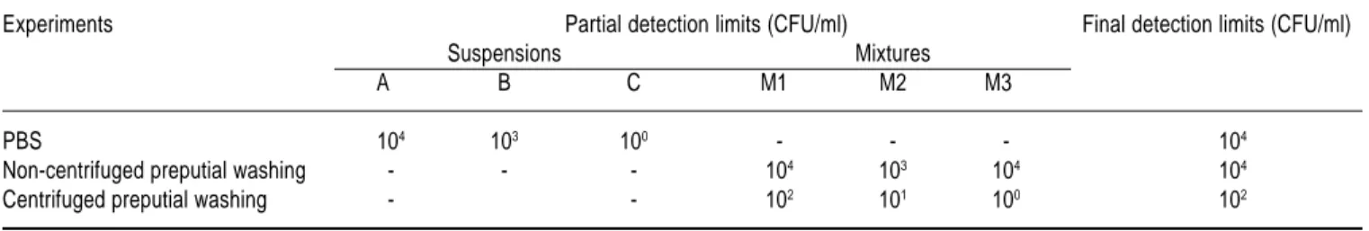
Limit of detection and observer effect – The limit of de-tection and the observer effect on DFAT were evaluated in three separated experiments: PBS inoculated with C. fetus subsp. venerealis strain NCTC 10354 (A, B and C suspen-sions) and preputial washings inoculated with C. fetus sub-sp. venerealis strain NCTC 10354 (M1, M2 and M3 mix-tures) with and without centrifugation (Fig. 1). The observer effect in each experiment was analyzed using the Cochran’s Q Test.22 The evaluation of the detection limit of DFAT was done in each experiment, considering the readings of all ob-servers. In order to find the final detection limit, the partial detection limit of each suspension was taken, i. e., the least concentration that all observers considered to be positive. Since three suspensions for each experiment were analyzed, three partial detection limits were taken. The higher concen-tration found among partial detection limits was regarded as the final detection limit because it indicated an agreement of all observers in all suspensions.
edigraphic.com
Specificity and sensitivity of DFAT – A total of 81read-ings of centrifuged inoculated preputial washread-ings (3 mix-tures x 9 dilutions x 3 observers) were used for sensitivity analysis and 9 readings of centrifuged but non-inoculated preputial washings (3 aliquots x 3 observers) were used for specificity analysis of DFAT (Fig. 1). The statistical analy-sis was done in accordance to Henken et al.,9 considering the inoculation of samples as the gold standard.
Specificity of the fluorescent conjugate – The specificity of the fluorescent conjugate was evaluated against refer-ence bacterial strains listed in Table 1 and bacteria from the preputial flora which were isolated from preputial washings that did not receive inocula. All bacteria were isolated on BHI agar supplemented with 10% equine blood for 48 h at 37ºC in aerobic and microaerobic atmospheres. After growth, these bacteria were ressuspended in PBS pH 7.2 at a concentration adjusted to MacFarland No. 3 stan-dard. From each bacterial suspension three serial ten-fold dilutions were made and 20 µl from each dilution were ex-amined in duplicate by DFAT.
RESULTS
The concentrations of nine dilutions from bacterial sus-pensions A, B and C varied from 108 to 100 CFU/ml and the concentrations of bacterial suspensions D, E and F and their first eight dilutions varied from 109 to 101 CFU/ml, respectively. The bacterial concentrations of nine dilutions of M1, M2 and M3 mixtures varied from 108 to 100 CFU/ ml, respectively.
The detection limits of DFAT in PBS, non-centrifuged and centrifuged preputial washings were 104, 104 and 102 CFU/ml, respectively (Table 2). The bacterial concentra-tion influenced the results in all experiments (Table 3) (PBS: χ2 = 7.78, df = 2, P<0.05; non-centrifuged prepu-tial washings: χ2 = 36.57, df = 2, P<0.001; centrifuged preputial washings: χ2 = 12.96, df = 2, P<0.01). In the three experiments analyzed no statistically significant dif-ference was found in the results of the observers’ read-ings (PBS: Q = 2.80, df = 2, P = 0.24; non-centrifuged preputial washings: Q = 2.88, df = 2, P = 0.23;
centri-Bacterial Suspension
A,B,C D,E,F
Nine ten-fold dilutions Eight ten-fold dilutions
Inoculation in preputial washing Evaluation of observer effect and
detection limit of DFAT in PBS
Preputial washing mixtures
M1, M2, M3
Each mixture gave 10 aliquots
1 aliquot without inoculum 9 aliquots with inoculum
No Centrifugation Centrifugation
Centrifugation
Evaluation the specificity of DFAT incentrifuged
preputial washings
Evaluation of the detection limit, the observer effect and
the sensitivy of DFAT in centrifuged preputial
washings
Evaluation of the detection limit, and the observer effect
of DFAT in non-centrifuged
Ferreira et al Evaluation of direct fluorescent antibody test for the diagnosis of Bovine Genital Campylobacteriosis
Rev Latinoam Microbiol 2002; 44 (3-4): 118-123
121
edigraphic.com
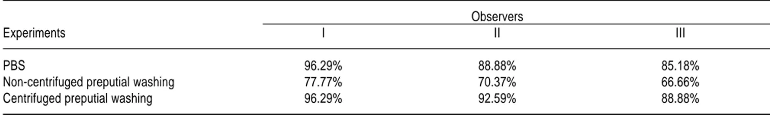
fuged preputial washings: Q = 4.66, df = 2, P = 0.09 ). However, in all experiments, observer I had a better per-formance on readings than the others, followed by the ob-server II and III (Table 4).
Among 81 readings from inoculated preputial washings, 75 were positive by DFAT and among 9 readings from non-inoculated preputial washings, 8 were negative by DFAT. Those results showed a sensitivity of 92.59% and a specificity of 88.89% of DFAT for C. fetus subsp. venerea-lis detection on preputial washings.
The specificity of the fluorescent conjugate against bac-terial reference strains and other bacteria from the preputial flora was the same for all observers. No fluorescent bacte-ria were seen in preparations from Campylobacter sp other than C. fetus, Helicobacter sp, Arcobacter sp, E. coli or bacteria from preputial flora. The conjugate prepared against C. fetus subsp. venerealis strain NCTC 10354 re-acted only with C. fetus subsp. venerealis strains ADRI 510, ADRI 528, ADRI 534 and ADRI 1832 and NCTC 10354 and with C. fetus subsp. fetus serotype A strains ADRI 1811 and ADRI 1812.
DISCUSSION
The detection limit is one of the most important points in test evaluation because it influences sensitivity. The de-tection limit of DFAT in PBS shows the ability of the tech-nique for C. fetus subsp. venerealis detection performed without contaminants and with the exact bacterial concen-tration of samples. Its results were similar to the ones found with preputial washings without centrifugation and only moderately high numbers of bacteria could be detected. Table 1. Reference bacterial strains used in the analysis of the specificity
of fluorescent conjugate against C. fetus subsp. venerealis.
Species Strains
Arcobacter skirrowii LMG1 6621
A. butzleri LMG 15919
Campylobacter coli NCTC2 11366T C. fetus subsp. fetus serotype B ATCC3 27374 T C. fetus subsp. fetus serotype B ADRI4 553 C. fetus subsp. fetus serotype B ADRI 1810
C. fetus subsp. fetus serotype A ADRI 1811
C. fetus subsp. fetus serotype A ADRI 1812
C. fetus subsp. venerealis NCTC 10354
C. fetus subsp. venerealis ADRI 510
C. fetus subsp. venerealis ADRI 528
C. fetus subsp. venerealis ADRI 534
C. fetus subsp. venerealis ADRI 1832
C. hyointestinalis subsp. hyointestinalis LCDC5 17398 (= LMG 12686) C. hyointestinalis subsp. lawsoni LMG 14432 T
C. jejuni subsp. doylei LMG 8843 T C. jejuni subsp. jejuni NCTC11351 T
C. lari NCTC 11352 T
C. sputorum biovar paraureolyticus LCDC 6577
C. sputorum biovar fecalis NCTC 11415
C. sputorum biovar paraureolyticus LCDC 6939
C. sputorum biovar sputorum6 LMG 6447 Escherichia coli ATCC 25922
Helicobacter fenneliae LMG 13306
T – Type Strain
1 – LMG – Laboratorium voor Microbiologie – Rijksuniversiteit Gent – Belgium 2 – NCTC – National Culture Type Collection – United Kingdom
3 - ATCC – American Type Culture Collection – United States of America 4 - ADRI – Animal Diseases Research Institute - Canada
5 – LCDC – Laboratory Center for Disease Control – Canada
6 – Formerly type strain of “C. sputorum biovar bubulus” (= old “C. bubulus”)
Table 3. Percentage of accurate readings in DFAT for C. fetus subsp. venerealis detection in different classes of bacterial concentrations.
Experiments Classes of Concentrations (CFU/ml)
108 to 106 105 to 103 102 to 1
PBS 100% 92.59% 77.77%
Non-centrifuged preputial washing 100% 85.18% 29.62%
Centrifuged preputial washing 100% 100% 77.77%
Table 2. Detection limits of DFAT for C. fetus subsp. venerealis detection in different experiments.
Experiments Partial detection limits (CFU/ml) Final detection limits (CFU/ml)
Suspensions Mixtures
A B C M1 M2 M3
PBS 104 103 100 - - - 104
Non-centrifuged preputial washing - - - 104 103 104 104
edigraphic.com
This was probably due to the lack of centrifugation steps inboth procedures. Moreover, in non-centrifuged preputial washings, cell debris could have impaired either the DFAT reaction or the reading by the observers.
DFAT readings in centrifuged preputial washings showed the best results due to high rate of accurate read-ings among different classes of bacterial concentration among observers and the lowest detection limit. These re-sults can be ascribed to the two step centrifugation process of samples that allow the elimination of cell debris and the concentration of bacteria in a small volume, improving the detection of C. fetus. The detection limit of DFAT found in centrifuged preputial washings in the present study was higher than that found by Eaglesome et al.4 using PCR for detection of C. fetus subsp. venerealis in bovine semen (3 CFU/ml). However, it was similar to the findings of Philpott18 that found a detection limit of DFAT in preputial washing of 50 to 100 CFU/ml.
The low subjectivity and detection limit of DFAT pro-vide some advantages of this test over isolation of C. fetus. Although the isolation of the C. fetus from preputial secre-tions is considered the gold standard for diagnosis of Bo-vine Genital Campylobacteriosis,10 low number of organ-isms per sample can impair the diagnosis due to the numer-ous contaminating bacteria which are normally present in preputial flora19,25 and which do not constitute a problem in DFAT as shown by the present study.
The fluorescent conjugate did react against neither E. coli nor other bacteria from preputial flora. That was simi-lar to the findings of Mellick et al.,15 Dufty3 and Philpott,18 but it was different from the findings of Philpott,19 who saw brilliantly stained diplococcus and rods in the samples. The specificity of the fluorescent conjugate used in DFAT against C. fetus demonstrates the efficiency of the technique, since some of bacteria evaluated in this study are very similar to C. fetus subsp. venerealis and they can be naturally found in preputial cavity of healthy bulls,8 aborted fetuses7 and in bovine intestinal tract.25
Moreover, the specificity of the fluorescent conjugate used in this study discloses other advantages of DFAT over the isolation technique: first, its high efficiency, even in the presence of contaminants19,20,26 and second, its ability to
Table 4. Percentage of accurate readings in DFAT for C. fetus subsp. venerealis detection by observers.
Observers
Experiments I II III
PBS 96.29% 88.88% 85.18%
Non-centrifuged preputial washing 77.77% 70.37% 66.66%
Centrifuged preputial washing 96.29% 92.59% 88.88%
identify and differentiate C. fetus from another Campylo-bacter sp without the use of biochemical tests.20,26
The absence of previous data on specificity and sensi-tivity of DFAT in the diagnosis of Bovine Genital Campy-lobacteriosis limits our inferences from the values found in this study. DFAT, however, was observed to be quite sen-sitive and specific for the diagnosis of C. fetus infection. The presence of false-negative results (lost of sensitivity) probably occurred due to the low bacterial concentration of the samples, since all false-negative readings were done in samples with bacterial concentration equal to or lower than 101 CFU/ml. However, the exactness of the observers readings (high rate of correct readings) confirms the pro-posal of Winter et al.26 in which the difficulties of prepara-tion of specimens and interpretaprepara-tion of findings can be overcome almost entirely by well trained technicians.
Just one false-positive reading (lost of specificity) was found and it occurred in only one of the duplicates. De-spite of the presence of few specimens for evaluation of the specificity of DFAT in this experiment, the value found, 88.89%, can be an estimate of the specificity of this technique in order to be discussed in future experiments.
Among all reference bacterial strains tested, the fluores-cent conjugate just reacted against C. fetus strains. This specificity was similar to that found by Mellick et al.,15 Dufty3 and Ruckerbauer et al.20 Moreover, in this study, it was found that the conjugate which was prepared against a C. fetus subsp. venerealis serotype A strain reacted just against C. fetus serotype A strains as mentioned by Dekey-ser2, recognizing all C. fetus subsp. venerealis strains but only C. fetus subsp. fetus serotype A strains.
The absence of reaction of the fluorescent conjugate against C. fetus subsp. fetus serotype B has no influence on the diagnosis of Bovine Genital Campylobacteriosis and on the epidemiological studies of C. fetus subsp. ve-nerealis, which is far more important than C. fetus subsp. fetus, due to its genital tropism.11 Besides, the C. fetus subsp. fetus infections are not considered to be endemic, but sporadic.17
corrobo-Ferreira et al Evaluation of direct fluorescent antibody test for the diagnosis of Bovine Genital Campylobacteriosis
Rev Latinoam Microbiol 2002; 44 (3-4): 118-123
123
edigraphic.com
rate the use of DFAT as an important and supportivetech-nique for the control of Bovine Genital Campylobacteriosis.
ACKNOWLEDGEMENTS
We thank Dr. Brian Brooks, Animal Diseases Research Institute, Canada, and Dr. Lawrence Price, Laboratory Cen-ter for Disease Control, Canada, for providing some of the reference bacterial strains. We are grateful to Dr. Misty Car-nes from Texas A&M University for reviewing the English and Dr. Ronnie Antunes Assis and Dr. Walter Motta Ferrei-ra for reviewing the Spanish. This study had the financial support of Fundação de Estudo e Pesquisa em Medicina Vet-erinária (FEP MVZ Coordenação Preventiva). We are in-debted to Coordenação de Aperfeiçoamento de Pessoal de Nível Superior – Capes (JFF) and Conselho Nacional de De-senvolvimento Científico e Tecnológico – CNPq (AOP, KLM and APL) for the fellowships.
REFERENCES
1. Clark, B.L. 1971. A review of vibriosis bovine. Aust. Vet. J. 47:103-107.
2. Dekeyser, J. 1984. Bovine Genital Campylobacteriosis, p. 181-191. In J.P. Butzler (ed). Campylobacter infection in man and animal, CRC Press, Boca Raton.
3. Dufty, J.H. 1967. Diagnosis of vibriosis in the bull. Aust. Vet. J. 43:433-437.
4. Eaglesome, M.D., M.I. Sampath & M.M. Garcia. 1995. A detection assay for Campylobacter fetus in bovine semen by restriction analy-sis of PCR amplified DNA. Vet. Res. Comm. 19: 253-263. 5. Eaglesome, M.D. & M.M. Garcia. 1997. Disease risks to animal
health from artificial insemination with bovine semen. Rev. sci tech. Off int. Epiz. 16:215-225.
6. Edwards, P.R. & W.H. Ewing. 1972. Identification of enterobacteri-aceae, p. 67-108. 3rd ed., Burgess Publishing Company, Minnesota. 7. Fernandez, H., X. Rojas & T. Gajardo. 1995. Primer aislamiento de
Arcobacter cryaerophilus a partir de un aborto bovino en Chile.
Arch. Med. Vet. 27:111-114.
8. Gill, K.P.W. 1988. Aerotolerant Campylobacter strain isolated from a bovine preputial sheath washing. Vet. Rec. 112:459.
9. Henken A.M., E.A. M. Graat & J. Casal. 1997. Measurement of dis-ease frequency, p. 63-98. In J.P.T.M. Noordhuizen, K. Frankena, C.M. van der Hoofd & E.A.M. Graat, Application of quantitative methods in Veterinary Epidemiology, Wageningen Pers, The Netherlands. 10. Hum, S., L.R. Stephens & C. Quinn. 1991. Diagnosis by ELISA of
bo-vine abortion due to Campylobacter fetus. Aust. Vet. J. 68:272-275. 11. Hum, S. & A. Mcinnes.1993. Bovine campylobacteriosis:
Bacteri-ology and antibody detection, p.1-8. In L.A., Corner & T.J. Bagust (ed). Australian standard diagnostic techniques for animal diseases, CSIRO, Australia.
12. Lage, A.P. & R.C. Leite. 2000. Campilobacteriose Genital Bovina. Pecuária de Corte, 10:50-54.
13. Leite, R.C., J.P.A. Haddad, G.M. Costa, A.O. Pellegrin & A.C.C.L. Ribeiro. 1995. Técnica modificada para coleta de lavado prepucial
de touros, para exame de Tricomonose e ou Campilobacteriose. In: CONGRESSO BRASILEIRO DE REPRODUAÇÃO ANIMAL, 11, 1995, Belo Horizonte. Anais – 11 Congresso Brasileiro de Re-produção Animal. Belo Horizonte: s.n. p.434.
14. Mckinney, M.M. & A. Parkinson. 1987. A simple non-chromato-graphic procedure to purify immunoglobulins from serum and as-citic fluid. J. Immunol. Methods. 96:271-278.
15. Mellick, P.W., A.J. Winter & K. Mcentee. 1965. Diagnosis of vibri-osis in the bull by the use of the fluorescent antibody technique. Corn. Vet. 55:280-294.
16. Miles, A.A. & S.S. Misra. 1938. The estimation of the bactericidal power of the blood. J. Hyg. 38:732-742.
17. Penner, J.L. 1988. The genus Campylobacter: a decade of progress. Clin. Microbiol. Rev. 1: 157-172.
18. Philpott, M. 1968. Diagnosis of Vibrio fetus infection in the bull. I – A modification of Mellick’s fluorescence antibody test. Vet. Rec.
82:424-426.
19. Philpott, M. 1968. Diagnosis of Vibrio fetus infection in the bull. II An epidemiological survey using a fluorescent antibody test and comparing this with a cultural method. Vet. Rec. 82:458-463. 20. Rucherbauer, G.M., K. Malkin, D. Mitchell & P. Baulanger. 1974.
Vibriosis: demonstration of Vibrio fetus and Vibrio bubulus organ-isms in preputial fluid by immunofluorescence and culture tech-niques. Can. J. Comp. Med. 38:321-327.
21. Sampaio, I.B.M. 1998. Estatística aplicada à experimentação
ani-mal. 221 pp Fundação de Ensino e Pesquisa em Medicina
Veter-inária, Belo Horizonte.
22. Siegel, S. 1975. Estatística não paramétrica. p.181-188. McGraw-Hill, São Paulo.
23. Stynen, A.P.R. 2000. Detecção de Campylobacter fetus em lavados prepuciais de touros pela PCR. Escola de Veterinária – UFMG, Belo Horizonte, 36 pp Dissertação (Mestrado em Medicina Veter-inária).
24. Wagenaar, J.A., Bergen, MAP-van; Guerin, B; van Bergen, M.A.P. 2000. Bovine genital Campylobacteriosis, p. 346-358. In: Manual
of standards for diagnostic tests and vaccines. List A and B diseas-es of mammals, birds and bediseas-es. Office International ddiseas-es Epizootidiseas-es;
Paris; France.
25. Wesley, V.I., S.J. Wells, K.M. Harmon, A. Green, L. Schroeder-Tucker, M. Glover & I. Siddique. 2000. Fecal shedding of
Campy-lobacter and Arcobacter spp. in dairy cattle. Appl. Environ. Micro-biol. 66:1994-2000.
26. Winter, A.J., J.D. Samuelson, & M. Elkana. 1967. A comparison of immunofluorescence and cultural techniques for demonstration of
Vibrio fetus. J. Am. Vet. Med. Assoc. 150:498-502.
Correspondence to:
Dr. Andrey Pereira Lage
Laboratório de Bacteriología Aplicada, Núcleo de Pesquisa em Saúde Animal,
Departamento de Medicina Veterninária Preventiva, Escola de Veterinária,
Universidade Federal de Minas Gerais Av. Antônio Carlos 6627,
Caixa Postal 567, 30123-970 Belo horizonte, Mg, Brazil.