Respiratory Physiology & Neurobiology174 (2010) 317–330
Contents lists available atScienceDirect
Respiratory Physiology & Neurobiology
j o u r n a l h o m e p a g e :w w w . e l s e v i e r . c o m / l o c a t e / r e s p h y s i o l
Review
A revisit to O
2
sensing and transduction in the carotid body chemoreceptors in
the context of reactive oxygen species biology
夽
C. Gonzalez
∗, M.T. Agapito, A. Rocher, A. Gomez-Ni ˜
no, R. Rigual, J. Casta ˜
neda, S.V. Conde, A. Obeso
Departamento de Bioquímica y Biología Molecular y Fisiología, Instituto de Biología y Genética Molecular y CIBER de Enfermedades Respiratorias, Universidad de Valladolid, Consejo Superior de Investigaciones Científicas e Instituto Carlos III, Facultad de Medicina, 47005 Valladolid, Spain
a r t i c l e i n f o
Article history:
Accepted 1 September 2010
Keywords:
Oxygen-sensing Transduction Sensor Transducer
a b s t r a c t
Oxygen-sensing and transduction in purposeful responses in cells and organisms is of great physiological and medical interest. All animals, including humans, encounter in their lifespan many situations in which oxygen availability might be insufficient, whether acutely or chronically, physiologically or pathologi-cally. Therefore to trace at the molecular level the sequence of events or steps connecting the oxygen deficit with the cell responses is of interest in itself as an achievement of science. In addition, it is also of great medical interest as such knowledge might facilitate the therapeutical approach to patients and to design strategies to minimize hypoxic damage. In our article we define the concepts of sensors and trans-ducers, the steps of the hypoxic transduction cascade in the carotid body chemoreceptor cells and also discuss current models of oxygen- sensing (bioenergetic, biosynthetic and conformational) with their supportive and unsupportive data from updated literature. We envision oxygen-sensing in carotid body chemoreceptor cells as a process initiated at the level of plasma membrane and performed by a hemopro-tein, which might be NOX4 or a hemoprotein not yet chemically identified. Upon oxygen-desaturation, the sensor would experience conformational changes allosterically transmitted to oxygen regulated K+ channels, the initial effectors in the transduction cascade. A decrease in their opening probability would produce cell depolarization, activation of voltage dependent calcium channels and release of neurotrans-mitters. Neurotransmitters would activate the nerve endings of the carotid body sensory nerve to convey the information of the hypoxic situation to the central nervous system that would command ventilation to fight hypoxia.
© 2010 Elsevier B.V. All rights reserved.
1. Introduction
1.1. Sensors and transducers
The words sensor and transducer are widely used in biology, frequently with identical meanings. Both words have Latin roots: sensor derives from the verbsentirewhich means to perceive or to feel, the suffix-orindicates the subject that performs the action of perceiving. Transducer is a compound word of the preposition trans(across, over, beyond) and the verbducere(to lead, to guide) with the suffix -ermeaning the one that performs the actions, the one thatleads across. Thus, from an etymological point of view both words would have different meanings. A sensor is a device, a molec-ular entity or even a more organized cellmolec-ular element capable of
夽This paper is part of a special issue entitled “Physiological Redox: Regulation
in Respiratory, Vascular, and Neural Cells”, guest-edited by Paul T. Schumacker and Jeremy P.T. Ward.
∗Corresponding author. Tel.: +34 983 42 30 89; fax: +34 983 42 35 88.
E-mail address:[email protected](C. Gonzalez).
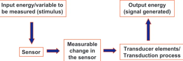
perceiving a change in its surroundings and generating a measur-able change.The sensor elements enter in contact with the stimulus, the measured variable of the surrounding milieu. For example, sens-ing of light intensity by photoreceptors consists in the absorption of photons energy by 11-cis-retinal and the genesis of 11-all-trans-retinal; i.e., the measurable change in light sensing is the genesis of 11-all-trans-retinal. In carotid body (CB) arterial chemorecep-tors we shall propose as a O2sensor a hemoprotein (vide infra),
which having a given molecular spatial structure when saturated with O2, acquires a new spatial configuration on O2desaturation;
this conformational change, much alike occurring in hemoglobin on its O2saturation–desaturation cycle, would be the measurable
signal generated by the O2-sensor. From there on, transduction
starts (Fig. 1).The transducer element(s) do(es) not enter in con-tact with the variable measured, it/they cannot directly “sense” the stimulus.Instead the transducer elements or the transduction pro-cess convey the measurable signal generated at the sensor to the effector, in a single or multi step process, transforming it in a new signal intelligible to the system. In sensory physiology the trans-duction process usually is a multistep process known as a whole as the transduction cascade, and the intelligible signal to the
sys-1569-9048/$ – see front matter© 2010 Elsevier B.V. All rights reserved.
Input energy/variable to be measured (stimulus)
Output energy (signal generated)
Sensor
Measurable change in the sensor
Transducer elements/ Transduction process
Fig. 1.Flow diagram showing the minimal operational steps in a sensor coupled to a transduction machinery. Note that “sensu stricto” in the basic functioning of the sensor, in the sensing process, there is a transduction. It might be a dilation or con-traction as it is the case in a thermometer of mercury or a conformational change of a protein as it is the case in the odorant receptor molecules. The transduction machin-ery captures the signal generated by the sensor and in a single or multistep process generates an output, a new signal that being intelligible to the system contains a representation of the parameters of the stimulus.
tem is electrogenesis (Grundfest, 1971), i.e., the genesis of action potentials with a frequency and interval distribution specific for the nature and intensity of the sensed signal. Taking another exam-ple from sensory physiology we can consider odorant sensing and odorant transduction cascade. Odorant sensing should be defined as the conformational change that occurs in the odorant receptor when an odorant molecule binds to it. The conformational change triggers the transduction cascade which is initiated by the activa-tion of specific G-proteins which in turn promote the activaactiva-tion of an adenylyl cyclase and the increase of cAMP levels; cAMP activates cationic channels which allow the movement of positive charged ionic species into the olfactory cilia causing their depolarization and subsequent triggering of conducted action potentials. These considerations not only apply to sensory physiology, as the action of hormones and growth factors in bodily cells, or antigen recog-nition by cell of the immune system, are initiated by sensing their presence by receptors (sensors). These receptors, upon binding of the signaling molecule, experience conformational changes which initiate the transduction cascade specific to the signal.
We have mentioned before that in sensory physiology, and the CB is a sensory receptor, the transduction cascade culminates in electrogenesis with specific codes of frequencies and intervals that are specific to the stimulus detected by the sensor. This assert is precise in primary sensory receptors (i.e., sensory receptors in which the sensing device and the transduction and electrogenesis machineries are located in the primary sensory neuron). However, in secondary sensory receptors the sensing device and transduction cascade reside in a cell that communicates with the cell responsible for the electrogenesis via a synapse. Therefore the transduction cas-cade culminates in a modification (usually an increase) of the rate of the release of neurotransmitters, being the stimulus-induced release of neurotransmitter which conveys the information to the cell responsible for the generation of the conducted electrogen-esis (Grundfest, 1971). Although it is not uncommon to include the synaptic transmission within the transduction cascade in sec-ondary sensory receptors, following a reductionist approach, we believe that it is more appropriate to consider the transduction cas-cade and the synaptic transmission as independent processes, both having specific tools and methodologies of investigation. Sensing and transduction process can even be more complex as it hap-pens for example in photoreceptors, where the conveying of the information contained in the stimulus to cells endowed with the capacity to generate action potentials and to conduct them to cen-tral nervous system (the retinal ganglion cells) is achieved through a complex net of cells and synapses.
1.2. The need for oxygen sensing and transduction cascades
Mammalian cells depend on oxygen for survival. In cell mitochondria the equivalents of reduction present in digestion-processed nutrients are transferred in an ordered sequence through the respiratory chain complexes down to molecular O2, which
represents the final acceptor of electrons to generate water. The stepwise transfer of electrons through the respiratory chain is accompanied by H+extrusion out the mitochondria. This H+
extru-sion, coupled to a near impermeability of internal mitochondrial membrane, generates a strong electrochemical gradient for H+with
the mitochondrial interior negative and alkaline in reference to cell cytoplasm. This H+gradient directed to the mitochondrial interior
is used to force a special ATPase to work in backwards mode and to synthesize ATP from ADP + Pi. In this manner, a significant part of the energy-content of nutrients (another significant part is lost as heat) is transferred to ATP, a compound that due to its chemical structure is capable of releasing such energy to be used by cells. The H+entering via mitochondrial ATPase react with reduced molecular
O2to form water. From this outline it is evident that O2
availabil-ity is nearly equivalent to ATP availabilavailabil-ity for cells to perform their functions (we are neglecting glycolytic anaerobic pathway which in most cells generates about 10% of ATP). These functions include the maintenance of their homeostasis and integrity, via de novo syn-thesis of continuously renewed cell constituents, the performance of external work.
The issue on this article is to discuss what happens when cells became hypoxic, i.e., when the PO2or O2 molar concentration in
the mitochondrial interior is low enough to be capable of accept-ing the amounts of electrons required to support an adequate rate of ATP synthesis. Do cells sense the O2-deficit? Which is (are) the
molecular identity(ies) of the O2-sensor(s)? How do cells transduce
the signal generated by the O2-sensor in teleologically meaningful
responses to cell and organism survival?
In this regard, we can distinguish in mammalian organisms two types or categories of cells. The first category is represented by the great majority of cells of the organism. Cells in this category are endowed with capacity to sense O2deficit with a high threshold, i.e.,
they detect intense hypoxias and only react to O2deficiency when
PO2is very low generating responses directed to their own survival.
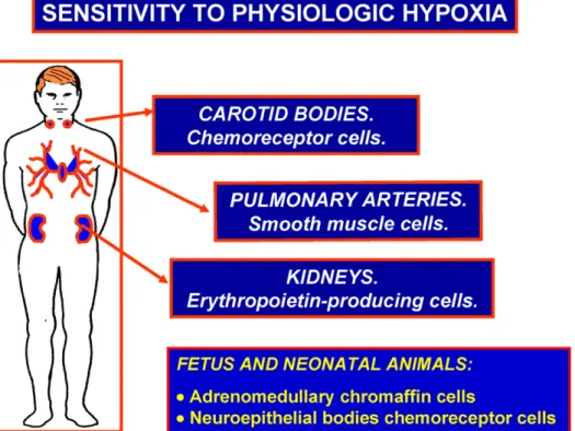
The second category (Fig. 2) is formed by small groups of special-ized cells that sense O2deficit with a low threshold, i.e., they detect
moderate decreases in PO2and, through regulatory loops, generate
responses aimed to restore or secure an adequate PO2for the entire
organism. This category of specialized cells includes: chemorecep-tor cells of the CB and aortic bodies chemorecepchemorecep-tors, smooth muscle cells of the pulmonary arteries and erythropoietin producing cells of the kidney. We can name these cells as cells endowed with sensi-tivity to physiological hypoxia (Gonzalez, 1998). Neonatal adrenal medulla chromaffin cells are also endowed with sensitivity to phys-iological hypoxia, however they lose their intrinsic O2-sensitivity
early in postnatal age when adrenal medulla becomes functionally innervated by splanchnic nerves (Seidler and Slotkin, 1985; Rico et al., 2005). Chemoreceptor cells of the airways neuroepithelial bod-ies also are oxygen sensitive in neonatal animals (Cutz et al., 2009), but become greatly atrophic in adult mammals (Cutz, 1997).
C. Gonzalez et al. / Respiratory Physiology & Neurobiology174 (2010) 317–330 319
Fig. 2.Cells with low threshold sensitivity to hypoxia. They detect moderate decreases in their surrounding PO2and, through regulatory loops, generate responses aimed to restore or secure an adequate PO2for the entire organism.
A recent review on oxygen-sensing (Ward, 2008) has a very important section entitled “matching the sensor to the sensed” in which it is nicely discussed the O2handling properties of the
pro-posed sensors (Kmor P50for O2utilization) with prevailing arterial
or ideally cytoplasmic or mitochondrial [O2] or PO2and the
func-tional properties or activity of the O2sensing organ. To his knowing
ideas on these and others aspects, such as the importance of the concentration of the putative sensor itself, as well as the great importance of the seldom considered concentrations of cofactors or co-substrates in the reactions involved in O2sensing, we want
to add some extra comment on these aspects pertaining to the CB (seeObeso et al., 1997).
The CB is the organ with the largest blood flow of the entire organism. Whether measured by gravidimetric or radioactive microsphere methods, CB blood flow oscillates between 1.5 and 2 l/100 g/min. Accordingly to this high blood flow, the density of capillaries in the CB represents 25–33% of the surface of histological sections obtained from organs perfused at normal (80–100 mmHg) pressure; just for comparative purposes we should mention that blood flow in rat brain is around 120 ml/100 g/min and the den-sity of capillaries in the cortex is <5% of the surface of the sections. O2 consumption in the CB measured by several
meth-ods (arteriovenous O2difference, kinetics of CB tissue PO2decay
after flow stopping and confinement methods) yields values of around 1.5 ml/100 g/min (i.e., comparatively it is 6–8 times lower than brain cortex). The high blood flow and relatively low O2
-consumption rate implies a very small arteriovenous O2difference.
Altogether these facts would predict a high tissue PO2. In fact,
mod-els for O2diffusion using the above set of values and morphometric
data obtained in cat CB showing that the distance from the center of most glomus cells to capillaries is between 10 and 20m, have allowed to calculate that less than 4% of CB tissue would have a PO2
below 40 mmHg, unless a high density of arteriovenous shunts or a high degree of plasma skimming occurs in CB circulation. Since morphological data do not support these particularities for the
cir-culation of the main arterial chemoreceptor organ the conclusion should be that CB tissue PO2is high. However, experimental data
obtained with O2 sensitive Clark type microelectrodesin situor
in artificially perfused organs with either blood or saline or with phosphorescent dyes have not always generated congruent results (Table 1). From the O2consumption rates, the calculations on O2
dif-fusion referred above, the data presented inTable 1and additional data ofWhalen and Nair (1976, 1983), we can summarize data of interest to discuss the possible nature of the O2sensor in
chemore-ceptor cells as follows: (a) venous PO2in the CB in normoxia would
be close to 90 mmHg; (b) mean tissue PO2in normoxia would be
around 75 mmHg; (c) threshold tissue PO2for the activation of the
afferent activity in the CSN oscillates between 50 and 65 mmHg; (d) tissue P50for afferent activity in the CSN has been measured
to be between 10 and 32 mmHg; (e) maximum activity in the CSN was obtained at tissues PO2 of 3–5 mmHg. Since as pointed out
byWard (2008)most commonly we refer our findings to arterial blood (or perfusate) PO2we could define the general behavior of
the CB in the following terms: PO2 threshold, an arterial PO2of
70–75 mmHg; P50, an arterial PO2of 40 mmHg; and PO2to obtain
maximum afferent CSN activity, 10 mmHg (Fidone and González, 1986; Gonzalez et al., 1994). Since even at PO2much higher than
physiological there is some activity in the CSN, threshold is as the arterial PO2on decreasing arterial PO2from >100 down to 0 mmHg
at which it occurs a change of slope in the activity of the CSN. As expected, in comparing both set of PO2values, CB tissue PO2and
arterial blood PO2, it is evident a drop in PO2 from capillaries to
tissue of around 15–20 mmHg at arterial blood PO2 higher than
60 mmHg, corresponding to the flat part of dissociation curve of HbO2, and smaller drops at arterial blood PO2below 60 mmHg, i.e.
in the steepest part of the HbO2curve.
These characteristics of blood flow, oxygen utilization and tis-sue PO2 in the CB conforms with the ideal for the location of an
O2-sensor, as described byHalperin et al. (2006)referring to
Table 1
Reported cat carotid body tissue PO2.
CB Perfusate Perfusate pressure Perfusate PO2 CB Tissue PO2 Author
Blood 120 95–113 25 Acker et al. (1971)Acker and Lübbers (1977)
Blood >70 39 20 Whalen and Nair (1976)
Blood >70 84 >65 Whalen and Nair (1983)
Saline 80 111±15 23±3 Rumsey et al. (1991)
Saline 80 131±12 74±11
Saline 80 109±5 59±13 Rumsey et al. (1992)
Blood 100 91 49±3
All values presented in the table refer to means (±SEM) as reported by the authors. In normal characters data obtained with microelectrodes, in italics data obtained with phosphorescent indicators that measure intravascular PO2, mostly microvascular PO2(Modified fromObeso et al., 1997).
(a) the tissue where the O2sensor is located extracts a small
pro-portion of the oxygen that is delivered by unit of blood, making the PO2 signal easier to recognize; (b) the high blood flow rate
improves O2 diffusion from capillaries to the O2 sensor, and; (c)
there is a constant ratio of the work performed to the CB blood flow rate (i.e., CB consumption of O2/CB delivery of O2). The two
initial properties are straight forward and easily understood for both renal cortex or CB. The third property makes reference in the case of the kidney to the fact that blood flow and glomeru-lar filtration rate are tightly matched (be aware that glomeruglomeru-lar filtration rate dictates the amount of Na+ to be reabsorbed and,
therefore, renal O2consumption and PO2). In the case of the CB it
is unknown if the relationship is so tight as it is in the kidney, but it is known than during acute hypoxia (arterial PO2close to P50)
when the CB activity is enhanced as it is responding to hypoxia, there is an increase in CB blood flow morphometrically verified by several authors, and estimated gravidimetrically in≈35% increase. Just to mention that this hyperemic response should be the sum of hypoxic (i.e., general metabolic factors involved in hypoxic hyper-aemia in any other tissue) and functional, as it is known that some of the neurotransmitters released during hypoxic activation of the CB (e.g., dopamine and nitric oxide) are powerful vasodilators. Even further, it is known that in chronic hypoxia, when the activity of the CB is permanently active, there is a marked enlargement of the CB which results mostly from the increase of CB capillaries (up to 10 times the control capillary volume) and a marked reduction in the mean distance from capillaries to the edge of chemorecep-tor cells (seeObeso et al., 1997for references). A fourth property thatHalperin et al. (2006)discuss in relationship with renal cor-tex blood flow and erythropoietin producing cells is that high renal cortical PCO2prevents an additional shift of the hemoglobin
disso-ciation curve by other factors from being a confounding variable. This property would not apply to CB as it has intrinsic sensitiv-ity to variations in arterial PCO2 which per se would probably
generate greater changes in CB afferent activity that those indi-rectly derived from the shifting of the hemoglobin dissociation curve.
2. O2-sensing and transduction
2.1. The putative nature of oxygen sensors
In the aforementioned article (Ward, 2008; see alsoEvans and Ward, 2009) distinguished two generic mechanisms of poten-tial O2 sensors: bioenergetic and biosynthetic. The former type
of sensors would signal perturbations of mitochondrial function and energy state, while the second would signal perturbations of O2-dependent synthesis or degradation of mediators, i.e., they
would signal the concentration of a metabolite whose synthesis or degradation requires oxygen; however, as Ward points out, in some places this distinction might become blurred. To these two categories we shall add a third generic mechanism that we will name conformational. In our view conformational O2
sen-sors would signal hypoxia as an internal molecular reorganization allosterically transmitted to the next (first) element in the trans-duction cascade (Lopez-Lopez and Gonzalez, 1992; Gonzalez et al., 1992, 1994, 2007; Riesco-Fagundo et al., 2001; Park et al., 2009).
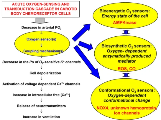
InFig. 3we show, to the left, a flow diagram of the transduction cascade for oxygen chemoreception as our laboratory proposed it in 1992 (Gonzalez et al., 1992). Circled in the flow diagram we include the O2 sensor and coupling mechanisms and, linked to them by
thick arrows, the three putative generic categories of O2sensors for
acute hypoxia in CB chemoreceptor cells. Each putative category of O2sensor is included in an ovoid that contains the principle of its
operation and tentative O2 sensor candidates. The double
arrow-head interconnecting the sensor categories intend to indicate that they might be interrelated as for example the main responsible for the bioenergetic status of the cells are mitochondria which in turn are potential sources of ROS, which in turn are the products of NOX4 (and other NOX isoforms). In the transduction cascade we present initalicsthe first known element of the transduction cascade, oxy-gen sensitive K+channels (Almaraz et al., 1986; Lopez-Barneo et al.,
1988); theitalicsintend to indicate, aside from the great diversity of O2sensitive K+channels and the variability of channels amongst
species, the discrepancies in the interpretation of the functional significance of some of the O2-sensitive channels, perhaps best
exemplified by maxi K (Wyatt et al., 1995; Buckler, 1997; Donnelly, 1997; Pardal et al., 2000; Gomez-Ni ˜no et al., 2009a,b; seeGonzalez et al., 2009). The rest of the steps of the transduction cascade are well accepted and present no major discrepancies except for neu-rotransmission which, although conflictive, has been recently and excellently reviewed (e.g.Iturriaga and Alcayaga, 2004; Iturriaga et al., 2009; Conde et al., 2009).
2.2. Bioenergetic O2sensors
In steady state conditions, the combined functioning of the respiratory (ventilatory) and cardiovascular systems secure and adequate supply of O2to the entire population of cells that
com-pose the complex mammalian organisms. In these conditions the amount of O2delivered to every cell supports an adequate rate of
ATP synthesis for cells to maintain their homeostasis, biosynthetic processes and to perform, in specialized cells, external work. Even in a resting mammalian organism the O2(and ATP) requirements
vary enormously, existing cells with a high rate of O2consumption
C. Gonzalez et al. / Respiratory Physiology & Neurobiology174 (2010) 317–330 321
Fig. 3.Oxygen transduction cascade and putative nature of O2sensors (see text for explanations).
A first and universal mechanism of dynamic adjustment is provided by cells themselves: the products of cell metabolism, which obviously increase in proportion to cell activity, are potent vasodilators making possible a diversion of blood to active tissues. Of course, if the mass of the active tissue is large (e.g. exercise comprising large muscular groups), it might also be required more integrated responses for the respiratory system and heart pumping to deliver enough O2to active cells.
A second, and probably also universal, mechanism is rep-resented by the 5-adenosine-monophosphate-activated protein kinase (AMPK). AMP-activated protein kinases are heterotrimeric complexes consisting of catalytic ␣-subunits and regulatory  -and ␥-subunits. Several isoforms of the three subunits exists in mammalians, and alternative splicing can enlarge the diversity of AMPK in different cells and tissues. The␣-subunits have ser-ine/threonine kinase domains at the N-terminus. One of these residues, Thr-172, is conserved in all isoforms and its phosphory-lation by upstream kinases (LKB1 serine-threonine kinase;Woods et al., 2003) is absolutely required for AMPK activation and phos-phorylation of downstream targets. Binding of AMP to AMPK (in fact to the␥subunit) promotes net phosphorylation of Thr-172 by inhibiting its dephosphorylation and allosterically activating the phosphorylated form of the AMPK. These two effects of AMP cause a multiplicative effect on the activity of AMPK, i.e., a small increase in cellular AMP levels cause a large effect on the kinase (Hardie et al., 1999; Hardie, 2008).
The cells “currency” to perform their normal functions is defined by the energy charge of cells defined by the equation:
energy charge= [ATP]+
1 2[ADP]
[ATP]+[ADP]+[AMP]
Energy charge of cells can take theoretical values between 1 and 0, all adenosine phosphates are as ATP or all are in the form of AMP, but most cells have steady state normal energy charges between 0.9 and 0.8. On decreasing PO2 energy charge decreases steadily
to a new lower value with a slope defined by the rate of ATP
gen-eration at the new PO2and the rate of energy expenditure in the
tissue or cell type under study; then, the slope of the decrease is higher the lower the PO2and final energy charge attained is lower
the lower the PO2. Molar concentrations of free ATP, ADP and AMP
vary among cells, with typical values for the three nucleotides, in the range of 5, 0.5 and 0.15 mM/l1intracellular water (or about half these values if expressed by kg of fresh tissues). Therefore, typical ADP/ATP and AMP/ATP ratios in normally oxygenated tissues are 0.15–0.1 and 0.06–0.03 (Ridge, 1972; Evans et al., 2005; Roepstorff et al., 2006). Most of ATP is used by cells in enzymatic reactions collectively known as orthophosphate cleavage reactions yielding ADP + Pi. The cleavage of the phosphate bond is energy-yielding and is coupled enzymatically to reactions that utilize the energy to run the cell functions. However, in some very important cellu-lar reactions (e.g., protein elongation) a pyrophosphate cleavage of ATP takes place yielding AMP + P∼P, and no doubt these reactions represent an important source of AMP in cells. Yet probably the highest amount of AMP in cells comes form the action of adenylate kinase, a system of several isoforms of enzymes that catalyze the reaction:
ADP+ADP⇒ATP+AMP
Although this reaction is reversible, in cells in periods of high activity when high amounts of ADP are generated or in situations of hypoxia (or shortage of energetic substrates) when the rate of ATP genesis is slow, the accumulation of ADP displaces the reaction to the right, as a fast strategy to maintain the availability of ATP. Obvi-ously in these circumstances the AMP/ATP ratio increases and this
ratio controls the activity of AMPK. Thus in a strict sense AMPK is not an O2-sensor but rather is a sensor of the energetic status of cells.
In these situations ofmetabolic crisis(Taylor, 2008), generated by the exaggerated ATP consumption during increased activity of cells or by insufficient ATP genesis due to hypoxia or a combination of both, it is needed a fundamental shift in the cellular metabolic strategy to facilitate the creation of an adaptive state which ulti-mately supports tissue survival. Insufficient ATP genesis due to insufficient substrate delivery to cells is a situation not encoun-teredin vivo, because the levels of glucose (the readily energetic substrate for cells) compatible with the life of the animals are high enough to keep the glucose utilizing machinery in cells working efficiently, otherwise animals will enter into hypoglycemia with serious risk to survive. However, in situations of ischemia a chronic decrease in substrate delivery and hypoxia are concurrent. In these situations, the action of AMPK goes beyond the regulation of fast post-translational events aimed to acutely maintain energy bal-ance (see below) and participates along with HIF-1␣and 2␣and sirtuins (particularly SIRT1) in an orchestrated control of differen-tial expressions of genes in the different cell types in the ischemic tissues directed to secure cell survival and blood vessel growth to solve the ischemic process (Cantó et al., 2009; Guarani and Potente, 2010; Ruderman et al., 2010). These and other aspects (Kahn et al., 2005) of chronic AMPK actions would not be considered further.
The point of interest in our context is which are the effects resulting from AMPK activation as a result of ametabolic crisis pro-duced by acute hypoxia and if these effects should be considered as mediators of oxygen-sensing and/or part of the transduction cas-cade in the chemoreceptor cells of the CB. In every cell studied it has been found that AMPK phosphorylates a great number of enzymes with the end result of promoting catabolic ATP-generating processes and inhibiting ATP-consuming anabolic processes and thereby tending to maintain the cell energy balance (Hardie, 2008; Taylor, 2008; Steinberg and Kemp, 2009). In the case of the CB, in an ample range of hypoxia down to very low arterial PO2
(25–30 mmHg;Biscoe et al., 1970; Nielsen et al., 1988) it would be, in a sense, inadequate talking ofmetabolic crisis because in spite of the intense hypoxia there was no decline in frequency of action potentials even at these low oxygen tensions, implying a very high rate of ATP synthesis to maintain the enhanced hypoxic activity. In other words, it would not be mitochondrial hypofunc-tion the responsible of the putative decrease in the energy charge of chemoreceptor cells, rather it would be an increase in the ATP consumption result of hypoxic activation (action potential gener-ation, ionic pumping, neurotransmitter release, etc.) which would determine a decrease in the energy charge of the cell and a pull for mitochondrial respiration and increase in ATP synthesis (Ereci ´nska and Wilson, 1982) to match ATP expenditure. Yet, in all probability AMPK plays a critical role in the maintenance of the required energy status by facilitating glucose uptake and oxidation by chemore-ceptor cells (Obeso et al., 1989, 1993) as well as transference of fatty acid and their oxidation in mitochondria. In fact in an vitro study on the levels of adenine nucleotides in the CB using normoxic air-equilibrated superfusatesvs.10% O2as hypoxic solutions, no
significant change was observed in ATP or ADP levels at either 4 or 30 min of hypoxia, but after 4 min there was an increase in AMP by a factor of 10 in the hypoxic CBs with no differences observed at 30 min (Verna et al., 1990) and in other study a modest decrease in ATP levels was observed on incubating the CBs with air vs. 100% O2equilibrated solutions (Obeso et al., 1985). Therefore, it appears
that the requirements for AMPK activation during hypoxic stimula-tion of the CB are met, as it is the case in pulmonary arteries smooth muscle lysates where is has been shown (Evans et al., 2005) that the AMP/ATP ratio rose from 0.040 under normoxia (155–160 mmHg, 2 h) to 0.083 under hypoxic conditions (16–21 mmHg, 1 h). In keep-ing with the CB,Wyatt et al. (2007)reported that in neonatal rat
AMPK co-localizes at the plasma membrane of chemoreceptor cells with maxiK, that the AMPK activator AICAR inhibited both O2
-sensitive currents expressed in these cells, maxiK and TASK-like channels carried currents, caused chemoreceptor cell depolariza-tion and augmented the afferent activity in the CSN. In a more recent studyDallas et al. (2009)showed using recombinant TASK channels that isoform sensitive to AICAR is TASK-3, a finding which in turn conforms recent findings ofKim et al. (2009)showing that the main O2 sensitive background current in rat chemoreceptor
cells is mostly carried by heteromeric TASK-1/TASK-3 channels and that in mouse TASK-3 is expressed at a higher level than TASK-1 (Ortega-Sáenz et al., 2010). In the study byWyatt et al. (2007)it was also shown that the AMPK antagonist compound C reversed the just described effects of hypoxia and AICAR on chemoreceptor cells. Although ironically, from a cynic point of view, it might be said that most of the effects seen on AMPK activation and inhibi-tion are epiphenomenal to O2-sensing, because maxiK inhibition
(Gomez-Ni ˜no et al., 2009a,b) and genetic elimination of TASK-1 and TASK-3 do not affect hypoxic behaviour of chemoreceptor cells (Ortega-Sáenz et al., 2010). It must be unambiguously stated that the data generated byWyatt et al. (2007)would indicate that AMPK dependent phosphorylation of K+ channels in CB chemoreceptor
cells mimic hypoxia and appears capable of eliciting integrated CB afferent activity, arguing in the favour that AMPK activation is a key element in the hypoxic transduction cascade. But the question on debate is if AMPK is the primary sensor of low PO2in chemoreceptor
cells or rather it is an amplifying element in the hypoxic trans-duction cascade which by phosphorylating channels amplifies their responsiveness to hypoxia. It is the authors opinion that AMPK is not the primary sensor. Data supporting our contention comes from several laboratories that using rabbit and rat chemoreceptor cells to record native O2 sensitive K currents and channels in isolated
membrane patches in the inside out configuration have evidenced that O2sensitivity of chemoreceptor cells is a membrane delimited
process that does not require any particular metabolic substrate or enzymatic reaction. The observations have also been extended to recombinant channels expressed in heterologous systems (see Section2.4).
2.3. Biosynthetic O2sensors
We are referring to sensors based in the measurement of the concentration of O2dependent enzymatically produced mediators
such as ROS and CO. In the last three years, since the previous review of our laboratory on the role ROS on oxygen chemoreception in the CB (Gonzalez et al., 2007) there have been substantial changes in the field, both in relation to mitochondrial derived ROS and more significantly on NADPH oxidase structure, subcellular location of potentiality of NADPH oxidase derived ROS for signalling.
2.3.1. Mitochondrial ROS
Literature on acute oxygen sensing and mitochondrial ROS has been dominated during almost the entire decade by opposite views on their significance, mostly centered in hypoxic pulmonary vasoconstriction (HPV) but including oxygen chemoreception in the CB (seeEvans and Ward, 2009). In the one side was Schumacker laboratory marshalling the case of hypoxia as trigger of increased ROS production at the complex III of the mitochondrial respiratory chain. Schumacker laboratory proposed that mitochondria func-tions astheO2sensors by increasing ROS which would represent
the coupling mechanisms with O2-sensitive K+ channels (or key
players in stabilizing HIF-1␣;Chandel et al., 1998, 2000; Waypa et al., 2001; Waypa and Schumacker, 2005, 2006, 2008); in their review of year 2000 they extended their model to O2
C. Gonzalez et al. / Respiratory Physiology & Neurobiology174 (2010) 317–330 323
Archer et al., 2008for representative reviews). In their article of 1995 Weir and Archerconcluded: “in pulmonary vascular smooth muscle as in the type I (chemoreceptor) cell of the carotid body, hypoxia causes a reduction in whole-cell potassium current. . .it is likely that the potassium current is controlled by changes in redox status, which could reflect the level of ROS generation by mitochondria or by an oxidase like NAD(P)H oxidase”. They also stated that K channels would be closed when reduced (R–SH SH–R) and open when oxidized (R–S–S–R). In1999 Archer et al. tested the hypothesis using gp91phox (i.e. NOX2) knockouts, and
found that animals have preserved HPV and a very low rate of ROS production in comparison to wild type controls. In additional experiments carried out in rat pulmonary arterial rings, they found that diphenyleneiodonium (DPI; an inhibitor of NADPH oxidase) dramatically decreased the normoxic rate of ROS production, indicating that most ROS in the rings in normoxic conditions are originated (within the specificity of DPI;Aldieri et al., 2008) at the NADPH oxidase level; they also observed that hypoxia reduced sig-nificantly ROS production to an identical level in control and in DPI treated rings, implying that hypoxia inhibits ROS production both at NADPH oxidase and mitochondria level. Conjugating findings in the knockout mice with the rat data they concluded that NADPH oxidase was not the sensor nor NADPH oxidase derived ROS were involved in O2-sensing, rather mitochondria would bethesensor
and the decrease of ROS levels produced by hypoxia the signals causing the reduced closed state of K+channels. The same notions
were defended in additional articles. For example inReeve et al. (2001)it was proposed that the increased basal PAP and the atten-uation of HPV after CH would be due to a loss of the inhibition of K+
channel activity by acute hypoxia which in turn would result from the inability of acute hypoxia to further modify the cytosolic redox status beyond the already reduced environment caused by CH itself. In another study (Michelakis et al., 2002) it was shown that while in PASMC acute hypoxia (36–40 mmHg) caused a decrease in ROS production, in renal artery smooth muscle cells (which exhibit a lower rate of normoxic ROS production) hypoxia increased ROS, being proposed that this difference would explain the opposite vasomotor effects of hypoxia in both arterial beds. Yet,Wu et al. (2007)compared PASMC and coronary artery smooth muscle cells and found that acute hypoxia PO2= 25–30 mmHg for 5–10 min
significantly and rapidly decreased ROS levels in both cell types. The putative significance of mitochondrial ROS in HPV, and by extension on CB chemoreception, has not concluded with these opposing views. In a very recent study Schumacker group (Waypa et al., 2010) based on data obtained with cell compartment tar-geted ROS indicators propose that hypoxia (superfusion of PASMC isolated cells with 1.5% O2-equilibrated solutions) augments in a
regulated manner ROS in the intermembrane mitochondrial space and by diffusion into the cell cytoplasm while lowering mito-chondrial matrix ROS levels; it would be the regulated increased cytoplasmic ROS level the trigger of HPV. It is the authors opin-ion that the slow time courses of ROS changes measured in present
experiments do not correlate with the fast contraction or Ca2+
tran-sients of PASMC in response to hypoxia observed both in vivo and in vitro preparations (seeEvans and Ward, 2009); additionally Waypa et al. observed that systemic arteries smooth muscle cells behaved alike PASMC (see above).
Quite recently other authors have made to intervene ROS of both origins, mitochondrial and NADPH oxidases, in the genesis of HPV, albeit in different manners (seeWeissmann et al., 2006; Rathore et al., 2006, 2008; Cogolludo et al., 2009and under con-sideration;Wang and Zheng, 2010; Daiber, 2010). Although these aspects surely will be covered in greater detail in other articles of this issue, we should mention thatCogolludo et al. (2009)found that neutral sphingomyelinase generated ceramide and PKC acti-vation are early and necessary events in the genesis of the HPV. In a recent article of Perez-Vizcaino laboratory (Frazziano et al., submitted) it is reported that PKCactivates phosphorylation of p47phoxand activation of NADPH oxidase with the ROS generated
playing a necessary role in controlling the open probability of Kv channels in PASMC; in addition they also present evidence sug-gestive that increased mitochondrial ROS production could play a critical role in the activation of neutral sphingomyelinase. In our laboratory, we have tested a wide variety of reducing and oxidizing agents (Sanz-Alfayate et al., 2001; Gonzalez et al., 2004a,b; Agapito et al., 2009) and we could not establish a relationship between the general redox status of the cell and the activity of chemorecep-tor cells. In a recent study (Gomez-Ni ˜no et al., 2009a,b) we aimed to manipulate mitochondrial rate of ROS production and redox status with the use of metabolic poisons and uncouplers in iso-lation and in the presence of N-acetylcysteine. As shown inTable 2 data indicate that mitochondrial alteration of the general status of cells is not linked to chemoreceptor activation. Thus, our findings would suggest that acute O2sensing in CB chemoreceptor cells and
PASMC do not follow the same paths. To further explore similar-ities between acute O2-sensing in PASMC and CB chemoreceptor
cells we have performed the group of experiments shown inFig. 4. Data indicate that the specific neutral sphingomyelinase inhibitor GW4869 at the same concentration (10M) that decreased by about 80% the tension developed by pulmonary artery segments in response to hypoxia did not alter the activity of chemoreceptor cells.
We do not want to conclude this section on mitochondrial ROS without stating some reflections made in previous reviews (Gonzalez et al., 2002, 2004b, 2007) and to add some additional comments. First, methods used to assess ROS concentrations or redox status are not reliable (see in additionJelic and Le Jemtel, 2008). Second, to rise conclusions on the significance of oxidative status on a given cell function based of antagonism of the observed effects by antioxidants might be marred by secondary effects of the antioxidant used (e.g.Sekiguchi et al., 2003). In this regard we want to make some additional reflections taken from (Armitage et al., 2009); for example antioxidants can in fact promote chain reac-tions initiated in their oxidized forms, and more importantly, if the
Table 2
Affects of Metabolic Poisons alone or in combination with N-acetylcysteine on the general redox status of cells, chemoreceptor cells catecholamine release and CB ATP levels.
Condition Control Rotenone
(1M)
3-NP (5 mM)
Antimicyn A (0,2g/ml)
Na Azide (5 mM)
DNP (100M) Parameter
EGSH(mV) −181/−186+ −175*/−179 −160**/−180 −165***/−178 −163**/−179 −180/−180 3H-CA release (% tissue content) 1.61/1.57 23.19/21.9 1.64/1.88 45.49/45.53 33.88/34.55 9.04/9.19
[ATP] (mol/g tissue) 1.46/1.46 0.50+++/0.51 1.54/1.54 0.72+++/0.81 0.60+++/0.48 1.46/1.46 0.80+++/073␣
0 25 50 75 100 125
Pulmanary
artery
contraction (
%
)
control PA + 10µM GW
0 20 40 60 80 100 120 140
0 500 1000 1500 2000 2500 3000
3 H-CA release
(dpm)
7% O2
7% O2+ 10 µM GW
7%O2 7%O2
GW
min
0,0 0,2 0,4 0,6 0,8 1,0 1,2 1,4
S2/S1 Evoked release
control PA
+ 10µM GW
Fig. 4.Effects of the GW4869, a specific inhibitor of neutral sphingomyelinase, on the activity of chemoreceptor cells in normoxia and hypoxia. The activity of chemoreceptor cells was measured as their release of CA. In a group of 10 CBs two hypoxic stimuli (10 min superfusion with a solution equilibrated with 7% O2instead of the normoxic 20% O2) and other group of equally 10 CBs was similarly stimu-lated, but prior and during the application of the second hypoxic stimulus 10M of GW4869 was present. In the upper figure it is shown the mean time course of the release evidencing that the presence of the sphingomyelinase inhibitor did not alter it. In the lower row of the figure at the left are shown the ratios of the evoked release in the second to the first stimulus in control and inhibitor treated organs. For com-parative purpose are shown mean effects of the same concentrations of GW4869 in pulmonary artery rings contraction; these last data are taken fromCogolludo et al. (2009).
antioxidant is fully effective the global removal of ROS may result in unwanted effects since ROS at many cell places (and not only at the one we want target) also regulate important physiological functions. Third, the compartimentalization of ROS generation and the likelihood of local signalling (D’Autréaux and Toledano, 2007; Fisher, 2009; Paulsen and Carroll, 2010; Lassègue and Griendling, 2010) would require in future years to develop reliable compart-ment specific indicators for the different ROS to truly understand the significance of ROS; in this regard the developments in the Ca2+
methodology and function understanding (Montero et al., 2000; Alvarez and Montero, 2002; Santodomingo et al., 2008) would be a path to emulate. Fourth, owed to the expression in mitochondria of superoxide dismutase and glutathione peroxidase as well as to their content in lipoic acid and glutathione, forces the teleologi-cal conclusion that mitochondria are capable of producing ROS as indeed has been proven under a great variety of conditions. The question as posed byNohl et al. (2005)is how much ROS mito-chondria produce in physiological conditions, being their view that physiological rate of ROS production is minimal (see below) and that the strong anti-ROS armamentarium that mitochondria pos-sess should be considered as a functional reserve to fight ROS in pathophysiological conditions. Fifth, from this conception we can ask how many ROS mitochondria would produce at the levels of hypoxia physiologically detected, both in CB chemoreceptor cells and in PASM which have comparable P50and shape of response
curves to hypoxia (Ward, 2008; see above).
Going behind an answer to this question we will refer to several considerations made byMurphy (2009)in his excellent review, as we consider they are germane in our context: (a) how much super-oxide anion (O2•−) do mammalian mitochondria produce in vivo?
Maximal rate of O2•−production by isolated mitochondria reaches
up to 2% of their O2 consumption and this occurs in conditions
uncertain to occur in living cells, i.e., when mitochondria are not synthesizing ATP, have a high electrochemical H+gradient and the
coenzyme Q is reduced. In these conditions most of O2•−produced
occurs at the complex I level, by a rotenone sensitive backflow of electrons from coenzyme Q to complex I. This value of 1–2% of res-piration going to O2•−has propagated through the literature, being
taken as an estimate of the rate mitochondrial O2•−productionin
vivo. (b)Murphy (2009)points out al least three reasons why this figure does not relate to the in vivo situation: one, backflow of elec-trons occurs with saturating concentration of succinate and when physiological concentrations of substrates are used the rate would drop to 0.12–0.15% of O2consumed; two, the most common
con-centration of O2used when working with isolated mitochondria is
that given by room air (≈200M) and in vivo the concentration is between 10 and 50M (in chemoreceptor cells and PASMC should be in the upper range), which would decrease the rate of production by a factor of at least four,2leaving the rate of production <0.1% of O2consumption; and three,in vivomitochondria are making ATP,
implying that the electrochemical gradient for H+is not maximal
and that neither NADH and CoQ pools are fully reduced. The pro-portion of time that mitochondria are in state 3 (i.e. actively making ATP)vs.state 4 (i.e. with low rate of ATP synthesis and high electro-chemical gradient) is not known, but for sure the active synthesis of ATP would further reduce the figures previously calculated for isolated mitochondria.
These concepts are extremely important for O2-sensing in the
context of ROS production, because in the three tissues endowed with physiological sensitivity to hypoxia (Fig. 2), hypoxia neces-sarily means increased activity, in the case of chemoreceptor cells and PASMC, without adaptation (i.e., sustained increased activity as long as hypoxia last) and therefore the rate of ATP synthesis at the mitochondria must be increased to support the increased activity (Obeso et al., 1993, 1997; see above). Then all factors, hypoxia itself, high rate of ATP synthesis and consequently a sub-maximal H+electrochemical gradient and a rather low fraction of
reduced components in the respiratory chain would imply a lower than normoxic mitochondrial rate of ROS production in cells with physiological sensitivity to hypoxia. In other words, active mito-chondrial ATP synthesis requires transport of ADP and inorganic phosphate and backflow of H+at the mitochondrial ATPase which
altogether decrease electrochemical gradient and direct electrons to fully reduce molecular O2 to water. Following this rationale it
is not possible to envision an increase in mitochondrial ROS pro-duction by hypoxia, unless some additional factor(s) enters into play to alter the parameters of the equation in footnote2. Nitric oxide (NO•) is one candidate to alter the values of the members of equation (Murphy, 2009). NO•can reach mitochondria from cyto-plasm NOS, can be synthesized by mitochondrial NOS or can even, in situations of extreme hypoxia, be synthesized by reduction of NO2−by cytochrome oxidase (Carreras and Poderoso, 2007).
How-ever, even if the NO•concentrations reached at the mitochondrial matrix are adequate to generate the inhibitory effects on respira-tion by inhibiting cytochrome oxidase and to increase the rate of
2The rate of O
C. Gonzalez et al. / Respiratory Physiology & Neurobiology174 (2010) 317–330 325
O2•−production by increasing the level in the reduction of
mito-chondrial components (Carreras and Poderoso, 2007), it is our view that cells whose function is to be fully active in hypoxia, and with greater activity the greater the intensity of hypoxia, this biological design would be purposeless. Yet, if several laboratories, includ-ing ourselves found that mitochondria of PASMC increase their rate of ROS production in hypoxia. Either the ROS measuring tools are faulty or we are missing the factor that without breaking ATP synthesis activates O2•− production. In this regard it should be
noted that according to some authors the potentially exist to have a reduced electron transport chain in an ample range of hypoxic levels well before theKmof cytochrome oxidase for O2is reached
and O2consumption is restricted (Ereci ´nska and Wilson, 1982; see
Guzy and Schumacker, 2006). Thus, according to the so-called near-equilibrium hypothesis of Ereci ´nska and Wilson (1982), hypoxia would intrinsically tend to augment the fractions of the different components of the respiratory chain that are in their reduced form, this factor tending to favour the production of ROS. Note, how-ever, that at the same the PO2 decrease would tend to diminish
ROS production.
2.3.2. NADPH oxidase derived ROS
In recent years have emerged several new aspects of the biology of NADPH oxidases, NOX, that are excellently compiled in recent reviews (Brown and Griendling, 2009; Fisher, 2009; Oakley et al., 2009; Ushio-Fukai, 2009; Lassègue and Griendling, 2010). Here we will summarize the basics necessary to follow recent endeavours on NADPH oxidase researches in the CB chemoreception. (1) Although of some NOX subtype (e.g. NOX 4) there have been described sev-eral isoforms, they are recognized seven NOX types (NOX1-5, DUX1 AND DUOX 2). (2) A given cell type may express several NOX isoforms, rising the possibility that different cell processes are reg-ulated by signals that modulate the activity of different NOX. (3) Cellular localization of NOX is variable, being the plasma membrane and the endoplasmic reticulum the most frequent locations; other common and specific locations for NOX expression include the nucleus, lipid-rafts-caveolae, cell matrix adhesions and cell to cell contacts, cell migrating edges and endosomes resulting from early endocytosis mediated by receptor resulting in specialized vesicu-lar compartments, redoxomes, where ROS (O2•−) are generated. (4)
The classical concept of NADPH oxidase as an unassembled multi-molecular complex that requires stimulus mediated assembling to be activated is still true for NOX1 (ubiquitously expressed, but is highly expressed in endothelial and vascular smooth muscle cells and thereby playing critical roles in vessel pathophysiology and in intestinal epithelium where its function would be mostly defen-sive), NOX2 (the classical phagocyte isoform but also expressed in many cell types but particularly in all cell types of vessels existing a direct correlation between NOX2 expression and hypertension [a high level of expression in vessel cells would lead to neutral-ization of the vasodilator action of NO• as it would react with the NOX derived O2•− generating ONOO− which is highly
reac-tive and damage propagating]) and NOX3 (expressed in the ear where it has been related to inner ear and vestibular function and lung where it has been associated to the development of emphy-sema by controlling the destruction of elastic fibers of the alveolar wall). The NOX4 isoform only requires subunit p22phoxto express
its full activity. NOX4 was originally described as Renox, being very abundant in kidney and in many cell types from skin to neurons including vessel cells and hepatocytes; it is constitutively active and its main regulation appears to be mediated at transcriptional level, although some regulation via p22phoxspecific ligands also
seems to exist. NOX5 is conformed by the single NOX subunit with its cytosolic N-terminal region being very reach in Ca2+binding to
EF-hand domains; it is expressed in lymphatic tissue, vessel cells and prostate, and appears to participate in the control of cell
pro-liferation, but interestingly it is not expressed in rodents. DUOX1 and DUOX2 activation involves Ca2+binding to EF-hand domains in
the cytosol generating directly H2O2by two electrons reduction of
molecular O2without the intermediate step of forming O2•−; they
are expressed in thyroid cells where they are critical to metabolism of iodine and thyroid hormone synthesis, being also abundant in the epithelial cells of the respiratory tract where they would play defence-type functions. (5) The above summarized recent findings provide an explanation to the postulate put forward bySaran and Bors (1994; see alsoGonzalez et al., 2002): “in order to properly convey a message from one cellular location to another, the mes-senger must get to the point of message reception in unaltered form or through a series of well-defined reproducible steps is of course trivial with respect to stable molecular messengers like hormones. Regarding radicals, which by their very nature are reactive species, the postulate is less trivial”. Aiming to clarify the feasibility of spe-cific signalling by ROS we should describe two situations. When NOX is expressed in a caveolae (invaginations with typical diame-ter of 80–120 nm and open pores to cell surroundings of 30–50 nm in diameter;Westermann et al., 1999), the O2•−would be released
into a compartment that, in the one hand, is specific for its con-tents and the abutting proteins and, in the other hand, taking into consideration diffusion coefficient and half life for O2•−(Saran and
Bors, 1994), it could be expected that many of the cell molecules exposed to the caveolar surface can be direct targets of significant fractions of the O2•−produced. Even in the absence of any special
constituent (transition metals, ascorbic acid, etc.) the O2•−
con-centration decay path is steep due to the spontaneous dismutation and a focal genesis of H2O2. This fact is very important because for
O2•− to signal it certainly must be there, but it is equally
neces-sary something to take the message (i.e., “a receptor” for O2•−).
In this regard it should be recalled that ROS chemistry dictates reactivity towards selective atomic targets in proteins, with O2•−
being active towards iron–sulphur clusters and H2O2targets are
reactive−SH of cysteine residues; i.e., it is the subcellular colo-calization of specific ROS and their targets what dictates signaling specificity (D’Autréaux and Toledano, 2007). At the extracellular surface of caveolae it is unlikely the presence of iron-sulfur clusters, but certainly many proteins likely have cysteine residues with dif-ferent reactivity with H2O2(Paulsen and Carroll, 2010) providing
specificity to signalling. When a redoxome is formed by receptor mediated endocytosis of a plasma membrane patch containing a NOX complex capable of generating O2•− inside the endosome,
superoxide production would stop unless O2•−processing enzymes
(SOD capable of generating freely diffusible H2O2) or chloride
chan-nels and/or anion exchangers capable of mediating O2•−export to
cell cytoplasm and maintaining membrane gradients required for NOX activity are present in the endosome membrane. It is obvious that receptor mediated redoxome formation is specific, as they are every other step capable of spilling ROS to cell cytoplasm (orienta-tion of anion channels or exchangers, aquaporin molecules focusing the exit of H2O2formed).
Departing from here we can ask how many NOX subtypes are expressed in the CB, and more specifically in chemoreceptor cells where they, via their enzymatic products, contribute to control O2-sensing and transduction cascade. Initial studies ofKummer
and Acker (1995)located p22phox, gp91phox, p47phox, and p67phox,
i.e. NOX2 in rat chemoreceptor cells, but a few years later Kum-mer’s laboratory (Dvorakova et al., 2000) using specific monoclonal antibodies against gp91phoxand additional markers concluded that
macrophages located in the rat CB represent the major location of NOX2. This finding in turn would give a full justification to the func-tional studies ofRoy et al. (2000)andHe et al. (2002)showing that gp91phoxKO mice have preserved normal K+currents and
magni-tude of hypoxic inhibition and normal hypoxic Ca2+transients in
to acute hypoxia. These findings as a whole would certify that NOX2 is not involved in acute O2-sensing or transduction cascade in mice
and probably also in the rat.
However, the very same year, Fidone’s laboratory (Sanders et al., 2002) showed that p47phoxknockout mice exhibited exaggerated
CSN discharges and ventilatory responses to acute hypoxic tests and normal levels of erythropoietin messenger expression after 72 h of hypoxia. In a further study of Fidone’s laboratory (He et al., 2005) in a comparative study of chemoreceptor cell responses to hypoxia in chemoreceptor cells of p47phoxKO and
correspon-dent wild type controls it was shown: (a) basal normoxic ROS production was comparable in control and KO cells; (b) hypoxia increased superoxide production (dihydroethidine oxidation) in control but not in KO cells; (c) hypoxic ROS production was sensi-tive to NADPH oxidase inhibitors; (d) sodium azide (a cytochrome oxidase inhibitor) increased O2•− production equally in control
and KO cells; (e) percentage K+current inhibition by hypoxia was
greater in KO than in control cells, and; (f) Ca2+transients elicited
by hypoxia were higher in KO than in control cells. These find-ings and additional northern blot data (seeHe et al., 2010) and the use of L-type Ca2+channel blocker (unpublished) allowed us
to conclude that NOX expressed in chemoreceptor cells was NOX4 (seeGonzalez et al., 2007), that NOX was not the oxygen sensor, that NOX derived ROS acted as negative modulators of the acute hypoxic transduction cascade, that Ca2+entering through L-type
channels was responsible for NOX activation during hypoxia and finally, that mitochondria did not increase their rate of ROS pro-duction in hypoxia as the rate of dihydroethidine oxidation was not altered by hypoxia in KO cells. Obviously these findings need to be reinterpreted at the light of new findings. Since we know today that p47phoxdoes not form part of NOX4 it is obvious that
chemorecep-tor cells must express at least one more NOX isoform requiring this subunit (i.e., NO1, NOX2 or NOX3) to explain the loss of chemore-ceptor cells capacity to produce ROS in the p47phox KO animals.
Since in pancreatic acinar cells Ca2+may mediate the activation of
NOX1 and thus ROS production (Yu et al., 2007) we would suggest that this is the p47phoxcontaining isoform present in
chemorecep-tor cells and activated as a result of Ca2+entering the cells by acute
hypoxic activation. Our suggestion is supported in part by the study ofPeng et al. (2009)showing that rat CB expresses NOX1. However, they also showed that the major isoform expressed in the CB, at least in terms of mRNA fold increase during intermittent hypoxia was NOX2 (but see above). They reported that the subcellular loca-tion in chemoreceptor cells was different as NOX2 (and probably NOX1) was cytoplasmic and NOX4 was perinuclear; no cellular or subcellular location was provided to NOX3, which is also expressed in the CB, but whose expression was not modified by intermittent hypoxia.Peng et al. (2009)showed additionally that neither apoc-inyn, AEBFS or DPI inhibited acute response to repetitive trains of hypoxic stimulation, conforming our previous conclusion that NOX were not the O2sensor and that NOX-derived ROS are not
neces-sary coupling factors in the transduction cascade; the three NADPH inhibitors prevented long term facilitation of sensory nerve dis-charge induced by repetitive stimulation that was also absent in NOX1 knockout mice.
2.4. Conformational O2sensors
We are referring to molecules that would bind O2in
concentra-tions proportional to their surrounding PO2, and as a result they
would experience conformational changes that would be alloster-ically transmitted to the first effectors of the transduction cascade, i.e., oxygen sensitive K+channels. An unknown hemoprotein, ion
channels themselves and NOX4 have been proposed as sensors of this category.
Leaving aside ion channels as putative O2sensors, there are two
concepts contained in our proposal of the conformational model astheO2 sensor for acute hypoxia in chemoreceptor cells. First,
that it should be a hemoprotein and second, that it must be located in the plasma membrane or strongly bound to some membrane component. In the remaining of this section we should intend to justify our views on the basis of existing literature.
Why a hemoprotein? First, the notion is not new as old CB lit-erature already contains the idea that the O2sensing device can be
a haemoglobin-like hemoprotein with a P50close to 40 mmHg and
a special Bohr effect (seeFidone and González, 1986for old refer-ences). Second, the idea returned when Lopez-Lopez and Gonzalez (1982) showed that in rabbit chemoreceptor recorded in whole-cell configurations CO prevented or reversed the inhibition of K+
cur-rents produced by hypoxia. A year laterLahiri et al. (1993)showed that CO at low concentrations inhibited the activity of the CSN elicited by hypoxia while at high concentrations (4–5×prevailing PO2) stimulated the activity in the CSN, being this last excitatory,
but not the inhibitory effect, sensitive to light, an unequivocal indi-cation that excitation was due to cytochrome oxidase inhibition, while the inhibitory effect was independent of CO actions at the mitochondrial level. Third, a survey to general biology books indi-cated that all proteins handling O2are metallo- or hemoproteins,
being also known that in biological systems CO is very unreactive, and as O2, CO is handled by metallo- or hemoproteins proteins
(Coburn and Forman, 1987). Fourth, the notion of channel gat-ing implied molecular rearrangements of the channel protein as to open the channel pore and allow the ion to pass through the selectivity filter. Fifth, It was also known that hemoproteins, as exemplified by myoglobin and particularly haemoglobin, on bind-ing and releasbind-ing O2suffer conformational changes in their tertiary
or quaternary structures. Sixth, putting together points fourth and fifth it is evident that a conformational change in a hemoprotein can be allosterically transmitted to the ion channels to cause important modifications in their kinetic properties if the physical relation-ships between both elements are the appropriated. Finally, asStreit et al. (2010)put it, heme is one of the nature’s most versatile cat-alytic building blocks as heme-binding proteins facilitate a range of redox, atom or electron-transfer, and small-molecule sensing and trafficking processes, with a specificity that is sometimes dif-ficult or impossible to reproduce synthetically. However, it should be added that specific kinetic parameters of hemoprotein functions are dependent on the protein moiety as exemplified by differences in O2vs.CO affinities binding in hemoglobin (CO affinity nearly 300
times higher than O2affinity)vs.cytochrome oxidase (CO affinity
about 5–10 times higher than O2affinity). Another example that we
want to bring here is the case of NOX2; in their classical review on NADPH oxidase (NOX2)Cross and Jones (1991)giveKmfor O2
oscil-lating between 5 and 30M, but according toIizuka et al. (1985) andParkos et al. (1988)it does not bind CO at all (but seeNakahira et al., 2006; Basuroy et al., 2009).
Why in the plasma membrane? Very early after the descrip-tion of the O2 sensitive K+ channels (Lopez-Barneo et al., 1988),
Ganfornina and Lopez-Barneo (1991)showed that isolated rab-bit chemoreceptor cell membrane patches recorded in the inside out configuration contained low unitary conductance K+
chan-nels that were reversibly inhibited by low PO2, implying that
the plasma membrane contained all elements to sense O2and to
couple the O2 sensor to the first line effectors in the
transduc-tion cascade, the K+ channels. A few years later, our laboratory
showed with inside out isolated patches obtained from HEK cells cotransfected with Kv4.2 + Kv1.2 regulatory subunits that the channels were O2-sensitive, being reversibly inhibited by hypoxia
and also were sensitive to CO in such a manner that CO pre-vented the low PO2 inhibition (Perez-Garcia et al., 1999). In an