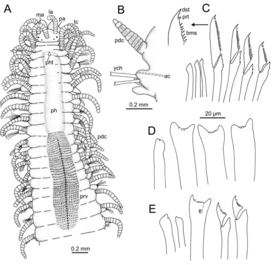
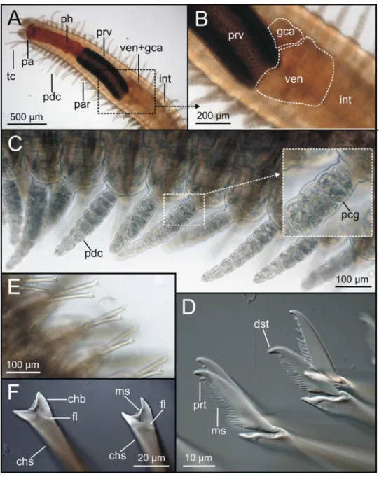
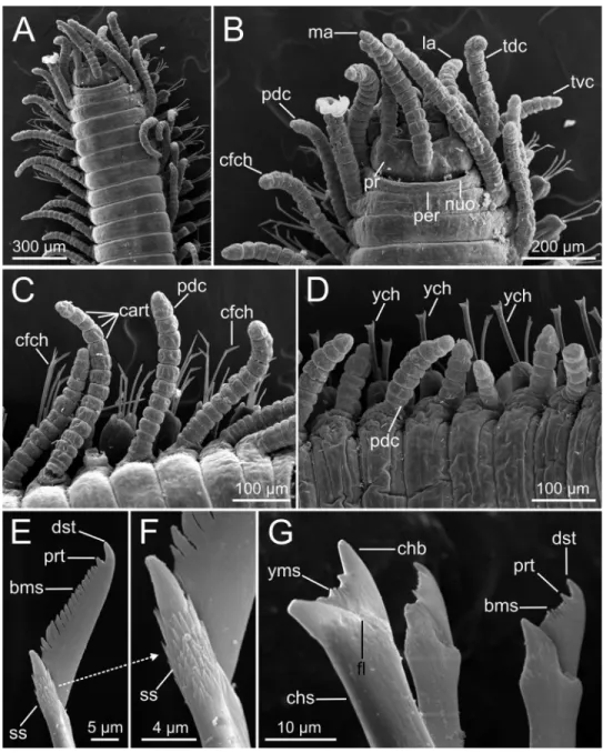
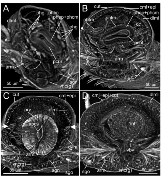
TítuloAn integrative approach to the anatomy of Syllis gracilis Grube, 1840 (Annelida) using micro computed X ray tomography
Texto completo
Figure


Documento similar
• Analysis of long-term variability of high-energy sources in the optical and X-ray bands, using INTEGRAL observations from IBIS (the gamma-ray imager), JEM-X (the X-ray monitor)
In addition to those studies the data used in this report has been used to obtain the monthly variability of clear and usable nights in a year, as well as relevant statistics for
A comparison between the radio and X-ray populations in Orion shows that the radio detections so far have been strongly biased to the brighter X-ray stars. This supports the
With the proposed QR factorization method and system solving using Out-Of-Core techniques we were able to reconstruct high- quality CT images using the minimum number of projections
Microbial loads (log CFU cm −2 ) of mandarins packaged within different packages types (ST, small tray; SB, small box; LT, large tray; LT+, large tray with alveoli tray; and LB,
In this chapter, an automated method for brain hematoma and edema segmentation, and volume measurement using computed tomography imaging is presented.. The method combines a
(7) Our study results differ in the disposition of the hepatic flexure of the colon as we identified differences between both groups, which have not been described in previous
We then show the findings of a recent approach to Faddeev equations using chiral unitary dynamics, where an explicit cancellation of the two body off shell amplitude with three
