Antibacterial Effect of Carbosilane Metallodendrimers in Planktonic Cells of Gram Positive and Gram Negative Bacteria and Staphylococcus aureus Biofilm
11
0
0
Texto completo
(2) Biomolecules 2019, 9, 405. 2 of 11. is facultative anaerobic, coagulase-positive, catalase-positive and oxidase-negative. It grows forming colonies in an optimum temperature range 30–40 ◦ C [2,3], and they can even grow in seawater and ferment glucose, lactose and maltose [1,4,5]. S. aureus is an opportunistic pathogen found in the normal human microbiota, at the skin of healthy individuals [6,7]. Between 30 and 50% of healthy adults are colonized, and between 10 and 20% remain persistently colonized [8–10]. Although anyone can develop staphylococcal infection, risk-populations include people with chronic conditions or weakened immune system, people who have had surgery and/or those who use a catheter (e.g., dialysis patients) [6,7,11–13]. S. aureus infections can affect the skin, the bloodstream, bone tissues or the eyes, leading to life-threatening diseases like endocarditis, pneumonia, toxic shock syndrome or keratitis. Most chronic and recurring infections, such as permanent medical device infections [14–18], are related to the production of bacteria biofilm [19,20]. Human bacterial infections are mainly produced by bacteria in a biofilm-mode of growth and not due to planktonically growing bacteria. A biofilm is a sessile community derived from microbes embedded in a matrix of extracellular polymeric substance which exhibit an altered phenotype with respect to growth [6,7,21]. Biofilm formation is divided into four distinct metabolic states: aerobic, fermentative, latent (including persistent very slow-growing cells) and dead cells [22,23]. The antimicrobial resistance of biofilms is explained by the stressed environment, which produces many cells with low metabolic rates, and its ability to act as a diffusion barrier that hinders the penetration of antimicrobial agents. In bacterial infection, biofilm matrix acts as a safe haven, protecting bacterial cells from antibiotics, immune cells and antimicrobial factors [24,25]. Apart from biofilm formation, other types of bacterial resistance can arise from spontaneous mutation or through the genes exchange between different strains or species of bacteria [26]. The evolution of antibiotic resistance is currently one of the main threats to public health security; the first warning came from Alexander Fleming, discoverer of penicillin. Only four years after penicillin introduction in clinic, 14% of S. aureus hospital strains were resistant, number that increased to 59% four years later. In the 1980s-90s, resistance exceeded 80% in communities and 95% in most hospitals [27]. Unfortunately, biofilm-bacteria are more resistant to conventional antimicrobials and require new approaches, such as those provided by nanotechnology [28–31]. Metal-based nanoparticles (NPs), carbon-based nanomaterials, as well as polymeric NPs, liposomes and dendrimers have been proposed as biofilm antimicrobials. These NPs not only possess antimicrobial properties of their own, but can also be used as drug delivery systems. Dendrimers are highly branched three-dimensional macromolecules whose structure is globular and monodisperse. These unique properties enabled their evaluation in multiple applications in the field of medicine, including drug carriers to increase bioavailability, gene carriers to protect nucleic acids, specific antitumor systems and broad-spectrum antiviral and antibacterial agents [32]. In the past years, successful dendrimer-based strategies to control microbial contamination and prevent biofilm formation have been proposed [33,34], mainly relying on peptide dendrimers, glycodendrimers, quaternary ammonium dendrimers and metallodendrimers. While exhibiting different modes of action, the antimicrobial activity ultimately is produced by the ability to bind to the negatively charge bacterial cell surface and/or membrane proteins and phospholipids that leads to cell membrane disruption. The presence of metal ions in the nanoparticle can further improve the antimicrobial action through the production of Reactive Oxygen Species (ROS) that induce DNA and mitochondria damage, cell membrane disruption and interruption of transmembrane electron transport [28]. Most antibacterial metallodendrimers reported in the literature rely on the presence of Ag(I) or Ag(0), well-known antimicrobial metal [35], and few examples include other metals such as Cu(II) and Zn(II) [36]. Although the functional groups in the dendrimer surface determine most of the properties of the nanoparticle, the nature of the scaffold also influences its biological activity. As an example, the hydrophobic, stable and flexible scaffold in carbosilane dendrimers enhances their interaction with biological membranes and enables a potent biological activity even with low generation dendrimers [37]. Our group has recently described the use of Schiff-base carbosilane dendrimers as promising delivery.
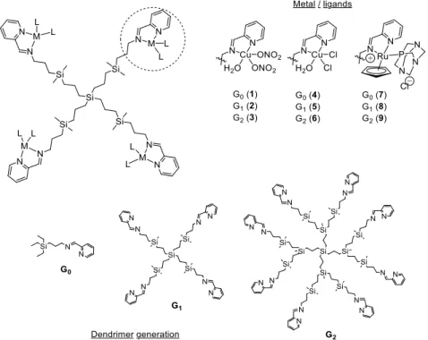
(3) Biomolecules 2019, 9, 405. 3 of 11. agents of Ru(II) and Cu(II) metallodrugs in cancer therapy [37–39]. These metallodendrimers produced a significant tumor size reduction in an in vivo mice model of resistant prostate cancer, with no signs of toxicity during the experiment. Herein, we present carbosilane metallodendrimers as an alternative to traditional antibiotics Biomolecules 2019, 9, x FOR PEER REVIEW 3 of 11 and evaluate the influence of different parameters—dendrimer generation, metal ion, ligand—in the biocide effect against planktonic cells and of S. aureus. bacteriostatic and bactericide biocide effect against planktonic cellsbiofilms and biofilms of S. Promising aureus. Promising bacteriostatic and activity and no hemolysis were found, especially for first-generation dendrimers. bactericide activity and no hemolysis were found, especially for first-generation dendrimers. 2. Materials and Methods 2. Materials and Methods 2.1.2.1. Metal Complexes Metal Complexesand andMetallodendrimers Metallodendrimers The selected comprised two families of copper (II) carbosilane complexes, The selectedmetal metal complexes complexes comprised two families of copper (II) carbosilane complexes, with with nitrate (1–3) and chloride (4–6) ligands, and a family of ruthenium (II) carbosilane complexes (7–9) nitrate (1–3) and chloride (4–6) ligands, and a family of ruthenium (II) carbosilane complexes (7–9) (Figure 1). These complexes were selected in order to study the influence of the metal, the ligand and (Figure 1). These complexes were selected in order to study the influence of the metal, the ligand and thethe generation onon their and Ru(II) Ru(II)mononuclear mononuclearcomplexes complexes and first generation theirantibacterial antibacterialactivity. activity. The The Cu(II) and and first and second-generation accordingtotopreviously previouslypublished published and second-generationmetallodendrimers metallodendrimers were were synthesized synthesized according protocols [37–39]. control included included Cu(NO Cu(NO )H protocols [37–39]. The The different different metal metal salts salts used used as as negative negative control 3)3 2· 2O, 2 ·H 2 O, CuCl 2·2H andRuCl RuCl 3·H O. CuCl and 2 ·2H 2 O2O 3 ·H 22O.. .. Figure 1. Chemical representation of the tested carbosilanecarbosilane metallodendrimers, highlighting Figure 1. Chemical representation of theSchiff-base tested Schiff-base metallodendrimers, thehighlighting structural parameters studied. the structural parameters studied.. Serial dilutions of the different biocides were prepared in sterile distilled water for copper Serial dilutions of the different biocides were prepared in sterile distilled water for copper complexes with nitrate 7–9due duetototheir theirgood goodsolubility, solubility, and complexes with nitrateligands ligands1–3 1–3and andruthenium ruthenium complexes complexes 7–9 and in in dimethyl sulfoxide (DMSO) concentration)for forchloride chloridecopper copper complexes dimethyl sulfoxide (DMSO): water : water(1:99 (1:99at atthe the highest highest concentration) complexes 4–6. The effect of DMSO at the different concentrations was evaluated in an independent study, ruling 4–6. The effect of DMSO at the different concentrations was evaluated in an independent study, outruling any possible toxicity for antibacterial assays. out any possible toxicity for antibacterial assays. 2.2.2.2. Bacterial Strains Bacterial Strains The microorganisms used in the assays werewere a strain of Escherichia coli (CECT 515, Gram-negative) The microorganisms used in the assays a strain of Escherichia coli (CECT 515, Gramand/or a strain of Staphylococcus aureus (CECT 240, Gram-positive) provided by the Type negative) and/or a strain of Staphylococcus aureus (CECT 240, Gram-positive) provided bySpanish the Spanish Culture Collection (CECT)(CECT) in lyophilized form. form. Type Culture Collection in lyophilized 2.3. Zeta Potential Evaluation Zeta potential was measured using a Photon Correlation spectrometer Zetasizer Nano ZS, Malvern Instruments (UK). Helmholtz-Smoluchowski’s equation was used to calculate the final.
(4) Biomolecules 2019, 9, 405. 4 of 11. 2.3. Zeta Potential Evaluation Zeta potential was measured using a Photon Correlation spectrometer Zetasizer Nano ZS, Malvern Instruments (UK). Helmholtz-Smoluchowski’s equation was used to calculate the final value. Five measurements in seven cycles of each sample were made. Compounds were measured in distilled water at a concentration of 30 µM. The data were analyzed using Zetasizer Software (version 7.11, Malvern Instruments Ltd., Malvern, UK). 2.4. In Vitro Antibacterial Activity Tests against Planktonic Cells The assay was based on the ISO 20776-1:2006 protocol. After inoculation, the microorganism was incubated with biocides and controls in sterile 96-well plates, at each of the 13 concentrations. All samples were evaluated in triplicate. The negative controls comprise the inoculum—sample without biocide—to test the correct growth of the microorganism; the biocide—sample without inoculum; and the culture medium, sample without inoculum and biocide. Negative controls were used to rule out any contamination or any additional effects which could affect the correct reading of the plate. The plates were incubated for 24 h at 37 ◦ C. Afterwards, the plates were analyzed using an Ultra Microplate reader (BIO-TEK Instruments, model ELx808, Winooski, Vermont, United States), using a wavelength of 630 nm. The results were collected to obtain the Minimum Inhibitory Concentration (MIC) of the biocide. Subsequently, 5 µL of one of the repetitions of each biocide concentration and of the controls were deposited on a petri dish containing solid medium. This test was performed in duplicate and incubated for 24 h to obtain the Minimum Bactericidal Concentration (MBC) values. For the tests of antibacterial activity using 96-well plates, medium Muller-Hinton (Scharlau, Madrid, Spain, ref. 02–136) was used as culture medium. For the growth of bacteria in petri dish, Plate Account Agar (PCA) (Scharlau, ref. 01–161) was used as culture medium. 2.5. In Vitro Antibacterial Activity Tests to Prevent S. aureus Biofilm Formation The assay was based on the ISO 20776–1:2006 protocol. Bacteria were cultured in PCA petri dish at 37 ◦ C for 24 h and then some colonies were taken and added to a tube containing Bacto Tryptic Soy Broth (Becton, Dickinson and Company, Franklin Lakes, NJ, United States, ref. 211825) until 0.5 units of McFarland scale was obtained. The tube was incubated at 37 ◦ C for 20 h. Afterwards, a dilution of 1:100 was made with the same medium (inoculum solution). An aliquot of 200 µL of inoculum solution was mixed with 50 µL of each of the 16 concentrations of the biocides and the controls in sterile 96-well plates and incubated for 10 h at 28 ◦ C. The different concentrations of biocide were evaluated by triplicate and controls of inoculum, biocide and culture medium were tested as well. Biofilm formation was measured as follows: first, the total absorbance of each well was measured using an Ultra Microplate reader (BIO-TEK Instruments, model ELx808). After that, the supernatant (planktonic cells) was removed and added to new 96-well plates and the absorbance was measured again, to determine the Minimum Inhibitory Concentration (MIC). In the first 96-well plate, remaining biofilms were stained with 1% violet crystal in water for 15 min. After removing excess dye with PSB (phosphate buffered saline, 10 mM, three gentle washing cycles), the plate was dried and 200 µL of acetic acid (33% water solution) was added to remove the dye inside the cells. The acetic acid solution was extracted from the well and deposited in another new 96-well plate in order to measure the absorbance of each well and determine the Minimum Biofilm Inhibitory Concentration (MBIC). In all cases, a 630 nm wavelength was used. The Minimum Bactericidal Concentration for Biofilms (MBC-B) was obtained using 5 µL of one of the replicates of each biocide concentration and controls for inoculating a petri dish with PCA medium. The plate was incubated for 24 h at 37 ◦ C, and the assay was performed in duplicate. 2.6. Hemolysis Evaluation The assay was adapted from the ISO 10993–4 protocol. Erythrocytes were isolated from lamb blood (RBC, Oxoid sheep erythrocytes) by centrifugation at 800× g for 10 min, washed three times with.
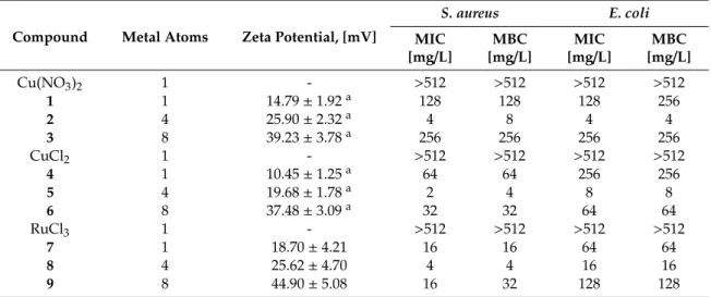
(5) Biomolecules 2019, 9, 405. 5 of 11. PBS 10 mM, pH 7.4 and finally resuspended to a final volume of 2 mL of PBS 10 mM. A 1:50 dilution in PBS 10 mM was used to analyze the hemolytic effect of the metallodendrimers. A 20 µL aliquot of the dendrimers in decreasing final concentrations from 32 to 0.25 mg/L was added to 180 µL of the erythrocytes solution. Then, the samples were incubated at 37 ◦ C for 2 h. Absorbance was measured at 540 nm using BioTek Epoch 2 spectrophotometer. Equation (1): H(%) =. [Abs(dendrimer) − Abs(negative control)] × 70 Abs(positive control). (1). The percentage of hemolysis was calculated using Equation (1). The negative control was PBS 10 mM (20 µL + 180 µL of erythrocyte solution) and the positive control was Triton X-100 1% (20 µL + 180 µL of erythrocyte solution). The latter control is considered to produce 70% hemolysis. The term “Abs(dendrimer)” is calculated by subtracting the absorbance of the compound alone to the absorbance of the compound-treated erythrocytes. 3. Results 3.1. Surface Charge of Metal Complexes and Metallodendrimers Zeta potential measurements provide information about the surface charge of the tested compounds. The metal complexes and metallodendrimers selected for this study exhibited cationic properties, according to the Z-potential measurements (Table 1). As expected, the surface charge on the dendrimers increased from G0 to G2 according to the increase in the number of branches and the number of positive charges in the periphery. Furthermore, nitrate-containing complexes 1–3 presented a higher positive charge than chloride-containing systems 4–6, with values in the range 14.79–39.23 and 10.45–37.48 mV, respectively [40]. This behavior is ascribed to the more labile properties of the nitrate ligands in solution that easily expose the copper charge. Regarding ruthenium complexes 7–9, the higher Z-potential values in the range 18.70–44.90 mV indicated an even higher overall cationic charge in the molecule. Table 1. Bacteriostatic (Minimum Inhibitory Concentration, MIC) and bactericide (Minimum Bactericidal Concentration, MBC) effect of carbosilane metallodendrimers in planktonic cells and comparative values of Z-potential. S. aureus. E. coli. Compound. Metal Atoms. Zeta Potential, [mV]. MIC [mg/L]. MBC [mg/L]. MIC [mg/L]. MBC [mg/L]. Cu(NO3 )2 1 2 3 CuCl2 4 5 6 RuCl3 7 8 9. 1 1 4 8 1 1 4 8 1 1 4 8. 14.79 ± 1.92 a 25.90 ± 2.32 a 39.23 ± 3.78 a 10.45 ± 1.25 a 19.68 ± 1.78 a 37.48 ± 3.09 a 18.70 ± 4.21 25.62 ± 4.70 44.90 ± 5.08. >512 128 4 256 >512 64 2 32 >512 16 4 16. >512 128 8 256 >512 64 4 32 >512 16 4 32. >512 128 4 256 >512 256 8 64 >512 64 16 128. >512 256 4 256 >512 256 8 64 >512 64 16 128. a. previously published results [40].. 3.2. Antibacterial Activity of Carbosilane Metallodendrimers on Planktonic Cells The antibacterial behavior of Cu(II) and Ru(II) metallodendrimers 1–9 was evaluated towards two different families of bacteria: Staphylococcus aureus as a model of Gram-positive bacteria, and Escherichia coli as a model of Gram-negative bacteria. Table 1 summarizes the obtained values for the.
(6) Biomolecules 2019, 9, 405. 6 of 11. Biomolecules 2019, 9, x FOR PEER REVIEW. 6 of 11. Minimum Inhibitory Concentration and thethat Minimum Bactericidal Concentrationare (MBC) for The data shown in Table (MIC) 1 indicates carbosilane metallodendrimers promising eachantibacterial of the synthesized complexes, as well as the metallic salts used as precursors. agents towards Gram-positive and Gram-negative bacteria. The different metal salts The data in Table 1 indicates that carbosilane metallodendrimers are promising antibacterial used as shown control—Cu(NO 3)2, CuCl 2 and RuCl3—showed no antibacterial effect. However, agents towards Gram-positive and Gram-negative bacteria. The different metal salts used as mononuclear complexes 1, 4 and 7 exhibited certain activity, especially higher towards S. aureus. control—Cu(NO ) , CuCl and RuCl —showed no antibacterial effect. However, mononuclear 3 2 2 the antibacterial 3 These results confirm that activity is ascribed to the metal complexation to the ligand. complexes 1, 4 and 7 exhibitedmainly certain activity, especiallysystems higher towards aureus. results confirm The metallodendrimers, first-generation 2, 5 andS. 8, with These 4 metal atoms in their thatperiphery, the antibacterial activity is ascribed to the metal complexation to the ligand. The metallodendrimers, are potent agents towards both types of bacteria, displaying MIC values in the range 2–8 mainly systems 2, 5 and 8, coli. withSecond-generation 4 metal atoms in their periphery, are potent agents mg/Lfirst-generation for S. aureus and 4–16 mg/L for E. counterparts revealed lower activity, towards both types of bacteria, displaying MIC values in the range 2–8 mg/L for S. aureus and 4–16 mg/L despite the increase in the amount of metal atoms in their structure. Overall, we can conclude that in for E. coli. Second-generation counterparts revealed lowerthe activity, increase in>the amount of planktonic cells the antibacterial activity follows trenddespite Ru(II)the complexes Cu(II) chloride metal atoms in their structure. Overall, we canwith conclude that in planktonicdendrimers cells the antibacterial activity complexes > Cu(II) nitrate complexes, the first-generation as the more potent follows the trend Ru(II) complexes > Cu(II) chloride complexes > Cu(II) nitrate complexes, with the members in each family. In fact, the antibacterial effect among first-generation systems is comparable. first-generation dendrimers as the more potent members in each family. In fact, the antibacterial effect among first-generation systems is comparable. 3.3. Antibacterial Activity of Carbosilane Metallodendrimers on Preventing S. aureus Biofilm Formation Considering the of involvement of S. aureus biofilms in numerous human infections, we selected 3.3. Antibacterial Activity Carbosilane Metallodendrimers on Preventing S. aureus Biofilm Formation the potent first-generation copper metallodendrimers 2 and 5 and the ruthenium counterpart 8 and Considering involvement of S.the aureus biofilms human infections, wesummarized selected evaluated theirthe capacity to prevent formation of in S. numerous aureus biofilms. The results are the in potent first-generation copper metallodendrimers 2 and 5 and the ruthenium counterpart 8 and Table 2 and Figure 2. evaluated their capacity to prevent the formation of S. aureus biofilms. The results are summarized in Table 2 and Figure 2. Table 2. Bacteriostatic effect (Minimum Inhibitory Concentration, MIC, and Minimum Biofilm Inhibitory Concentration, MBIC) of first-generation metallodendrimers in preventing S. aureus Table 2. Bacteriostatic biofilm formation.effect (Minimum Inhibitory Concentration, MIC, and Minimum Biofilm Inhibitory Concentration, MBIC) of first-generation metallodendrimers in preventing S. aureus biofilm formation.. S. aureus Biofilms S. aureus Biofilms MIC MBIC MIC [mg/L] [mg/L] MBIC [mg/L] [mg/L] 8 8 4 4 8 8 8 8 32 32 32 32. Metal Atoms CompoundCompound Metal Atoms 2 5 8. 2 5 8. 4 4 4. 4 4 4. B. A. MBC-B= 8 mg/L. C. MBC-B= 16 mg/L. MBC-B= 128 mg/L. Figure 2. Effect of first-generation metallodendrimers in preventing the formation S. aureus Figure 2. Effect of first-generation metallodendrimers in preventing the formation of S. aureus of biofilms. (A)2, Compound 2, withcomplex; Cu(II) nitrate complex; 5,(B) compound 5, with Cu(II) chloride (A) biofilms. Compound with Cu(II) nitrate (B) compound with Cu(II) chloride complex; and complex; and (C)Ru(II) compound 8, with The Ru(II) Cp/PTABactericidal complex. Concentration The Minimum Bactericidal (C) compound 8, with Cp/PTA complex. Minimum for Biofilms (MBC-B) representsfor theBiofilms minimal(MBC-B) concentration of the metallodendrimer that can kill Concentration represents the minimal concentration of the microorganism metallodendrimer in the conditions used to produce a in biofilm. that can kill the microorganism the conditions used to produce a biofilm.. TheThe MICMIC represents the the minimal concentration thatthat inhibits the the growth of the microorganisms represents minimal concentration inhibits growth of the microorganisms while the MBIC indicates the minimal concentration that inhibits the formation of the biofilm while the MBIC indicates the minimal concentration that inhibits the formation of although the biofilm not although the growth the microorganisms. Therefore, MBIC valueMBIC is equal (e.g., compounds 5 and 8) 5 notofthe growth of the microorganisms. Therefore, value is equal (e.g., compounds or lower compound 2) than MIC value. The MBC-B the minimal concentration that and 8)(e.g., or lower (e.g., compound 2) than MIC value. Theindicates MBC-B indicates the minimal concentration that kills the microorganisms and it is equal (e.g., compound 2) or higher (e.g., compounds 5 and 8) than MIC value..
(7) Biomolecules 2019, 9, 405. 7 of 11. Biomolecules 2019, 9, x FOR PEERand REVIEW kills the microorganisms it is equal. 7 of 11 (e.g., compound 2) or higher (e.g., compounds 5 and 8) than MIC value. The resultsindicate indicate that carbosilane metallodendrimers areeffective also effective inhibitors of the The results that carbosilane metallodendrimers are also inhibitors of the formation formation of S. aureus biofilms. Copper metallodendrimer 2, with nitrate ligands, kept the potent of S. aureus biofilms. Copper metallodendrimer 2, with nitrate ligands, kept the potent bacteriostatic and bacteriostatic andobserved bactericide effect observed in planktonic cells (MIC(planktonic) MBIC = 4 mg/L bactericide effect in planktonic cells (MIC(planktonic) = MBIC = 4 mg/L and=MBC(planktonic) and MBC(planktonic) = MBC-B = 8 mg/L). The ligand exchange, from nitrate to chloride, produced a = MBC-B = 8 mg/L). The ligand exchange, from nitrate to chloride, produced a decrease in the decrease in the bacteriostatic and bactericide effect when S. aureus are prompt to form biofilms (MBIC bacteriostatic and bactericide effect when S. aureus are prompt to form biofilms (MBIC = 8 mg/L =and 8 mg/L and= MBC-B = 16 mg/L), and happened the same happened for exchange, the metal from exchange, from copper to MBC-B 16 mg/L), and the same for the metal copper to ruthenium ruthenium (MBIC = 32 mg/L and MBC-B = 128 mg/L). It is not surprising that the value of MIC in the (MBIC = 32 mg/L and MBC-B = 128 mg/L). It is not surprising that the value of MIC in the conditions conditions of biofilm production are higher than in the case of planktonic cells, because of biofilm production are higher than in the case of planktonic cells, because the concentrationthe of concentration of microorganisms is higher in the former case. Overall, the best activity was found microorganisms is higher in the former case. Overall, the best activity was found with compound 2 with compound 2 because MIC are andcoincident MBC values coincident (8 mg/L). In the case5of compounds 5 because MIC and MBC values (8 are mg/L). In the case of compounds and 8, the MBC and 8, the MBC value is higher than the MIC value, twice and four times, respectively, thus increasing value is higher than the MIC value, twice and four times, respectively, thus increasing the potential the potential of the metallodendrimer. toxicity of thetoxicity metallodendrimer.. 3.4. 3.4. Hemolysis Hemolysis Positively-charged toxicity through the the interaction with with cells cells Positively-chargedmolecules moleculesfrequently frequentlyexhibit exhibit toxicity through interaction membrane TheThe interaction withwith erythrocyte membranes produces membrane and andsubsequent subsequentdestruction. destruction. interaction erythrocyte membranes produces the other components, components,and andcan canbe beused usedto tomeasure measurethe thetoxicity toxicityofofa new the release release of hemoglobin, among other adrug. new The drug. The hemolysis is a common a first screening ruletoxicity out any hemolysis assay isassay a common test usedtest as aused first as screening to rule outtoany for the toxicity for use the of therapeutic use of The a compound The by hemolysis by selected therapeutic a compound [41]. hemolysis [41]. produced selected produced metallodendrimers after 2 h metallodendrimers after h incubation is depicted in Figure 3. incubation is depicted in2Figure 3.. Figure 3.3.Erythrocyte Erythrocyte hemolysis induced by selected carbosilane metallodendrimers 2 (copper Figure hemolysis induced by selected carbosilane metallodendrimers 2 (copper nitrate nitrate complex) and 8 (ruthenium Cp/PTA complex) after 2 h incubation. The concentration ranged complex) and 8 (ruthenium Cp/PTA complex) after 2 h incubation. The concentration ranged 0.25-32 0.25-32The mg/L. The Minimum Inhibitory Concentration (MIC) MinimumBiofilm Biofilm Inhibitory Inhibitory mg/L. Minimum Inhibitory Concentration (MIC) andand thethe Minimum Concentration (MBIC) for both compounds are highlighted. Results are mean ± S.E.M (standard error Concentration (MBIC) for both compounds are highlighted. Results are mean ± S.E.M (standard error of the mean). of the mean).. The assay results showed an increase in hemolytic behavior when increasing dendrimer The assay results showed an increase in hemolytic behavior when increasing dendrimer concentration. At all concentrations tested, ruthenium system 8 produces a substantially lower concentration. At all concentrations tested, ruthenium system 8 produces a substantially lower hemolysis, i.e., at the higher concentration tested it exhibited 5% hemolysis compared to the hemolysis, i.e., at the higher concentration tested it exhibited 5% hemolysis compared to the 27% 27% produced by the copper counterpart 2. Chloride-containing copper metallodendrimer 5 has produced by the copper counterpart 2. Chloride-containing copper metallodendrimer 5 has been been reported to exhibit higher hemolytic behavior than the nitrate analogue [40]. In any case, reported to exhibit higher hemolytic behavior than the nitrate analogue [40]. In any case, the the metallodendrimers produce low hemolysis at the MIC value in planktonic cells (4 mg/L) and in metallodendrimers produce low hemolysis at the MIC value in planktonic cells (4 mg/L) and in biofilm-forming cells (4 mg/L for 2, 32 mg/L for 8). biofilm-forming cells (4 mg/L for 2, 32 mg/L for 8). 4. Discussion Schiff-base carbosilane dendrimers are promising carriers of metallodrugs. Our previous studies confirmed that the resultant metallodendrimers, containing Cu(II) and Ru(II) complexes, displayed.
(8) Biomolecules 2019, 9, 405. 8 of 11. 4. Discussion Schiff-base carbosilane dendrimers are promising carriers of metallodrugs. Our previous studies confirmed that the resultant metallodendrimers, containing Cu(II) and Ru(II) complexes, displayed promising antitumoral activity in both in vitro and in vivo assays [37–39]. Furthermore, the different structural parameters—dendrimer generation, metal ion, ligands—influenced the biological activity of the final metallodendrimers. Herein, we discuss the bacteriostatic and bactericide properties of selected carbosilane metallodendrimers and the influence of these structural parameters on the antimicrobial activity (Figure 1). The metal complexes and metallodendrimers selected for this study exhibit cationic properties, according to the Z-potential measurements (Table 1) [40]. Therefore, they can interact with the negatively charged bacterial membranes through electrostatic interactions and disrupt the membrane. This mechanism has been previously described for other cationic dendrimers [33]. The antibacterial effect of carbosilane metallodendrimers was evaluated by measuring the bacteriostatic (MIC) and bactericide (MBC) properties towards Gram-positive and Gram-negative bacteria (Table 1). From this assay, several conclusions were drawn: 1) the antibacterial effect arises from the metal complexation to the ligand, according to the inactivity of the metal salts used as control. Furthermore, previous studies have demonstrated that the Cu(II) and Ru(II) complexes herein reported are completely stable and no metal release has been observed due to the chelating effect of the iminopyridine ligands. Conversely, most antibacterial metallodendrimers reported in the literature rely on the complexation of silver or other metals and the subsequent release of metal ions to disrupt the protein structure [33]. 2) In first-generation dendrimers the dendritic effect becomes evident, with potent antibacterial activity. Surprisingly, mononuclear and second-generation complexes showed similar antibacterial activities and ruled out a direct relationship between antibacterial activity and number of metal atoms. This unique behavior of first-generation carbosilane metallodendrimers has been previously reported [37–39] and differs from most of other types dendritic scaffolds. As an example, polyamide Pt(II) and Pd(II) metallodendrimers display an increasing antibacterial effect when increasing generation, being second-generation dendrimer with 12 active groups the most effective with MIC values of 70 µg/mL (PdG2) and 78 µg/mL (PtG2) against E. coli [42]. Conversely, four active groups in the first-generation carbosilane dendrimer 2 are enough to reach MIC values of 4 mg/L towards E. coli. This is translated into a resources saving—reagents, time, money—when preparing the antibacterial agent. 3) Carbosilane metallodendrimers are potential broad-spectrum antibiotics, with potent activity in both S. aureus (Gram-positive) and E. coli (Gram-negative) bacteria. Other dendrimers, such as poly(amidoamine) (PAMAM) on titanium substrates described by Wang et al. [43] inhibited Gram-negative bacteria and to less extent Gram-positive S. aureus. The authors indicated that the negatively charged lipopolysaccharide in Gram-negative bacteria facilitates dendrimer binding to the membrane and subsequent disruption, while dendrimers barely disrupt the crosslinked peptidoglycan in Gram-positive bacteria. 4) The cationic charge increases with generation, unlike the antibacterial properties. It becomes evident that the antibacterial properties are not directly related to the cationic charge at the dendrimer surface and other mechanisms may be involved related to the presence of the metal ions. Silver and other metal nanoparticles (e.g., ZnO, CuO, SiO2 ) show good antimicrobial effects on drug-resistant strains as well as prevention of biofilm formation and eradication. They have been reported to use antimicrobial mechanisms involving (1) toxic metal ion release, (2) bacterial membrane disruption and (3) reactive oxygen species production [28]. 5) As shown in Table 1, the overall antibacterial activity in planktonic cells follows the trend Ru(II) complexes > Cu(II) chloride complexes > Cu(II) nitrate complexes, with the first-generation dendrimers being the more potent members in each family and exhibiting comparable activity. The potent antibacterial effect of first-generation dendrimers was subsequently tested for preventing the formation of S. aureus biofilms (Table 2, Figure 2). Most human infections produced by S. aureus are due to its biofilm-mode of growth, which is highly resistant to traditional antibiotics. In this case, Cu(II) metallodendrimers still keep the potent activity, with subtle differences between nitrate.
(9) Biomolecules 2019, 9, 405. 9 of 11. and chloride complexes that point to a higher activity in nitrate systems. Indeed, metallodendrimer 2 exhibited the lowest and coincident MIC and MBC values (8 mg/L) for biofilm formation, thus decreasing the potential toxicity of the antibacterial agent. A similar effect towards cancer cells has been previously reported [37]. Using Electron Paramagnetic Resonance (EPR) analysis, we confirmed that the change of the Cu(II) counter-ion—from nitrate to chloride—produced an increased relative amount and strength of interaction of the dendrimer with model membranes. Interestingly, the stabilization effect observed in chloride dendrimers produced a lower toxicity towards cancer cells. Furthermore, in water solution, the NO3 − groups are more labile than the Cl− ligands and are easily released, increasing the overall positive charge in the metallodendrimer. The higher cationic charge and the lower membrane stabilization may explain the more potent biocide activity of nitrate metallodendrimer 2 in the present experiments. The ruthenium counterpart 8 showed a potent activity in planktonic cells, lower for biofilm mode-of-growth planktonic cells. The significant morphological and physiological differences between planktonic cells in “normal” and biofilm-forming modes of growth could be responsible of the different behavior of our metallodendrimers. Finally, and considering the cationic nature of the tested metallodendrimers, we evaluated the hemolysis of first-generation derivatives (Figure 3). Carbosilane metallodendrimers did not produce hemolysis at the MIC concentrations and can be safely used as antibacterial agents. The hemolytic trend was 8 < 2 < 5, confirming the influence of both the metal ion and the ligands on the interaction with erythrocytes membrane. Despite the similar Z-potential between copper complex 2 and ruthenium counterpart 5, especially low hemolysis was observed for the Ru(II) derivative which can be ascribed to macromolecules aggregation that decrease the number of available charges. These results had been also observed in a longer exposure (24 h) using erythrocytes from healthy human donors, obtaining around 5% hemolysis at these concentrations [40]. 5. Conclusions Nanotechnology opens new avenues in the treatment of resistant bacteria infections. In particular, the unique properties of dendrimers—monodispersity and multivalency—enable the accurate design of effective treatments by establishing an exact structure-to-activity relationship. A wise selection of dendritic scaffold, generation, metal complex and ligand can lead to potent broad-spectrum antibiotics that can overcome the current limitations of traditional therapies. For example, herein we found that metallodendrimer 2 is the most promising system among those tested. It is stable, water-soluble and exhibits potent bacteriostatic and bactericide effect in planktonic S. aureus and E. coli. Furthermore, it prevents the formation of S. aureus biofilms at a low concentration. Importantly, at the working concentrations, it is not hemotoxic. Further studies to gain insight into the mechanism of action and the in vivo activity are currently under way. Author Contributions: Methodology, S.G.-G. and P.O.; investigation, N.S.d.O. and C.L.; resources, N.S.d.O.; writing—original draft preparation, S.G.-G. and J.L.C.-P.; writing—review and editing, F.J.d.l.M.; visualization, F.J.d.l.M. and J.L.C.-P.; supervision, R.G. and J.S.; project administration, S.G.-G.; funding acquisition, F.J.d.l.M. and J.L.C.-P. Funding: This research was funded by grants from CTQ2017–86224–P (MINECO), Consortiums IMMUNOTHERCAN-CM B2017/BMD–3733 and NANODENDMED–II B2017/BMD–3703 (CAM), project SBPLY/17/180501/000358 JCCM and the CAM Research Talent Attraction Program 2017–T2/IND–5243. CIBER-BBN is an initiative funded by the VI National R&D&I Plan 2008–2011, Iniciativa Ingenio 2010, Consolider Program, CIBER Actions and financed by the Instituto de Salud Carlos III with assistance from the European Regional Development Fund. N. S. O. wishes to thank JCCM for a predoctoral fellowship. Conflicts of Interest: The authors declare no conflict of interest..
(10) Biomolecules 2019, 9, 405. 10 of 11. References 1. 2. 3.. 4.. 5. 6. 7. 8. 9. 10.. 11. 12. 13. 14. 15. 16. 17. 18. 19.. 20. 21. 22. 23. 24.. Kloss, W.E.; Schleir, K.H.; Goirtz, F. The genus Staphylococcus. In The Prokaryotes, 2nd ed.; Balows, A., Truper, H.G., Dwoekin, M., Eds.; Springer: New York, NY, USA, 1992. Vivoni, A.M.; Moreira, B.M. Application of molecular techniques in the study of Staphylococcus aureus clonal evolution—A review. Mem. Inst. Oswaldo Cruz. 2005, 100, 693–698. [CrossRef] [PubMed] Compernolle, V.; Verschraegen, G.; Claeys, G. Combined use of Pastorex Staph-Plus and either of two new chromogenic agars, MRSA ID and CHROMagar MRSA, for detection of methicillin-resistant Staphylococcus aureus. J. Clin. Microbiol. 2007, 45, 154–158. [CrossRef] [PubMed] Kuroda, M.; Ohta, T.; Uchiyama, I.; Baba, T.; Yuzawa, H.; Kobayashi, I.; Cui, L.; Oguchi, A.; Aoki, K.; Nagai, Y.; et al. Whole genome sequencing of meticillin-resistant Staphylococcus aureus. Lancet 2001, 357, 1225–1240. [CrossRef] Kloss, W.E.; Bamerman, T.L. Staphylococcus and Micrococcus. In Manual of Clinical Microbiology, 6th ed.; Murra, P.R., Baron, E.J., Pfaller, M.A., Eds.; ASM Press: Washington DC, USA, 1995. Lowy, F.D. Staphylococcus aureus infections. N. Engl. J. Med. 1998, 339, 520–532. [CrossRef] [PubMed] Moreillon, P.; Que, Y.; Glauser, M. Staphylococcus aureus. In Principles and Practice of Infectious Diseases, 6th ed.; Mandell, G.L., Bennett, J.E., Olin, R., Eds.; Churchill Livingston: Philadelphia, PA, USA, 2005. Kluytmans, J.; van Belkum, A.; Verbrugh, H. Nasal carriage of Staphylococcus aureus: Epidemiology, underlying mechanisms, and associated risks. Clin. Microbiol. Rev. 1997, 10, 505–520. [CrossRef] [PubMed] Dall’Antonia, M.; Coen, P.G.; Wilks, M.; Whiley, A.; Millar, M. Competition between methicillin-sensitive and resistant Staphylococcus aureus in the anterior nares. J. Hosp. Infect. 2005, 61, 62–67. [CrossRef] [PubMed] Verhoeven, P.O.; Gagnaire, J.; Botelho-Nevers, E.; Grattard, F.; Carricajo, A.; Lucht, F.; Pozzetto, B.; Berthelot, P. Detection and clinical relevance of Staphylococcus aureus nasal carriage: An update. Expert Rev. Anti-infect. Ther. 2014, 12, 75–89. [CrossRef] [PubMed] Lowy, F.D. Antimicrobial resistance: The example of Staphylococcus aureus. J. Clin. Investig. 2003, 111, 1265–1273. [CrossRef] [PubMed] Hiramatsu, K.; Cui, L.; Kuroda, M.; Ito, T. The emergence and evolution of methicillin-resistant Staphylococcus aureus. Trends Microbiol. 2001, 9, 486–493. [CrossRef] Crossley, K.B.; Jefferson, K.K.; Archer, G.; Fouler, V.G. Staphylococci in Human Disease, 2nd ed.; John Wiley & Sons: Chichester, UK, 2009. Lew, D.P.; Waldvogel, F.A. Osteomyelitis. Lancet, 2004, 364, 369–379. [CrossRef] Anwar, S.; Prince, L.R.; Foster, S.J.; Whyte, M.K.; Sabroe, I. The rise and rise of Staphylococcus aureus: Laughing in the face of granulocytes. Clin. Exp. Immunol. 2009, 157, 216–224. [CrossRef] [PubMed] Akiyama, H.; Torigoe, R.; Arata, J. Interaction of Staphylococcus aureus cells and silk threads in vitro and in mouse skin. J. Dermatol. Sci. 1993, 6, 247–257. [CrossRef] Costerton, J.W.; Montanaro, L.; Arciola, C.R. Biofilm in implant infections: Its production and regulation. Int. J. Artif. Organs 2005, 28, 1062–1068. [CrossRef] [PubMed] Heitz-Mayfield, L.J.; Lang, N.P. Comparative biology of chronic and aggressive periodontitis vs. peri-implantitis. Periodontology 2000, 53, 167–181. [CrossRef] [PubMed] Tong, S.Y.C.; Davis, J.S.; Eichenberger, E.; Holland, T.L.; Fowler, V.G. Staphylococcus aureus infections: Epidemiology, pathophysiology, clinical manifestations, and management. Clin. Microbiol. Rev. 2015, 28, 603–661. [CrossRef] [PubMed] Dastgheyb, S.; Parvizi, J.; Shapiro, I.M.; Hickok, N.J.; Otto, M. Effect of biofilms on recalcitrance of staphylococcal joint infection to antibiotic treatment. J. Infect. Dis. 2015, 211, 641–650. [CrossRef] [PubMed] Winn, W.; Allen, S.; Janda, W.; Koneman, E.; Procop, G. Schreckenberger, P. and Woods, G. Koneman’s Color Atlas and Textbook of Diagnostic Microbiology, 6th ed.; Lippincott Williams & Wilkins: Philadelphia, PA, USA, 2005. O’Toole, G.; Kaplan, H.B.; Kolter, R. Biofilm formation as microbial development. Annu. Rev. Microbiol. 2000, 54, 49–79. [CrossRef] [PubMed] Denyer, S.P.; Gorman, S.P.; Sussman, M. Microbial Biofilms: Formation and Control; Wiley-Blackwell Scientific Publications LTD: London, UK, 1993. Proctor, R.A.; Balwit, J.M.; Vesga, O. Variant subpopulations of Staphylococcus aureus as cause of persistent and recurrent infections. Infect. Agents Dis. 1994, 3, 302–312..
(11) Biomolecules 2019, 9, 405. 25. 26. 27. 28.. 29.. 30. 31. 32. 33. 34. 35. 36.. 37.. 38.. 39.. 40.. 41.. 42. 43.. 11 of 11. Kraus, D.; Peschel, A. Staphylococcus aureus evasion of innate antimicrobial defense. Future Microbiol. 2008, 3, 437–451. [CrossRef] [PubMed] Stryjewski, M.E.; Corey, G.R. Methicillin-resistant Staphylococcus aureus: An evolving pathogen. Clin. Infect. Dis. 2014, 58, S10–S19. [CrossRef] [PubMed] Heymann, D.L. , Prentice, T. and Reinders, L.T. The World Health Report 2007: A Safer Future: Global Public Health Security in The 21st Century; WHO: Geneva, Switzerland, 2007. Liu, Y.; Shi, L.; Su, L.; van der Mei, H.C.; Jutte, P.C.; Ren, Y.; Busscher, H.J. Nanotechnology-based antimicrobials and delivery systems for biofilm-infection control. Chem. Soc. Rev. 2019, 48, 428–446. [CrossRef] [PubMed] Nguyen, T.-K.; Lam, S.J.; Ho, K.K.K.; Kumar, N.; Qiao, G.G.; Egan, S.; Boyer, C.; Wong, E.H.H. Rational design of single-chain polymeric nanoparticles that kill planktonic and biofilm bacteria. ACS Infect. Dis. 2017, 3, 237–248. [CrossRef] [PubMed] Taylor, E.N.; Webster, T.J. The use of superparamagnetic nanoparticles for prosthetic biofilm prevention. Int. J. Nanomed. 2009, 4, 145–152. Chung, P.Y.; Toh, Y.S. Anti-biofilm agents: Recent breakthrough against multi-drug resistant Staphylococcus aureus. Pathog. Dis. 2014, 70, 231–239. [CrossRef] [PubMed] Tomalia, D.A.; Christensen, J.B.; Boas, U. Dendrimers, Dendrons and Dendritic Polymers: Discovery, Applications and the Future; Cambridge University Press: Cambridge, UK, 2012. Zarena, A.S.; Shubha, G. Dendrimer a new dimension in targeting biofilms. Mini-Rev. Med. Chem. 2013, 13, 1448–1461. [CrossRef] [PubMed] Mintzer, M.A.; Dane, E.L.; O’Toole, G.A.; Grinstaff, M.W. Exploiting dendrimer multivalency to combat emerging and re-emerging infectious diseases. Mol. Pharm. 2012, 9, 342–354. [CrossRef] [PubMed] Balogh, L.; Swanson, D.R.; Tomalia, D.A.; Hagnauer, G.L.; McManus, A.T. Dendrimer−silver complexes and nanocomposites as antimicrobial agents. Nano Lett. 2001, 1, 18–21. [CrossRef] Grabchev, I.; Vasileva-Tonkova, E.; Staneva, D.; Bosch, P.; Kukeva, R.; Stoyanova, R. Impact of Cu(II) and Zn(II) ions on the functional properties of new PAMAM metallodendrimers. New J. Chem. 2018, 42, 7853–7862. [CrossRef] Sanz del Olmo, N.; Carloni, R.; Bajo, A.M.; Ortega, P.; Fattori, A.; Gómez, R.; Ottaviani, M.F.; García-Gallego, S.; Cangiotti, M.; de la Mata, F.J. Insight into the antitumor activity of carbosilane Cu(II)–metallodendrimers through their interaction with biological membrane models. Nanoscale 2019, 11, 13330–13342. [CrossRef] Sanz del Olmo, N.; Maroto-Díaz, M.; Gómez, R.; Ortega, P.; Cangiotti, M.; Ottaviani, M.F.; de la Mata, F.J. Carbosilane metallodendrimers based on copper (II) complexes: Synthesis, EPR characterization and anticancer activity. J. Inorg. Biochem. 2017, 177, 211–218. [CrossRef] De la Mata, F.J.; Gómez, R.; Ortega, P.; García-Gallego, S.; Sanz del Olmo, N.; Triguero Rubio, A.; Maroto-Díaz, M. Metalodendrímeros de naturaleza carbosilano conteniendo rutenio y cobre coordinados a ligandos base de Schiff, su preparación y sus usos. PCT/ES2019/070416, 14 June 2019. Holota, M.; Magiera, J.; Michlewska, S.; Kubczak, M.; Del Olmo, N.S.; García-Gallego, S.; Ortega, P.; de la Mata, F.J.; Ionov, M.; Bryszewska, M. In vitro anticancer properties of copper metallodendrimers. Biomolecules 2019, 9, 155. [CrossRef] [PubMed] Tramer, F.; Da Ros, T.; Passamonti, S. Screening of fullerene toxicity by hemolysis assay. In Nanotoxicity Methods in Molecular Biology (Methods and Protocols); Reineke, J., Ed.; Humana Press: Totowa, NJ, USA, 2012; Volume 926. Ahamad, T.; Mapolie, S.F.; Alshehri, S.M. Synthesis and characterization of polyamide metallodendrimers and their anti-bacterial and anti-tumor activities. Med. Chem. Res. 2012, 21, 2023–2031. [CrossRef] Wang, L.; Erasquin, U.J.; Zhao, M.; Ren, L.; Zhang, M.Y.; Cheng, G.J.; Wang, Y.; Cai, C. Stability, antimicrobial activity, and cytotoxicity of poly(amidoamine) dendrimers on titanium substrates. ACS Appl. Mater. Interfaces 2011, 3, 2885–2894. [CrossRef] [PubMed] © 2019 by the authors. Licensee MDPI, Basel, Switzerland. This article is an open access article distributed under the terms and conditions of the Creative Commons Attribution (CC BY) license (http://creativecommons.org/licenses/by/4.0/)..
(12)
Figure


Documento similar
To make a precise evaluation of the influence of incorporating nanoparticles into the structure of a dental restorative material on their antibacterial capacity, three
Scarcity: The resources available Albani, are not very difficult to achieve, since with regard to human resources, with a good selection and training of its employees
In this paper, we observed that 7-O-glycosyl moieties did not enhance the inhibitory activity of the aglycone NGE.. against seven Gram-positive and three Gram-negative
The performance and durability of PEM fuel cells significantly depend on variations in the concentrations of hydrogen and oxygen in the gas channels, water activity in the
Considering the recognized antibacterial activity of Zn 2+ and the potential of MOFs in the progressive release of metals, the potential antimicrobial properties of these
Since melatonin inhibits the growth of C6 cells, and this indoleamine has been demon- strated to be capable of decreasing aromatase expression and activity in these cells, the aim
Many experiments have evaluated the effect of different levels of DLI on growth, development, and plant quality on a wide range of crops [98,99] and the effect on consumption
After observation of the central role of ERK and AKT phosphorylation in the cytotoxic effect of HT, HT-Ac and HT-Et treatment in 22Rv1 cells, the effect in AKT and ERK activation
