Safety of the oral methionine load test:
effects on the clinical performance and laboratory tests
Abraham Majluf-Cruz,* Manuel Moreno-Hernández,*
José Antonio Alvarado-Moreno,* Irma Isordia-Salas,* Rodolfo Guardado-Mendoza,**
Karim Majluf-Cruz,* Erika Coria-Ramírez,* Jesús Hernández-Juárez*
*Unidad de Investigación Médica en Trombosis, Hemostasia y Aterogénesis, Instituto Mexicano del Seguro Social. **Facultad de Medicina, Universidad de Guanajuato.
ARTÍCULO ORIGINAL
ABSTRACT
Introduction. Hyperhomocysteinemia is a prothrombotic risk factor. Homocysteine is evaluated during fasting and after an oral methionine load (OML). Aim. To determine the safety of the OML test according to the general performance status and clinical laboratory tests. We studied healthy nonsmoking volunteers and patients with several thrombotic conditions. Before and after receiving an OML, blood samples were obtained to perform several laboratory tests. We also evaluated acute and subacute adverse effects and 30-day associated morbidity and mortality. Of 353 individuals, three were eliminated because they did not tolerate the OML. We studied 175 healthy individuals and 175 patients without age differences. After OML, mild to moderate clinical abnormalities were recorded in 78 subjects (22.1%): nausea (n = 69; 88.5%), dizziness (n = 13; 16.7%) and decreased or increased blood pressure (n = 8; 10.2%). Nausea always disappeared after breakfast in affected individuals. Prevalence of complications was similar in patients and controls. No patient required hospitalization and there was no mortality during the 30-day study period. In conclusion, OML test had no significant undesirable effects on the clinical status or the general laboratory tests of patients and healthy controls. Some mild and moderate symptoms associated with OML tests were observed, and OML test did not negatively affect general laboratory tests. OML test is a safe diagnostic procedure in patients with previous thrombotic events (and with the consequent associated risk factors such as diabetes mellitus or dyslipidemia) and in healthy subjects.
Key words. Hyperhomocysteinemia. Methionine load. Laboratory tests. Thrombosis. Post-oral methionine load test.
Seguridad de la prueba de
carga oral de metionina: efectos sobre el estado clínico y pruebas de laboratiorio
RESUMEN
Introducción. La hiperhomocisteinemia es un factor de ries-go trombótico. La homocisteína se evalúa en ayuno y lueries-go de una carga oral de metionina (COM). Objetivo. Establecer la seguridad de la prueba de COM en términos del estado clíni-co general y las pruebas generales de laboratorio. Material y métodos. Se estudiaron adultos de ambos sexos, sanos y no fumadores, y pacientes con diversas condiciones trombóticas. Antes y después de recibir la COM se obtuvieron muestras de sangre para realizar las pruebas de laboratorio. También se evaluaron los efectos adversos agudos y sub-agudos y la mor-bilidad y mortalidad asociadas a los 30 días. De 353 indivi-duos, tres fueron eliminados ya que no toleraron la COM. Se estudiaron 175 individuos sanos y 175 pacientes sin diferen-cias significativas en términos de edad. Luego de la COM se encontraron algunas alteraciones clínicas leves y moderadas en 78 individuos (22.1%): náusea (n = 69; 88.5%), mareo (n = 13; 16.7%) y aumento o disminución de la presión arterial (n = 8; 10.2%). La náusea siempre desapareció luego del desa-yuno. La prevalencia de las complicaciones fue similar en los pacientes y en los controles. Ningún paciente requirió hospita-lización y la mortalidad en los 30 días del periodo de estudio fue cero. Conclusión. La COM no induce efectos indeseables significativos sobre el estado clínico o las pruebas de labora-torio en pacientes y en voluntarios sanos. La COM se asocia con algunos efectos clínicos leves a moderados, pero no afecta ninguna prueba de laboratorio significativamente. Por lo tan-to, la COM es una prueba diagnóstica segura en pacientes con eventos trombóticos previos que son portadores de factores de riesgo asociados como diabetes mellitus o dislipidemia, así como en controles sanos.
It is likely that an excess of methionine administe-red to an individual and the subsequent increase in Hcy plasma levels may acutely affect individual clini-cal status, overall metabolism or endothelial func-tion. Therefore, our objective was to determine the safety of the OML test in terms of the general perfor-mance status but more importantly in regard to the general clinical laboratory tests because this analysis has not been previously reported in the literature.
MATERIAL AND METHODS
General characteristics of the study
This was a prospective, longitudinal, nonran-domly assigned study of adult Mexicans of both gen-ders who required an OML test. Between January 2010 and January 2012 we included two groups of subjects: healthy nonsmoking volunteers with no history of thrombotic diseases or with several condi-tions predisposing to thrombosis. Volunteers were blood donors, patient caregivers at our hospital, and health services personnel working at our hospital. The second group was comprised of patients with se-veral thrombotic conditions who required the quan-tification of Hcy. All participants were instructed about the nature of the study and received precise instructions in regard to the OML test. We followed one of the most commonly accepted protocols.19 Briefly, we take a blood sample in the morning in or-der to obtain a basal sample to measure fasting Hcy. Subjects must carry out their normal daily activities and, after dinner, were asked to take me-thionine (100 mg/kg of body weight) (L-methinone, Sigma Aldrich, St. Louis, MO, USA), which was diluted in 250-500 mL orange juice. Patients were asked to drink the methionine during a 20 min period and 8 h later a blood sample was obtained. Patients were instructed to continue with any required medications.
Blood sample collection
Before and after the OML test we obtained 3 mL of blood from each patient in vacuum plastic tubes added with sodium citrate 0.109 M (9:1, vol:vol) (Na Citrate, BD Vacutainer, Franklin Lakes, NJ, USA); 5 mL was drawn in a vacuum glass tube with EDTA (K3 EDTA, BD Vacutainer) and 10 mL in two glass tubes without anticoagulant (SST, BD Vacutainer). All samples were centrifuged at 2,000 g for 15 min in order to obtain platelet poor plasma and serum.
INTRODUCTION
Hyperhomocysteinemia (HHC) is a prothrombotic risk factor that has gained importance during recent years. It has been demonstrated that HHC is an in-dependent risk factor for venous and arterial throm-boembolic diseases.1,2 Depending on the plasma levels of homocysteine (Hcy), HHC may be classified as mild (16-30 µmol/L), moderate (31-100 µmol/L), and severe (> 100 µmol/L).3,4
Hcy can be quantified in serum and plasma using several methods including high-performance liquid chromatography (HPLC), gas chromatography with mass spectrophotometry, and amino acid analysis, among others. Hcy may be evaluated in fasting state and after administering the patient an oral methio-nine load (OML) using a single dose of 100 mg/kg of methionine, namely, the OML test, a widely used diagnostic tool to detect abnormal metabolism of Hcy in patients with various conditions. An oral load with L-methionine was first reported in homo-cysteinuric patients5 and was subsequently adapted as a test to identify heterozygous deficiency of the enzyme cystathionine b synthase (CBS).6-10 Howe-ver, it may also detect the homozygous state for the C677T mutation of the methylenetetrahydrofolate reductase (MTHFR) enzyme, which induces a selec-tive defect in the remethylation pathway of the Hcy metabolism.11 When Hcy metabolism is normal, fol-lowing the OML test the level of this amino acid in-creases an average 20 µmol/L higher than basal levels but returns to normal levels during the next few hours. Hcy is quantified 2 to 8 h after OML and is considered abnormal when its concentration is > 2 standard deviations above basal fasting Hcy level.10-14 The OML test also identifies patients with HHC who have normal fasting Hcy levels, a fact that allows the identification of > 50% of patients with HHC. Therefore, the OML test is considered the most sensitive test for diagnosing moderate HHC.15
Acute and subacute adverse clinical effects and 30-day morbidity and mortality
associated with the OML test
To ascertain any possible subjective complications, patients were asked how they felt prior to the test and 30 min and 8 h after the test began. The inter-val could be proportionally shortened for subjects reporting any problems. Blood pressure and heart rate were measured and recorded before the OML test, 30 min after the test, and 8 h after the OML test. In case of any complaints, the partici-pants were evaluated more frequently. To determine whether the OML test could result in a possible subacute or lethal complication, we gathered infor-mation from direct interviews and from the clinical chart of the patients during the 30 day period after performance of the OML test. Our objective was to determine any clinical worsening of pre-existing conditions or novel occurrence of abnormal clinical data. Mortality within 30 days after the OML test was analyzed in all participants in this study using interviews and searching in the death registry of our hospital.
Laboratory tests
Blood cell count was performed using Cell-Dyn 3700 equipment (Abbott Park, IL, USA). Erythrocyte sedimentation rate and blood group analysis were performed according to worldwide accepted techni-ques. Glucose, urea, creatinine, triglycerides, total cholesterol, HDL-cholesterol (HDL-c), alkaline phosphatase, lactate dehydrogenase, alanine transa-minase, aspartate transatransa-minase, total bilirubin, di-rect bilirubin, total serum protein, albumin, globulins, and gamma-glutamyl transferase levels were assayed using Synchron LX20 equipment (Beckman Coulter, Fullerton, CA, USA); high-sensi-tivity C-reactive protein (hsCRP) was evaluated with a AXSYM System analyzer (Abbott Park, IL, USA). Prothrombin time (PT), activated partial thromboplastin time (APTT), and fibrinogen level (Clauss technique) were evaluated using commercial-ly available tests (Neoplastin Plus, PTT Reagent, STA Thrombin, Fibrinogen Reagent, Stago, Asnie-res, France) in STA Compaq equipment (Stago).
Ethics
Each patient received information in regard to the study characteristics. Informed written consent was obtained from all subjects before study
enroll-ment. The study protocol conformed to the ethical guidelines of the 1975 Declaration of Helsinki and was accepted by the Local Ethics Committee and the Investigation Research Board of our hospital.
Statistical analysis
For analysis of the general patient characteristics and clinical abnormalities, we used descriptive sta-tistics. Continuous data are expressed as means and ranges, whereas categorical data are presented as percentages. The significance of the differences bet-ween continuous variables was determined by paired
t test. Results of laboratory tests are expressed as medians and percentiles 2.5-97.5. To determine the presence of significant differences between basal and after OML test laboratory values, paired t test or Wilcoxon test was used according to the distribution of the results. To analyze the results of the PT and APTT tests, we used χ2 test. For these two tests, normal values were obtained from pooled plasmas obtained from healthy individuals; p value < 0.05 was considered significant. All statistical analyses were performed using SPSS (Statistical Package for the Social Sciences) software (version 16; SPSS Inc., Chicago, IL, USA).
RESULTS
Three hundred and fifty-three individuals were originally enrolled in the study and all of them were analyzed in terms of clinical performance changes: 176 healthy individuals (72 females and 104 males) and 177 patients (92 females and 85 males) with a mean age of 43.0 years (range: 18-93 years) and 46.5 years (range: 18-91 years), respectively. When analyzing both groups separately and as a single group, there were no significant differences between females and males in terms of age.
Clinical complications of the OML test
It is worth noting that nausea and vomiting always disappeared after breakfast in all affected indivi-duals. There was no correlation between vomiting and nausea with dizziness and abnormalities in blood pressure according to age or gender. Symptoms and/or signs were isolated in 65 indivi-duals, whereas in 16 persons a combination of symptoms/signs was present and all experienced nausea. The prevalence of complications was not significantly different between females and males: 42 (51.8%) vs. 39 (48.1%) (p = 0.07), respectively. The prevalence of complications was similar in patients and controls: 41 (50.6%) vs. 40 (49.4%) (p = 0.52), respectively. The OML test did not lead to signifi-cant changes in systolic or diastolic blood pressure in both groups. No cases were reported of serious worsening of the overall clinical condition requiring hospitalization.
In terms of subacute adverse effects and mortality within the 30-day period after the OML test, none of
the subjects experienced clinical worsening of the pre-existing disease or novel clinical abnormalities. Finally, there was no mortality during the 30-day study period.
Effects on laboratory tests
As previously mentioned three patients experien-ced intense vomiting and were excluded from the analysis of laboratory changes. Therefore, the analysis about changes in the laboratory tests was performed in only 175 healthy individuals (71 fema-les and 104 mafema-les) and 175 patients (91 femafema-les and 84 males) with a mean age of 42.5 years (range: 18-93 years) and 46.5 years (range: 18-91 years), res-pectively. Results of all metabolic tests performed before and after the OML test are shown in table 1. Except for basal total cholesterol levels, all results obtained before and after the OML test had an ab-normal distribution. Descriptive analysis showed
l 1 l 1 Table 1.
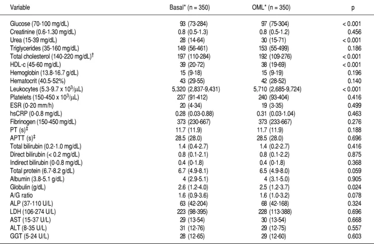
Table 1. Changes in general laboratory tests before and after OML test.
Variable Basal* (n = 350) OML* (n = 350) p
Glucose (70-100 mg/dL) 93 (73-284) 97 (75-304) < 0.001 Creatinine (0.6-1.30 mg/dL) 0.8 (0.5-1.3) 0.8 (0.5-1.2) 0.456 Urea (15-39 mg/dL) 28 (14-64) 30 (15-71) < 0.001 Triglycerides (35-160 mg/dL) 149 (56-461) 153 (55-499) 0.186 Total cholesterol (140-220 mg/dL)† 197 (110-284) 192 (109-276) < 0.001 HDL-c (45-60 mg/dL) 39 (20-72) 38 (19-69) < 0.001 Hemoglobin (13.8-16.7 g/dL) 15 (9-18) 15 (9-19) 0.196 Hematocrit (40.5-52%) 43 (29-55) 42 (28-52) 0.140 Leukocytes (5.3-9.7 x 10³/μL) 5,320 (2,837-9,431) 5,710 (2,685-9,724) < 0.001 Platelets (150-450 x 103/μL) 237 (91-412) 240 (93-404) 0.416 ESR (0-20 mm/h) 20 (4-34) 19 (3-35) 0.499 hsCRP (0-0.8 mg/dL) 0.28 (0.03-0.88) 0.31 (0.03-1.04) 0.463 Fibrinogen (150-450 mg/dL) 373 (230-667) 373 (233-667) 0.276 PT (s)‡ 11.7 (11.9) 11.7 (11.9) 0.188 APTT (s)‡ 28.5 (28.0) 28.5 (28.0) 0.696 Total bilirubin (0.2-1.0 mg/dL) 1.4 (0.4-2.7) 1.4 (0.2-2.7) 0.416 Direct bilirubin (< 0.2 mg/dL) 0.8 (0.1-2.1) 0.8 (0.1-2.2) 0.875 Indirect bilirubin (0-0.8 mg/dL) 0.4 (0-1.8) 0.4 (0-1.8) 0.368 Total protein (6.7-8.2 g/dL) 6.7 (4.9-8.1) 6.5 (4.9-8.0) 0.059 Albumin (3.8-5.1 g/dL) 4 (2.9-5.1) 4 (3.1-5.0) 0.905 Globulin (g/dL) 2.6 (1.2-4.0) 2.5 (1.2-3.7) 0.024 A/G ratio 1.6 (0.9-3.6) 1.6 (1.0-3.2) 0.078 ALP (37-110 U/L) 63 (42-204) 68 (42-168) 0.324 LDH (106-274 U/L) 223 (98-395) 228 (113-388) 0.696 AST (15-37 U/L) 29 (13-54) 30 (13-54) 0.668 ALT (8-35 U/L) 31 (12-76) 29 (12-75) 0.557 GGT (5-24 U/L) 28 (12-65) 29 (12-60) 0.603
that means for four basal variables were not bet-ween the reference values: gamma-glutamyl transfe-rase, HDL-c, total bilirubin, and direct bilirubin. We found that six variables were significantly modified after the OML test as compared with basal levels. Glucose and urea levels as well as leukocyte count showed higher levels than the corresponding basal levels. Moreover, levels of total cholesterol, HDL-c, and serum globulins decreased after the OML (Table 1). Except for HDL-c, mean values observed for all the variables analyzed were always between the nor-mal reference ranges.
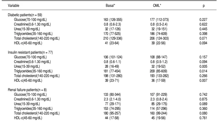
Because the study population included several cli-nical disorders, laboratory abnormalities could be completely different and may have different degrees of impact on the patients. Therefore, we performed a specific analysis considering only diabetic, insulin resistant, and chronic renal failure patients (Table 2). Except for basal total cholesterol levels, all results obtained before and after the OML test had an abnormal distribution. Descriptive analysis showed that there were not significant changes in all varia-bles analyzed in diabetic and renal patients. In insu-lin resistant patients, three variables significantly changed as compared with basal levels: urea, trigly-cerides, and HDL-c. Except for triglytrigly-cerides, mean
values observed for all the variables analyzed were always between the normal reference ranges.
DISCUSSION
Thrombotic disease represents the most frequent cause of morbidity and mortality worldwide. During the last decades, advances in regard to the traditio-nal atherothrombotic risk factors (diabetes, high blood pressure, dyslipidemia, obesity, and smoking) have grown exponentially. However, we are aware that these risk factors cannot explain all cases with atherothrombotic diseases because in up to 25% of patients with premature vascular disease there is no well-established risk factor.20 In 1996, the 27th Be-thesda Conference described the so-called new car-diovascular risk factors including HHC.20,21 It is currently well known that HHC is a risk factor as-sociated with venous thromboembolic disease and atherothombosis. As a consequence, a body of infor-mation has been published during recent years about the importance of HHC, mainly in human thrombotic disease.
Following reports on abnormal Hcy metabolism in patients with atherosclerosis.22 the OML test was used to determine heterozygosity for cystathionine
.
Table 2. Changes in laboratory tests before and after OML test in patients with diabetes, insulin resistance, and chronic renal failure.
Variable Basal* OML* p
Diabetic patients(n = 59)
Glucose(70-100 mg/dL) 163 (126-355) 177 (112-373) 0.227 Creatinine(0.6-1.30 mg/dL) 0.8 (0.6-2.3) 0.8 (0.5-2.4) 0.622 Urea(15-39 mg/dL) 32 (17-126) 32 (19-151) 0.445 Triglycerides(35-160 mg/dL) 170 (77-525) 186 (74-609) 0.398 Total cholesterol(140-220 mg/dL) 210 (129-336) 206 (124-303) 0.071 HDL-c(45-60 mg/dL) 41 (23-64) 39 (22-56) 0.094
Insulin resistant patients(n = 77)
Glucose(70-100 mg/dL) 106 (101-124) 108 (88-147) 0.157 Creatinine(0.6-1.30 mg/dL) 0.8 (0.6-1.1) 0.8 (0.5-1.2) 0.094 Urea(15-39 mg/dL) 28 (16-49) 32 (19-52) 0.005 Triglycerides(35-160 mg/dL) 181 (77-454) 209 (85-609) 0.014 Total cholesterol(140-220 mg/dL) 198 (131-280) 193 (133-282) 0.266 HDL-c(45-60 mg/dL) 39 (23-71) 36 (17-59) 0.007
Renal failure patients(n = 8)
Glucose(70-100 mg/dL) 133 (80-344) 107 (91-229) 0.742 Creatinine(0.6-1.30 mg/dL) 2.3 (2.1-4.0) 2.3 (0.8-2.4) 0.875 Urea(15-39 mg/dL) 77 (29-171) 85 (29-175) 0.089 Triglycerides(35-160 mg/dL) 153 (74-295) 114 (57-296) 0.360 Total cholesterol(140-220 mg/dL) 180 (95-257) 160 (99-244) 0.080 HDL-c(45-60 mg/dL) 44 (17-58) 45 (19-56) 0.761
beta-synthase (CBS) deficiency in patients with pre-mature atherosclerosis.7,23,24 Subsequently, this test was accepted as a regular procedure in patients with venous and arterial thrombosis, especially at a young age because elevated HHC was found in 60% of patients in fasting plasma samples, whereas in the remaining 40% of patients HHC was apparent only in the post-load specimens.12 It is currently sugges-ted that the OML test should be performed in per-sons at high risk of atherosclerosis or venous thrombotic disease or with a history of thrombosis and normal fasting Hcy concentration.25
Methionine, a normal component of alimentary pro-teins, is a harmless compound. The dose of methionine used in the OML test is almost 2.5-4 times the amount of a regular Western diet containing 1.6-2.8 g/day.26 Although higher doses of methinone (70-300 mg/kg) have been safely used in patients with several condi-tions requiring an OML test, incidence of adverse events increases with these high doses.27-29 Therefore, an appropriate methionine dose should always be indi-cated in a patient requiring an OML test.
The endothelium is perhaps the main target for the HHC-induced vascular damage.30-32 Some endo-thelial abnormalities have been reported following acute administration of methionine as happens in the OML test: endothelial dysfunction,31,33,34 impai-red endothelium dependent relaxation of larger arte-ries,30,35-37 impaired nitric oxide activity without change of oxidative status,32 and endothelial cell desquamation.38 Other abnormalities associated with acute HHC include renal metabolic dysfunc-tion,39 reduction of cerebral blood flow,40 changes in pulmonary vascular function,41 increased lipid pe-roxidation,42 elevated oxidative stress status,43 arte-rial smooth muscle dysfunction, disturbances in plasma lipid profile and in blood coagulation tests,44,45 elevated acute endogenous fibrinolytic ca-pacity,46 increased plasma oxidation markers,44 and transient abnormalities in perception and vigilance without effects on the vasculature.17 Of course, all these functional disturbances may be especially im-portant in individuals with pre-existing endothelial dysfunction40,47 as occurs in patients with a history of venous or arterial thrombotic events. Because the possibility of impairment of vascular function secon-dary to an acute rise in Hcy plasma levels may oc-cur after an OML test, especially in individuals with a prothrombotic status, reports in regard to the cli-nical safety of the test are always useful.
In our 30-day study interval, only three indivi-duals had an immediate severe complication (vomi-ting in all of them), and there was no mortality
related to the OML test. In fact, all symptoms ap-pearing after the OML disappeared immediately after breakfast (none of the adverse clinical effects persis-ted > 4 h after the meal), a fact that suggests that these symptoms may be attributed to a local effect of the OML. Indeed, because nausea was the most common complication of the OML test, we feel that this reaction may be caused by a direct reaction of the stomach secondary to the methionine itself, as strongly suggested by the immediate improvement after breakfast. Finally, vomiting and nausea may also be related to the intake of large amounts of fruit juice.
Only insulin resistant patients showed statistical differences before and after the OML test and, as previously described for the whole group of indi-viduals studied, most of these changes appear clinical-ly irrelevant. Finalclinical-ly, we feel that the sample size of our study provides us with a realistic view of the safety of the OML both in patients and controls.
CONCLUSION
We found that the OML test had no significant un-desirable effects on clinical status (as previously de-monstrated) or general laboratory tests of patients and healthy controls. Although some symptoms asso-ciated with the OML test were observed, none of the subjects experienced a worsening of the primary di-sease during the 30-day study period. Moreover, the OML test did not negatively affect general laboratory tests. Therefore, the OML test can be considered a safe diagnostic procedure in patients with a previous thrombotic event (and with the consequent associa-ted risk factors such as diabetes mellitus or dyslipide-mia), as well as in healthy subjects.
ACKNOWLEDGMENTS
The authors acknowledge the participation of Sharon Morey, Executive Editor, Scientific Commu-nications, for editorial assistance in the preparation of the manuscript.
REFERENCES
1. Finch JM, Joseph J. Homocysteine, cardiovascular inflamma-tion, and myocardial remodeling. Cardiovasc Hematol Disord Drug Targets 2010; 10: 241-5.
2. Lippi G, Plebani M. Hyperhomocysteinemia in health and di-sease: where we are now, and where do we go from here? Clin Chem Lab Med 2012 [Epub ahead of print].
3. Maron BA, Loscalzo J. Homocysteine. Clin Lab Med 2006; 26: 591-609.
4. Zepeda-Gómez S, Montano-Loza A, Zapata-Colindres JC, Var-gas-Vorackova F, Majluf-Cruz A, Uscanga L. Oral challenge with a methionine load in patients with inflammatory bowel di-sease: a better test to identify hyperhomocysteinemia. Inflamm Bowel Dis 2008; 14: 383-8.
5. Carson NAJ, Cusworth DC, Dent CE, Field CMB, Neill DW, Westall RG. Homocystinuria: A new inborn error of metabo-lism associated with mental deficiency. Arch Dis Child 1963; 38: 425-36.
6. Fowler B, Sardharwalla IB, Robins AJ. The detection of hete-rozygotes for homocystinuria bye oral loading with L-methio-nine. Biochem J 1971; 122: 23-4.
7. Lievers KJA, Kluijtmans LAJ, Heil SG, Borres GHL, Verhoef P, van Oppenraay-Emmerzaal D, et al. A 31 bp VNTR in the cystathionine b-synthase (CBS) gene is associated with reduced CBS activity and elevated post-load homocysteine levels. Eur J Human Gen 2001; 9: 583-9.
8. de Jong SC, Stehouwer CDA, van den Berg M, Kostense PJ, Alders D, Jakobs C, et al. Determinants of fasting and post-me-thionine homocysteine levels in families predisposed to hyper-homocysteinemia and premature vascular disease. Arterioscler Thromb Vasc Biol 1999; 19; 1316-24.
9. Atkinson W, Elmslie J, Lever M, Chambers ST, George PM. Dietary and supplementary betaine: acute effects on plasma be-taine and homocysteine concentrations under standard and postmethionine load conditions in healthy male subjects. Am J Clin Nutr 2008; 87: 577-85.
10. Gerdes VEA, Kremer Hovinga HA, Ten Cate H, MacGillavry MR, Leijte A, Reirsma PH, et al. Homocysteine and markers of coagulation and endothelial cell activation. J Thromb Haemost
2004; 2: 445-51.
11. Maron BA, Loscalzo J. The treatment of hyperhomocysteine-mia. Annu Rev Med 2009; 60: 39-54.
12. Ganji V, Kafai MR. Demographic, health, lifestyle, and blood vitamin determinants of serum total homocysteine concentra-tions in the third National Health and Nutrition Examination Survey, 1988-1994. Am J Clin Nutr 2003; 77: 826-33. 13. Sassi S, Cosme B, Palareti G, Legnani C, Grossi G, Musolesi S,
et al. Influence of age, sex and vitamin status on fasting and post-methionine load plasma homocysteine levels. Haematolo-gica 2002; 87: 957-64.
14. Refsum H, Smith AD, Ueland PM, Nexo E, Clarke R, McPart-lin J, Johnston C, et al. Facts and recommendations about total homocysteine determinations: an expert opinion. Clin Chem
2004; 50: 3-32.
15. de Jonge R, Griffioen PH, van Zelst B, Brouns RM, Visser W, Lindemans J. Evaluation of a shorter methionine loading test.
Clin Chem Lab Med 2004; 42: 1027-31.
16. Hart SR, Mangoni AA, Swift CG, Jackson SH. Effect of me-thionine loading on pulse wave analysis in elderly volunteers.
Postgrad Med J 2006; 82: 524-7.
17. Krupková-Meixnerová L, Veselá K, Vitová A, Janosíková B, Andel M, Kozich V. Methionine-loading test: evaluation of ad-verse effects and safety in an epidemiological study. Clin Nutr
2002; 21: 151-6.
18. Hart SR, Mangoni AA, Swift CG, Jackson SH. Lack of signifi-cant effects of methionine loading on endothelial function in elderly volunteers. Heart Lung Circ 2006; 15: 358-61. 19. Silberger J, Dudman N. Methionine loading. In: Carmel R,
Ja-cobsen DW (eds.). Homocysteine in health and disease. Cam-bridge University Press: The Press Syndicate of the University of Cambridge; 2001, p. 212-9.
20. Pasternak RC, Grundy SM, Levy D, Thompson PD. 27th Be-thesda Conference: matching the intensity of risk factor mana-gement with the hazard for coronary disease events. Task Force 3. Spectrum of risk factors for coronary heart disease. J Am Coll Cardiol 1996; 27: 978-90.
21. Harjai KJ. Potential new cardiovascular risk factors: left ventricu-lar hypertrophy, homocysteine, lipoprotein(a), triglycerides, oxi-dative stress, and fibrinogen. Ann Intern Med 1999; 131: 376-86. 22. Alemán G, Tovar A, Torres N. Metabolismo de la homocisteí-na y riesgo de enfermedades cardiovasculares: importancia del estado nutricio en ácido fólico, vitaminas B6 y B12. Rev Inv Clin 2001; 53: 141-51.
23. Boers GH, Smals AG, Trijbels FJ, Fowler B, Bakkeren JAJM, Schoonderwaldt HC, et al. Heterozygosity for homocystinuria in premature peripheral and cerebral occlusive arterial disease.
N Engl J Med 1985; 313: 709-15.
24. Clarke R, Daly L, Robinson K, Naughten E, Cahalane S, Fowler B, et al. Hyperhomocysteinemia: an independent risk factor for vascular disease. N Engl J Med 1991; 324: 1149-55.
profes-sionals from the Nutrition Committee, American Heart Asso-ciation. Circulation 1999; 99: 178-82.
26. Shoob HD, Sargent RG, Thompson SJ, Best RG, Drane JW, Tocharoen A. Dietary methionine is involved in the etiology of neural tube defect-affected pregnancies in humans. J Nutr
2001; 131: 2653-8.
27. Floyd JC Jr., Fajans SS, Conn JW, Knopf RF, Rull J. Stimula-tion of insulin secreStimula-tion by amino acids. J Clin Invest 1966; 45: 1487-502.
28. Perry TL, Hardwick DF, Dixon GH, Dolman CL, Hansen S. Hypermethioninemia: a metabolic disorder associated with ci-rrhosis, islet cell hyperplasia, and renal tubular degeneration. Pediatrics 1965;36:236-250.
29. Cohen SM, Nichols A, Wyatt R, Pollin W. The administration of methionine to chronic schizophrenic patients: a review of ten studies. Biol Psychiatry 1974; 8: 209-25.
30. Korokin MV, Pokrovskiy MV, Novikov OO, Gudyrev OS, Gu-reev VV, Denisyuk TA, et al. A model of hyperhomocysteine-induced endothelial dysfunction in rats. Bull Exp Biol Med
2011; 152: 213-5.
31. Munjal C, Tyagi N, Lominadze D, Tyagi SC. Matrix metallo-proteinase-9 in homocysteine-induced intestinal microvascular endothelial paracellular and transcellular permeability. J Cell Biochem 2012; 113: 1159-69.
32. Chao CL, Kuo TL, Lee YT. Effects of methionine-induced hy-perhomocysteinemia on endothelium-dependent vasodilation and oxidative status in healthy adults. Circulation 2008; 101: 485-90.
33. Cheng Z, Jiang X, Kruger WD, Praticò D, Gupta S, Mallilanka-raman K, et al. Hyperhomocysteinemia impairs endothelium-derived hyperpolarizing factor-mediated vasorelaxation in transgenic cystathionine beta synthase-deficient mice. Blood
2011; 118: 1998-2006.
34. Doshi S, McDowell I, Goodfellow J, Stabler S, Boger R, Allen R, et al. Relationship between S-adenosylmethionine, S-adenosyl-homocysteine, asymmetric dimethylarginine, and endothelial function in healthy human subjects during experimental hyper-and hypohomocysteinemia. Metabolism 2005; 54: 351-60. 35. Chambers JC, McGregor A, Jean-Marie J, Obeid OA, Kooner
JS. Demonstration of rapid onset vascular endothelial dysfunc-tion after hyperhomocysteinemia: an effect reversible with vi-tamin C therapy. Circulation 1999; 99: 1156-60.
36. Usui M, Matsuoka H, Miyazaki H, Ueda S, Okuda S, Imaizumi T. Endothelial dysfunction by acute hyperhomocyst(e)inaemia: restoration by folic acid. Clin Sci (Lond) 1999; 96: 235-9. 37. He L, Zeng H, Li F, Feng J, Liu S, Liu J, et al. Homocysteine
impairs coronary artery endothelial function by inhibiting te-trahydrobiopterin in patients with hyperhomocysteinemia. Am J Physiol Endocrinol Metab 2010; 299: E1061-E1065.
38. Hladovec J, Sommerová Z, Písarikova A. Homocysteinemia and endothelial damage after methionine load. Thromb Res
1997; 88: 361-4.
3 9 . Herrmann W, Stanger O, Knapp JP, Semmelrock HJ, Lem-merer M, Rigler B. Post-methionine-load hyperhomocystei-nemia and increased lipoprotein(a) are associated with renal metabolic dysfunction: a hypothesis. Metabolism 2002; 51: 1235-40.
40. Chao CL, Lee YT. Impairment of cerebrovascular reactivity by methionine-induced hyperhomocysteinemia and amelioration by quinapril treatment. Stroke 2000; 31: 2907-11.
41. O’Dochartaigh C, Ong H, Lovell S, Donnelly R, Hanratty C, Riley M, et al. Changes in pulmonary vascular function after acute methionine loading in normal men. Clin Sci (Lond)
2004; 106: 413-9.
42. Domagala TB, Libura M, Szczeklik A. Hyperhomocysteinemia following oral methionine load is associated with increased li-pid peroxidation. Thromb Res 1997; 87: 411-6.
43. Tousoulis D, Bouras G, Antoniades C, Marinou K, Miliou A, Papageorgiou N, et al. The activation of endothelin-1 pathway during methionine-induced homocysteinemia mediates endo-thelial dysfunction in hypertensive individuals. J Hypertens
2010; 28: 925-30.
44. Ventura P, Panini R, Verlato C, Scarpetta G, Salvioli G. Peroxi-dation indices and total antioxidant capacity in plasma during hyperhomocysteinemia induced by methionine oral loading.
Metabolism 2000; 49: 225-8.
4 5 . Selhub J, Jacques PF, Wilson PW, Rush D, Rosenberg IH. Vitamin status and intake as primary determinants of homo-cysteinemia in an elderly population. JAMA 1993; 270: 2693-8.
46. Labinjoh C, Newby DE, Wilkinson IB, Megson IL, MacCallum H, Melville V, et al. Effects of acute methionine loading and vitamin C on endogenous fibrinolysis, endothelium-dependent vasomotion and platelet aggregation. Clin Sci (Lond) 2001; 100: 127-35.
47. De Caterina R. Endothelial dysfunctions: common denomina-tors in vascular disease. Curr Opin Clin Nutr Metab Care 2000; 3: 453-67.
Correspondence and reprint request:
Abraham Majluf-Cruz A.P. 12-1100, México, D.F. Tel./Fax: 5255 5574-5626 E-mail: [email protected]